Septum resection surgery can treat poor breeding and restore normal reproductive performance and fertility in laboratory mice affected by a longitudinal vaginal septum.
Yöntem makalesi
Vaginal Septum Resection Technique for Optimizing Fertility in Mice
Bu makalede
Özet
Özet
A vaginal septum is a congenital anomaly in which a fibrous tissue band divides the vaginal canal longitudinally. Humans diagnosed with vaginal septum may suffer negative effects on fertility and childbirth. Similarly, mice with a vaginal septum may exhibit poor fertility, prolonged breeding times, and labor dystocia. High rates of vaginal septum in some mouse colonies, with reports as high as 38%, can delay breeding programs and increase animal housing costs. To address this problem, we developed a surgical approach to remove the septum and restore reproductive function in affected mice. Female mice from our colony were screened at 6–8 weeks of age and assigned to three groups: septum resection, septum without resection, and nonseptate controls. Septal tissue was excised under general anesthesia using microscissors and a dissection microscope. Compared with septate mice that did not undergo surgery, mice that underwent resection had higher copulatory plug rates (82% vs. 46%, p < 0.05), higher pregnancy rates after 30 days of breeding (95% vs. 36%, p < 0.0001), higher pregnancy rates after 75 days of breeding (100% vs. 72%, p < 0.01), and a shorter time to pregnancy (p < 0.01). Mice that underwent septum resection showed normalization of reproductive outcomes, including copulatory plug rates and pregnancy rates, to levels comparable to the nonseptate control mice. This suggests that resection of a longitudinal vaginal septum can restore reproductive performance in affected mice while reducing breeding delays, resource use, and animal waste in laboratory colonies.
Giriş
Vaginal septum is a condition that presents with a band of fibrous connective tissue bisecting the vaginal canal. Several species may be affected by vaginal septum, including humans, rodents, dogs, cats, and horses (Figure 1)1,2,3,4,5,6. The occurrence of this phenomenon within mammals varies greatly, affecting an estimated 1 in every 70,000 women, or as many as 38% in certain mouse colonies7,8. While genetic factors contribute to the formation of this congenital anomaly, the exact genetic mechanisms remain poorly understood, and many cases occur sporadically without a clear hereditary pattern1,9.
In humans, a vaginal septum may cause dyspareunia, hematocolpos, difficulty with tampon use, and labor dystocia6,10. Due to the adverse impacts on their reproductive health, patients with vaginal septum can undergo septum resection surgery. This procedure has been shown to lead to symptom resolution and normalization of reproductive outcomes in affected women9,10.
We observed a very high incidence of longitudinal vaginal septum in one of our mixed-background (Swiss Webster, C57BL/6J) inbred mouse colonies (n = 51/149, 34%) that was associated with poor breeding11. Previous studies reported detrimental effects on both financial resources and research time due to maintaining colonies of laboratory mice affected by vaginal septum12. Some studies suggested training technicians to identify female mice with vaginal septum and removing them from the breeding colony to prevent transmission of the trait1. While it would be most ideal to breed this trait out of the gene pool, vaginal septum has been found to have a polygenic inheritance pattern that is subject to genetic drift, thus making it difficult to eliminate or target the underlying causes of this congenital abnormality12,13. Removing these mice from the breeding colony may reduce the time and financial resources required, but would also increase animal waste. Such an approach creates a conflict with the 3Rs principle (replacement, refinement, and reduction) as it relates to laboratory animal use14.
These reasons led us to seek a surgical solution that would correct the anatomic abnormality and normalize reproductive outcomes in the affected colony. We hypothesized that vaginal septum resection would normalize fertility and reproductive outcomes in affected mice, as it does in humans with vaginal septum.
Protokol
Procedures involving animal subjects have been approved by the Institutional Animal Care and Use Committee (IACUC) at Yale University (protocol #2023-20496). Mice were housed in a facility with temperatures between 20 °C and 26 °C and a 12 h light/dark cycle (0700–1900), and were assessed daily by Yale Animal Resources Center (YARC) technicians to ensure the availability of food, water, and clean bedding.
1. Evaluation for vaginal septa
- Evaluate female mice at least 6 weeks of age (after vaginal opening) for longitudinal vaginal septum (Figure 1). While holding the mouse restrained in a supine position, use a glass Pasteur pipette to apply gentle pressure to the external aspect of the vaginal wall for inspection of the vaginal orifice (Figure 2).
NOTE: A non-septate mouse has a completely unobstructed vaginal canal, whereas a mouse with a vaginal septum presents a band of tissue longitudinally bisecting the canal, as shown in Figure 2.
2. Preoperation and anesthesia
- Prepare mice with vaginal septum for septum resection surgery at 7–8 weeks of age.
- Weigh selected mice to determine the correct analgesic dosage to be delivered according to body weight (Figure 3A).
- Prepare a preoperative area (Figure 3B) to include lubricant eye ointment to prevent eye dryness, 5% lidocaine ointment to be applied to the vagina before the operation, and meloxicam analgesic according to dosage determined by body weight (1 mg/kg body weight).
- Prepare the surgical area (Figure 3C) to include a stereomicroscope with a sterile surgical drape over the stage; proper lighting that is positioned within the field of view of the microscope; a nose cone in the surgical area to deliver anesthesia throughout the procedure; and sterilized tools, including gloves, gauze, forceps, and microscissors.
- Anesthetize the mouse in an induction chamber with a continuous flow of oxygen and isoflurane (Figure 4A). Once anesthetized, remove the mouse from the chamber and transfer it to the preoperative area in a prone position (Figure 4B).
- Apply lubricant eye ointment to the eyes quickly, then position the mouse in the nose cone, making any necessary height adjustments using the hinge on the side of the apparatus to properly secure its face (Figure 4B,C). Employ a toe-pinch technique to ensure the mouse maintains anesthetic depth following the transfer.
- Administer the predetermined dose of Meloxicam (1 mg/kg) by subcutaneous injection (Figure 4D).
- Apply 5% lidocaine ointment to the external orifice of the vagina using a sterile cotton-tipped applicator (Figure 4E). Allow a few moments for the lidocaine ointment to take effect, then transfer the mouse over to the operative area.
NOTE: While not always necessary, accessory tools for the procedure include an electric shaver to remove excess hair from the surgical area, and 4.0 Vicryl sutures in the event of damage to the vaginal wall.
Vaginal surgery is a clean procedure. Sterile preparation of the vagina is not possible without chemically damaging the vaginal mucosa and is not necessary. To keep the procedure as clean as possible, sterile surgical gloves and instruments are used.
3. Septum resection surgery
- In the surgical area, position the mouse within the field of view of the stereomicroscope (Figure 4F).
NOTE: A surgical assistant is necessary to maintain the animal’s position during the resection surgery, keeping the vaginal orifice in view by using one hand to grasp the tail and support the back of the mouse. - Identify the vaginal septum within the field of view of the stereomicroscope and grasp the septal tissue using sterile forceps (Figure 5A).
- Use sterile microscissors to make the first incision at the dorsal aspect of the longitudinal vaginal septum, while using the forceps to apply traction on the septal tissue (Figure 5B).
- Continue cutting along the dorsal plane of the septum, moving toward the cervix (Figure 5C).
- Create another incision at the ventral aspect of the septum and excise the septal tissue along this plane (Figure 5D).
- While dissecting the septum, apply traction and countertraction using forceps and microscissors to anchor the septal tissue and cut along the septal planes.
- Using a dynamic approach, alternate between resecting the dorsal and ventral aspects of the septum to ensure a complete resection (Figure 5E,F).
NOTE: The procedure is completed once the septal tissue can be removed in its entirety. A residual visible tissue bridge crossing the vaginal canal after excision should be considered evidence of incomplete resection and should prompt continued careful dissection only if it can be done without damaging the vaginal wall.
4. Postoperative care
- Transfer the mouse into a recovery cage and place it gently alone atop a warm heating pad (Figure 6A).
- Once the mouse is awake and active, return it to its original cage with access to food and water (Figure 6B).
- Administer meloxicam (1 mg/kg) by subcutaneous injection 24 h after the surgery.
- Continue to monitor for a clean surgical site, hydration, and normal activity for a total duration of 48 h.
- Allow for at least 7 days of recovery before placing the mouse for breeding.
NOTE: For studies focused on immune responses, wound healing, reproductive tract biology, or local tissue remodeling, exposure to anesthesia, analgesics, and vaginal tissue manipulation should be considered potential experimental variables, and recovery intervals may need to be adapted to the study design.
Sonuçlar
Representative outcomes following this septum resection protocol are summarized below to illustrate expected breeding outcomes after successful resection. Virgin septate female mice and their non-septate littermates were divided into three groups: septum surgery, septum-no-surgery, and no-septum control (no surgery). All surgeries were performed without significant complications or mortality, and all animals recovered well in the postoperative period. Table 1 includes potential problems that may arise during surgery and their solutions. A week following septum resection surgery, mice from all groups were bred with proven male breeders at 1600 h and checked for copulatory plugs the following morning at 0800 h. Mice were evaluated for the presence of copulatory plugs for 10 days, or until the entire cohort had been identified as having copulatory plugs. At the end of the observation period, there was a significantly increased copulatory plug rate for septum surgery mice (82%) compared to septum-non-surgery mice (46%) (Figure 7A, p < 0.05). The percentage of mice that formed copulatory plugs over the 10-day observation period is shown in a Kaplan-Meier curve (Figure 7B).
To evaluate the effects of septum resection surgery on mice fertility, septum surgery, septum-no-surgery, and no-septum control mice were placed for breeding with proven male breeders and weighed every 2 days from the onset of breeding. Pregnancy was defined as an increase in weight of >8 g from baseline. After 30 days of breeding, septum surgery mice had a significantly increased pregnancy rate (95%) compared to septum-no-surgery mice (36%) (Figure 8A, p < 0.0001). The no-septum control group (63%) also showed significantly higher pregnancy rates compared with the septum-no-surgery group (36%) (Figure 8A, p < 0.05). At the end of the breeding trial (75 days), the septum surgery mice demonstrated significantly increased pregnancy rates (100%) compared to the septum-no-surgery mice (72%) (Figure 8B, p < 0.01), while exhibiting pregnancy rates comparable to the no-septum control group (100%). A Kaplan-Meier curve shows that the septum surgery mice had a comparable time to pregnancy to the no-septum controls and a significantly shorter time to pregnancy compared to the septum-no-surgery group when evaluated over a total breeding duration of 75 days (Figure 8C, p < 0.01).
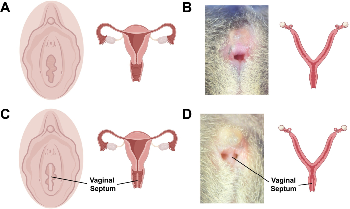
Figure 1. Vaginal opening with longitudinal septum. External anatomy (left) and reproductive tract cross-section (right) of normal, non-septate vagina depicted in (A) humans and (B) mice. External anatomy (left) and reproductive tract cross-section (right) of vagina with longitudinal vaginal septum depicted in (C) humans and (D) mice. Please click here to view a larger version of this figure.
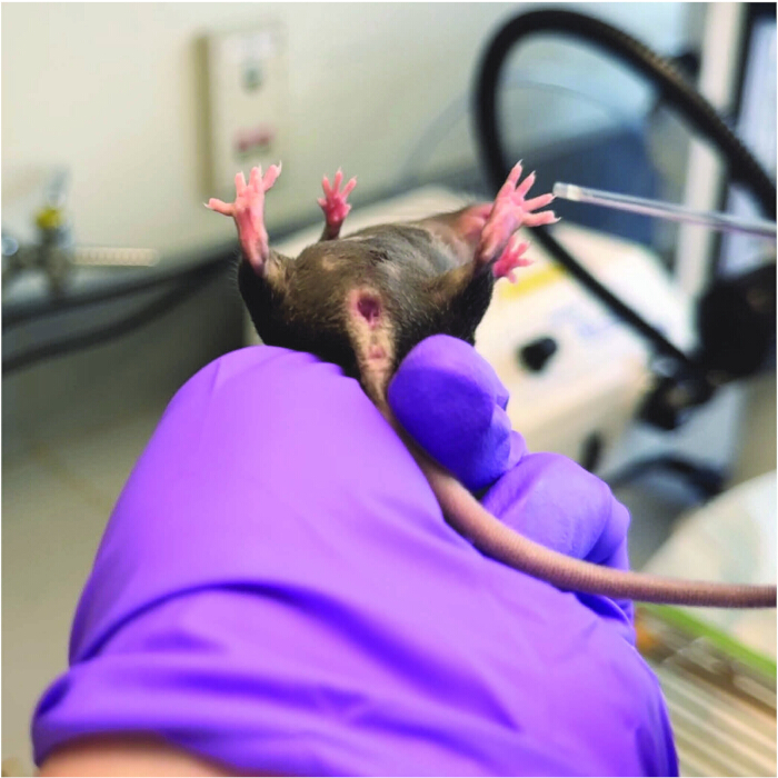
Figure 2. Evaluating mice for vaginal septum. A mouse aged 6–8 weeks is gently restrained by hand, and a glass Pasteur pipette is used to gently apply pressure on the vaginal wall to inspect the vaginal orifice. Please click here to view a larger version of this figure.
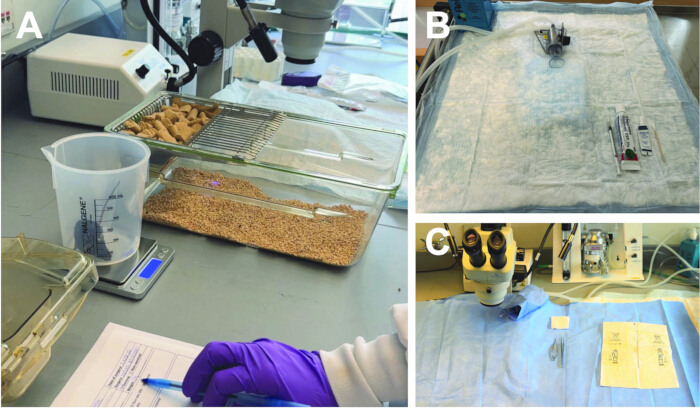
Figure 3. Preoperative procedures and setup. (A) The mouse is weighed on a small scale, and the weight is recorded in a surgery log. (B) Preoperative area setup includes a nose cone breather, meloxicam at the appropriate dose, 5% lidocaine ointment, and lubricant eye ointment. (C) The surgical area setup includes a stereomicroscope, sterile gloves, forceps, microscissors, and gauze. Please click here to view a larger version of this figure.
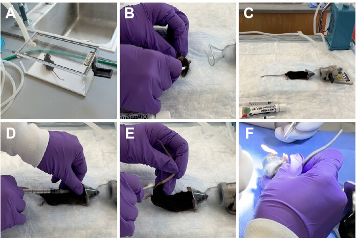
Figure 4. Anesthesia and surgery preparation procedures. (A) The mouse is anesthetized in a small chamber supplied with a continuous flow of oxygen and isoflurane. (B) Lubricant eye ointment is applied to both eyes before transferring the mouse to the nose cone. (C) The anesthetized mouse is placed in the preoperative area in a prone position with anesthetic depth maintained via a nose cone. (D) Subcutaneous injection of meloxicam is administered. (E) A 5% lidocaine ointment is applied to the external vagina. (F) The mouse is transferred back to the surgery area and placed in a prone position within view of the stereomicroscope by a surgical assistant in preparation for surgery. Please click here to view a larger version of this figure.
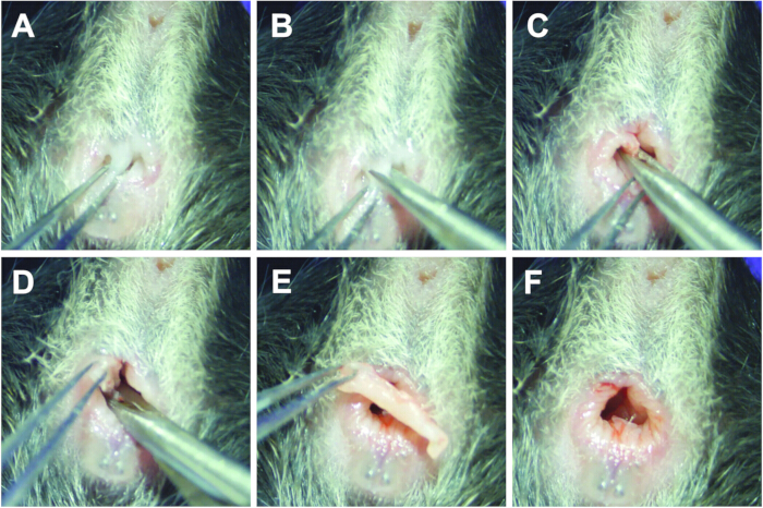
Figure 5. Septum resection surgery. (A) The longitudinal vaginal septum is identified and grasped with forceps. (B) Traction is applied to the septal tissue using forceps while the first incision is made at the dorsal aspect of the septum. (C) Excision of the septum continues along the dorsal plane. (D) The septum is excised along the ventral plane. (E) Vaginal orifice with fully excised septal tissue shown. (F) Vaginal orifice following septum excision. Please click here to view a larger version of this figure.
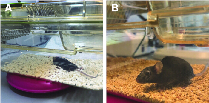
Figure 6. Postoperative recovery. (A) The mouse is placed in a recovery cage on a warm heating pad until it is alert and active, at which point (B) it is returned to its original cage for monitoring. Please click here to view a larger version of this figure.
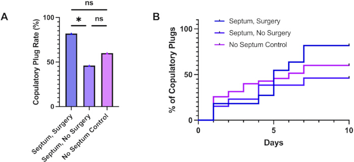
Figure 7. Copulatory plug rates. (A) Plug rates in septum surgery (n = 11), septum no surgery (n = 13), and no septum control (n = 35) mice. (B) Kaplan-Meier curve analysis of the percentage of mice achieving a copulatory plug over time. *p < 0.05. The figure was reproduced with permission from Solomon et al.11. Please click here to view a larger version of this figure.
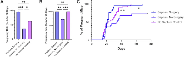
Figure 8. Pregnancy rates. Mice in septum surgery (n = 21), septum no surgery (n = 22), and no septum control (n = 38) were evaluated (A) after 30 days of breeding and (B) at the end of the breeding period, after 75 days of breeding. (C) Kaplan-Meier curve analysis of the percentage of pregnant mice over the breeding period. Data are shown as mean ± standard error of the mean (S.E.M.). *p < 0.05, **p < 0.01, ***p < 0.001. The figure was reproduced with permission from Solomon et al.11. Please click here to view a larger version of this figure.
| Problem | Cause | Solution |
| Reduced visibility of vaginal orifice | Fur surrounding the vaginal opening may make it difficult to visualize the vagina/septum during surgery | Before applying lidocaine ointment, use small animal trimmer to remove excess fur prior to the beginning of the surgery |
| Bleeding during septum resection | Some septal tissue may have more vascular infiltration than others, resulting in bleeding encountered as the septal tissue is cut away | Grasp a sheet of sterile gauze using forceps and apply with gentle pressure to the external vagina. Hold for a few moments, then gently wipe away blood to clear the surgical area |
| Damage to vaginal wall | Although uncommon, it is possible for the microscissors to cut the vaginal wall while removing the septal tissue | Use absorbable 4.0 vicryl suture to repair damage to vaginal wall |
Table 1: Possible procedural challenges and solutions.
Tartışma
Vaginal septum is a congenital anatomical abnormality characterized by a fibrous band of connective tissue that blocks the vaginal canal. It may present in several orientations (transverse, longitudinal, or oblique) and can partially or completely obstruct the vaginal orifice6,10. This anomaly can be found in several species, including humans and mice1,6. Furthermore, several studies have reported a wide range of incidence in several strains of mice. A study performed at Jackson Laboratories found a high incidence of vaginal septum in BALB/c mice (38%), and a lower incidence in C57BL/6J mice (4%)7. In 2004, a study by Gearhart reported an increased rate of vaginal septum in their colony of C57BL/6J mice (11.3%), along with a 1% incidence of vaginal septum in their C57BL/6N colony12. A more recent study described a 14.2% rate of longitudinal vaginal septum in their colony of BALB/cByJNarl mice1. These findings, combined with our own, indicate a wide range of variability in septum rates across several strains of laboratory mice, making the issue of vaginal septum and our findings relevant to the wider research community dealing with the breeding of laboratory mice. The critical elements of the protocol are accurate identification of the septum after vaginal opening, maintenance of a clear surgical field under the stereomicroscope, gentle traction and countertraction during excision, and adequate postoperative analgesia and recovery.
A high incidence of vaginal septum in a mouse colony can be particularly detrimental for laboratory research. Several studies have observed significant detrimental effects on fertility and reproductive outcomes in affected mice used for breeding1,11,12. A study by Chang et al. reported a significant reduction in copulatory plug rates in septate mice (51%) compared to nonseptate mice (72%), as well as a significantly decreased pregnancy rate for septate mice (27%) compared to nonseptate mice (54%)1. Therefore, the aim of this study was to develop a septum resection surgery technique in laboratory mice and determine whether it could optimize fertility and reproductive outcomes in affected mice. Herein, we developed a surgical procedure that would allow these mice to continue to breed effectively while also saving time and laboratory resources and preventing animal waste.
Mice require a thorough examination of the vaginal orifice for the diagnosis of a longitudinal vaginal septum. In our experience, by examination with the aid of a glass Pasteur pipette to apply gentle pressure to the vaginal opening, we found that the septum should be easily visible to the naked eye. However, the sensitivity of external inspection for smaller, thinner, or anatomically variant septa across different mouse strains was not systematically evaluated in this study; therefore, careful inspection after vaginal opening, with gentle pressure applied using a glass Pasteur pipette, is recommended. It is widely accepted that vaginal opening, an apoptosis-mediated event, occurs at ~4–5 weeks of life15,16. Since the vaginal septum cannot be visualized prior to this event, we chose to evaluate our female colony at 6 weeks of age to ensure that the process of vaginal opening had been completed. This timeline allowed us to perform septum surgery at 7 weeks of age and then breed the mice after a week of recovery, coinciding with reaching sexual maturity at 8 weeks. We did not evaluate whether resection at earlier post-vaginal-opening ages or in smaller mice affects technical ease, completeness of resection, or postoperative recovery. Surgery was not scheduled according to the estrous cycle stage. In our observations, septum visibility did not appear to vary by estrous cycle stage, although this was not formally tested as part of the present protocol.
The septum resection surgery, modeled closely after the procedure in women, is both brief and relatively uncomplicated. A basic understanding of how to handle mice and provide anesthesia is required. Experience performing mouse surgery is necessary, although this procedure does not require advanced surgical skills. Proper pain management is also required for the septum resection surgery. Application of meloxicam and lidocaine ointment prior to the first incision, and a second meloxicam injection 24 h after the operation, is necessary to minimize postoperative discomfort. In our hands, this procedure was not associated with any intraoperative complications and achieved a 100% survival rate.
There are several critical steps in performing a successful septum resection. The first step is to ensure a clear, unobstructed view of the septum within the stereomicroscope's field of view. This can be achieved with the aid of a surgical assistant who holds the mouse and maintains its position throughout the procedure (Figure 4F). When developing this technique, we did not identify any other methods that were as successful at holding the mouse as securely as the surgical assistant's hand. Another important point is the use of traction and countertraction during septum resection. For this, properly supporting the mouse's body with an assistant allows the surgeon to use forceps to easily manipulate the septal tissue and create tension to facilitate cutting along the tissue's natural planes. This technique fosters a clear surgical field, provides better access to the target tissue, and directs precise incisions to allow for complete resection of the vaginal septum.
In our study, female virgin septate mice were divided into two categories: septum surgery and septum, no surgery. Following the brief procedure and a 7 day recovery period, both groups were bred to proven male breeders. Another group of non-septate mice served as controls. Successful mating activity was assessed by the presence of copulatory plugs, revealing a significant increase in copulatory plug rates for mice that underwent resection surgery as compared to septate mice that did not receive surgery11. Moreover, mice with septum resection had a shorter time to pregnancy and a significantly higher pregnancy rate compared with septate mice without surgery, normalizing their reproductive outcomes to the level of the non-septate control mice11. These findings indicate that mouse breeding is adversely affected by a longitudinal vaginal septum and that septum resection can optimize reproductive capacity and pregnancy outcomes in affected mice. Interestingly, the pregnancy rate in the septum surgery group was significantly higher than that of the no septum control group over the initial 30 days of breeding (Figure 8A). A likely explanation for this initial increased pregnancy rate can be an increased drive toward copulatory activity in the septum surgery group following the removal of the physical barrier—the vaginal septum. Indeed, this is supported by our copulatory plug data, which showed a trend towards an increase in the copulatory plug rate in septum-surgery mice compared to the no-septum control groups. Importantly, this difference in pregnancy rates appears to be a transient finding observed within the first month of breeding, since our results show comparable values for pregnancy rates between the septum surgery and the no septum control group at the end of the 75 day breeding trial (Figure 8B).
While there is a significant detriment to breeding caused by vaginal septum in mice, data from our breeding trial revealed that a subset of females with longitudinal vaginal septum are still capable of copulatory activity (Figure 7) as well as achieving pregnancy (Figure 8). According to our observations and previous literature, this is likely due to rupture of the septum during copulation, which effectively removes the physical barrier that can impede copulation. In a study by Gearhart, it was found that 50% of the mice previously identified to have a longitudinal vaginal septum had ruptured their septum while checking for copulatory plugs12. At the end of the breeding trial in this study, Gearhart reports that 86% of females that had ruptured their septum during breeding had successful pregnancies, whereas only 14% of females with an intact septum had a successful pregnancy12. Thus, in mice, the functional impact of a longitudinal vaginal septum may be variable and depend in part on whether the septum remains intact or ruptures during copulatory activity. Direct comparison of infertility severity across mice, humans, and other species is limited by differences in anatomy, mating physiology, diagnosis, and clinical presentation.
Prior studies demonstrate that mice with a longitudinal vaginal septum have extended breeding times compared with non-septate littermates1. As prolonged breeding periods can delay project timelines and waste financial resources allocated to housing and maintaining the breeding mice, several authors have suggested removing septate females from the breeding colony to prevent further propagation of congenital anomaly1. While this may be an effective technique, it may not be feasible to remove septate females with a desired phenotype or genetic lineage, especially if the incidence of septa in this cohort is high. Additionally, this solution may result in a significant amount of animal waste, which conflicts with the “reduction” tenet of the 3Rs principles for animal research12,14. Given these considerations, performing septum resection surgery would allow affected females to breed effectively and efficiently with male mice, achieving experimental goals without detrimental effects on time or finances.
Another proposed solution to avoid the detrimental effects associated with colonies affected by a longitudinal vaginal septum is to use structured breeding schemes to eliminate the trait from the gene pool1,12,13. While this solution would allow for the continued propagation of the colony and the elimination of the problematic condition, there is, unfortunately, little information on the genetic nature of the longitudinal vaginal septum to target specific genes. Chang et al. used a strict brother-sister mating scheme to reduce trait frequency, but their findings indicate a polygenic recessive inheritance pattern that is susceptible to genetic drift among breeding sublines1. Investigations into the genetics of vaginal septum formation suggest defects in the vaginal apoptotic program or in metalloproteinases that mediate extracellular matrix (ECM) remodeling in the developing vagina15,16,17,18,19,20,21. However, without specific knowledge of the underlying genetic basis of longitudinal vaginal septum, utilizing structured breeding to eliminate this phenotype from a particular colony has been ineffective. In conclusion, septum resection surgery is a technique that can eliminate the time and financial burdens associated with longitudinal vaginal septa in mice while also reducing animal waste, in accordance with the 3Rs principle. It is a brief surgical procedure with minimal risks that can be easily performed by researchers in the lab without advanced surgical skills.
Açıklamalar
The authors have no conflicts of interest to declare.
Teşekkürler
This study was supported by funding from NIH/NICHD grant 1R01HD109326 (RT).
Malzemeler
| Ad | Şirket | Katalog numarası | Yorumlar |
|---|---|---|---|
| 2" x 2" Gauze | Medicom | 4522-5 | |
| 5% Lidocaine Ointment | TARO | 51672-3020 | |
| Absorbable 4-0 vicryl suture | ETHICON | VCP315H | Only necessary if vaginal wall repair is needed. |
| ACCU-SCOPE Stereo Microscope | ACCU-SCOPE | 3075-LED | |
| BD 1 mL Syringe, Tuberculin Slip Tip | Becton, Dickinson and Company | 309659 | |
| BD PrecisionGlide Needle, 27 G x 1/2" | Becton, Dickinson and Company | 305109 | |
| Bench Cover Underpad Tendersorb 23" x 24" | Kendall Healthcare | 7134 | |
| Cotton-Tipped Applicators | McKesson | 24-103 | |
| Dumont #5 Fine Forceps | Fine Science Tools | 11254-20 | |
| Excelis HD Camera | ACCU-SCOPE | AU-600-HD | |
| E-Z Single Animal Anesthesia System | E-Z Systems | EZ-SA800 | |
| Fiber-Lite MI-LED Illuminator | Dolan-Jenner Industries | NC0756326 | |
| Heating Pad | SnuggleSafe | 5650-6250 | |
| MAXUS Digitial Gram Scale | MAXUS | D126 | For weighing mice |
| Medline Half Drape Demi-champ | Medline | DYNJP2411 | |
| Meloxicam (5 mg/mL) | Covetrus | 68110 | |
| Nalgene PMP Griffin Low-Form Plastic Beakers | Nalgene | 1201-0600 | For weighing mice |
| Novaplus Gammex Non-Latex PI Micro Surgical Gloves | novaplus | V20685970 | |
| Purple Nitrile Powder-Free Exam Gloves | McKesson General Medical | 55082 | |
| Refresh P.M. Lubricant Eye Ointment | REFRESH | 351842 | |
| Vannas Spring Scissors - 3MM cutting edge | Fine Science Tools | 15000-00 | |
| Wahl Professional Animal Pocket Pro Trimmer, Purple | Wahl | 9861-930 |
Kaynaklar
- Chang TK, Ho P, Liang CT, Yu CK. Effects of vaginal septa on the reproductive performance of BALB/cByJNarl mice. J Am Assoc Lab Anim Sci. 2013;52(5):520-3.
- Hernandez SP, Ponthier J, Deleuze S. A case of longitudinal vaginal septum in a mare. Reprod Domest Anim. 2013;48:112-3.
- Lezmi S, et al. Spontaneous metritis related to the presence of vaginal septum in pregnant Sprague Dawley Crl:CD(SD) rats: impact on reproductive toxicity studies. Vet Pathol. 2011;48(5):964-9.
- Nomura K, Koreeda T, Kawata M, Shiraishi Y. Vaginal atresia with transverse septum in a cat. J Vet Med Sci. 1997;59(11):1045-8.
- Root MV, Johnston SD, Johnston GR. Vaginal septa in dogs: 15 cases (1983-1992). J Am Vet Med Assoc. 1995;206(1):56-8.
- Ludwin A, Lindheim SR, Bhagavath B, Martins WP, et al. Longitudinal vaginal septum: a proposed classification and surgical management. Fertil Steril. 2020;114(4):899-901.
- Cunliffe-Beamer TL, Feldman DB. Vaginal septa in mice: incidence, inheritance, and effect on reproductive performance. Lab Anim Sci. 1976;26(6 Pt 1):895-8.
- El Abbassi I, Bensouda ME, Jalal M, Lamrissi A, et al. Surgical management of a transverse vaginal septum: about a rare case. Int J Surg Case Rep. 2023;110:108682.
- Akcaoglu T, Atilgan AE. Surgical management of longitudinal vaginal septum: a case report and literature review. Eur J Obstet Gynecol Reprod Biol. 2025;310:113992.
- Wu YC, Li PC, Ding DC. Vaginal septum in women: a review of diagnosis, management, and obstetric outcomes. Tzu Chi Med J. 2026;38(1):40-7.
- Solomon AE, et al. Surgical resection of longitudinal vaginal septum improves fertility and reproductive capacity in mice. Reprod Fertil. 2026;7(2):RAF260035.
- Gearhart S, Kalishman J, Melikyan H, Mason C, et al. Increased incidence of vaginal septum in C57BL/6J mice since 1976. Comp Med. 2004;54(4):418-21.
- Shire JG. Studies on the inheritance of vaginal septa in mice, a trait with low penetrance. J Reprod Fertil. 1984;70(1):333-9.
- Russell WMS, Burch RL. The principles of humane experimental technique. London: Methuen; 1959. 238 p.
- Lin X, Wang C, Zhang Q, Pan YH, et al. ADAMTS18 regulates vaginal opening through influencing the fusion of Mullerian duct and apoptosis of vaginal epithelial cells in mice. Reprod Biol. 2021;21(3):100537.
- Rodriguez I, Araki K, Khatib K, Martinou JC, et al. Mouse vaginal opening is an apoptosis-dependent process which can be prevented by the overexpression of Bcl2. Dev Biol. 1997;184(1):115-21.
- Ito T, et al. Estrogen-dependent proteolytic cleavage of semaphorin 4D and plexin-B1 enhances semaphorin 4D-induced apoptosis during postnatal vaginal remodeling in pubescent mice. PLoS One. 2014;9(5):e97909.
- Ito T, et al. Semaphorin 4D induces vaginal epithelial cell apoptosis to control mouse postnatal vaginal tissue remodeling. Mol Med Rep. 2015;11(2):829-36.
- Ren D, et al. BID, BIM, and PUMA are essential for activation of the BAX- and BAK-dependent cell death program. Science. 2010;330(6009):1390-3.
- Lindsten T, et al. The combined functions of proapoptotic Bcl-2 family members Bak and Bax are essential for normal development of multiple tissues. Mol Cell. 2000;6(6):1389-99.
- Ataca D, et al. Adamts18 deletion results in distinct developmental defects and provides a model for congenital disorders of lens, lung, and female reproductive tract development. Biol Open. 2016;5(11):1585-94.
Yeniden basım ve izinler
Bu makale yayımlandı
Video yakında