To describe a stepwise strategy combining urgent PTCD and peroral cholangioscopic laser lithotripsy for safe, effective clearance of impacted CBD stones in super-elderly patients with prior Billroth II gastrectomy, avoiding open surgery.
Yöntem makalesi
PTCD and Peroral Electronic Cholangioscopy with Holmium Laser Lithotripsy for CBD Stones in a Super-Elderly Patient Post-Billroth II Gastrojejunostomy
* These authors contributed equally
Bu makalede
Özet
Özet
The management of impacted common bile duct stones in patients with prior Billroth II gastrectomy poses unique challenges. Altered gastrointestinal anatomy renders duodenoscope passage and biliary cannulation during conventional ERCP exceedingly difficult, resulting in a low success rate. Super‑elderly (over 90 years old) patients often have multiple comorbidities such as cardiovascular diseases and require rapid relief of biliary obstruction and infection control to create a subsequent therapeutic window. Meanwhile, traditional open biliary surgery and general anesthesia carry prohibitive risk, along with prolonged preoperative preparation and extensive trauma. Although percutaneous transhepatic choledochoscopic lithotripsy (PTCSL) is unaffected by gastrointestinal reconstruction, it often depends on a large mature biliary sinus tract, necessitating a multi‑stage procedure, entails a risk of hemorrhage, and has difficulty in managing stones, especially papillary impacted stones. To overcome these difficulties, an innovative stepwise strategy was devised comprising “urgent PTCD for rapid decompression followed by multimodal lithotripsy under direct visualization with a peroral electronic cholangioscope.” A 92‑year‑old male had a history of multiple surgeries, including Billroth II gastrectomy, cholecystectomy, and colon resection, along with coronary heart disease and atrial fibrillation. Emergency PTCD was performed first, promptly controlling infection and relieving jaundice. After preoperative preparation, the tract was dilated to 10 Fr, and a peroral electronic cholangioscope was introduced through the sheath. Under direct visualization, the common bile duct stones were efficiently fragmented using a holmium laser, initially in a fragmentation mode and then in a dusting mode. Post‑procedural cholangiography confirmed complete stone clearance. The PTCD tube was subsequently removed uneventfully, and the patient recovered well without complications. This strategy successfully circumvents the anatomical alterations, integrates biliary drainage, direct intra‑ductal visualization, and lithotripsy, providing a minimally invasive treatment strategy for patients with bile duct stones after gastrointestinal reconstruction, especially for super‑elderly and other high‑risk individuals.
Giriş
Patients with prior gastrointestinal reconstruction who develop impacted common bile duct stones pose a uniquely challenging management problem. The surgically altered upper gastrointestinal anatomy disrupts the conventional endoscopic route: the afferent limb is often lengthy and acutely angulated, and the native papilla must be approached from a reversed direction. Consequently, during conventional endoscopic retrograde cholangiopancreatography (ERCP), duodenoscope passage and selective biliary cannulation become exceedingly challenging, with success rates heavily dependent on the expertise of the primary operator1. Moreover, total gastrectomy, gastrojejunostomy, and the use of forward-viewing endoscopes are independent risk factors for cannulation failure2. In elderly patients with multiple comorbidities, rapid relief of biliary obstruction and effective control of infection are urgent priorities for creating a window for subsequent intervention.
Percutaneous transhepatic choledochoscopic lithotripsy (PTCSL) is, in theory, unaffected by gastrointestinal reconstruction3. However, PTCSL requires additional time for tract dilation and maturation, and may necessitate repeated procedures4. Furthermore, even after tract maturation, dilation to 16–18 Fr is still required to accommodate the choledochoscope; the larger the tract, the greater the trauma and the associated risk. To overcome these dual barriers—the anatomical challenges of ERCP and the protracted timeline of conventional PTCSL—an innovative stepwise strategy was therefore developed in which emergency percutaneous transhepatic cholangiography and drainage (PTCD) is performed first to achieve immediate decompression; subsequently, a peroral electronic cholangioscope is introduced through a PTCD sheath after single-stage dilation to only 10 Fr, enabling multimodal laser lithotripsy under direct visualization.
This paper aims to describe in detail the procedural steps of emergency PTCD combined with peroral electronic cholangioscope-assisted laser lithotripsy, and to demonstrate its successful application in a 92-year-old male patient with impacted common bile duct stones following Billroth II gastrectomy.
Protokol
The procedure was performed in accordance with institutional ethical guidelines. Following review by the Medical Ethics Committee of Guangzhou Red Cross Hospital of Jinan University (Approval No. AF/SC-10/1.2; approval date: June 26, 2026), the project was determined to meet the criteria for exemption from institutional review board approval. Written informed consent was obtained from the patient for publication.
Refer to the Table of Materials for details of all materials and equipment used in this protocol.
1. Patient selection and preparation
- Select elderly patients with a history of Billroth II gastrectomy who present with impacted common bile duct stones and acute cholangitis and in whom conventional ERCP has failed.
- Exclude patients with coagulopathy, massive ascites, absence of a safe percutaneous access window, or eligibility for conventional open surgery.
- Obtain a pre-procedural contrast-enhanced CT scan to plan the percutaneous access route. Inform the patient and family of the associated risks and obtain written informed consent.
2. Emergency PTCD
- Under fluoroscopic guidance, puncture a peripheral intrahepatic bile duct with a 22 G needle. Introduce a 0.018-inch guidewire, dilate the transhepatic tract to 8 Fr, and place an 8 Fr pigtail drainage catheter in the common bile duct.
- Maintain external drainage and administer concurrent antibiotic therapy until cholangitis is controlled. Complete the remaining preoperative workup, such as transthoracic echocardiography.
3. Working sheath establishment
- Insert a 0.018-inch guidewire through the indwelling PTCD catheter. Remove the drainage catheter over the guidewire, serially dilate the tract to 10 Fr, and advance a 10 Fr peel-away sheath over the guidewire into the common bile duct. Confirm the sheath position with contrast injection.
4. Peroral cholangioscopy and holmium laser lithotripsy
- Connect the peroral electronic cholangioscope system. Introduce the cholangioscope through the 10 Fr sheath and advance it to the site of stone impaction under direct visualization. Maintain continuous saline irrigation to ensure a clear visual field.
- Insert a 200 µm holmium laser fiber through the working channel, allowing the fiber tip to protrude 2–3 mm beyond the scope.
- Apply laser energy at 3.0 J and 10 Hz (30 W, narrow pulse width), with the fiber tip in direct contact with the stone, for fragmentation.
- Switch to 0.8 J and 30 Hz (24 W, wide pulse width) to pulverize the residual fragments into fine powder. Use irrigation flow to flush out the stone dust, and retrieve larger fragments with a stone extraction basket if necessary.
- Inspect the entire biliary tree thoroughly by cholangioscopy to confirm complete stone clearance.
- Obtain a completion cholangiogram through the sheath. If no filling defects are observed, exchange the peel-away sheath for a 10 Fr temporary drainage catheter.
5. Post-procedural assessment and catheter removal
- Maintain gravity drainage for 2–3 days, and then repeat the cholangiogram. If the findings are normal, clamp the catheter for 24 h.
- Monitor serial bilirubin and other laboratory parameters at regular intervals. Once the laboratory values have normalized, discharge the patient with the clamped catheter in place.
- At approximately 1 month after discharge, readmit the patient and repeat the cholangiogram. Remove the PTCD catheter once unimpeded contrast flow into the duodenum is confirmed.
Sonuçlar
A 92-year-old male with a history of Billroth II gastrectomy, atrial fibrillation, and bladder stones presented with acute exacerbation of abdominal pain. Contrast-enhanced computed tomography revealed a distal common bile duct stone with upstream biliary dilatation, consistent with acute cholangitis (Figure 1A). After multidisciplinary assessment, conventional ERCP was deemed infeasible, and the patient was enrolled in a staged treatment protocol. On hospital day 4, emergency PTCD was performed: an 8 Fr pigtail drainage catheter was placed for external drainage, and broad-spectrum antibiotics were initiated concurrently. During the interval required for cholangitis control, the preoperative workup, including transthoracic echocardiography, was completed. Once the patient was stabilized, a cholangiogram obtained through the PTCD catheter confirmed a filling defect measuring approximately 10 × 5 mm in the mid common bile duct (Figure 1B). The cholangiogram also revealed partial retrograde filling of the main pancreatic duct. Contrast injection was performed gently under fluoroscopic guidance, and the contrast volume was minimized to confirm successful biliary puncture; the pancreatic duct opacification was therefore considered transient. Although contrast entry into the pancreatic duct may carry a risk of post-procedural pancreatitis, the patient was closely monitored after the procedure, remained free of clinical pancreatitis, and postoperative serum amylase levels remained normal. The transhepatic tract was dilated to 10 Fr, and a 10 Fr peel-away sheath was advanced into the common bile duct. Under direct visualization with a peroral electronic cholangioscope, a yellowish-brown stone measuring approximately 8 × 8 mm was observed in the mid common bile duct, and a dark-green stone measuring approximately 3 × 3 mm was identified at the orifice of the distal common bile duct (Figure 2A,C). Holmium laser lithotripsy was applied until no large stone fragments remained in the bile duct (Figure 2B,D), after which the procedure was concluded. During fragmentation, a stone fragment lodged in the peripapillary recess required repeated adjustments of the laser fiber angle and energy settings before complete pulverization. A 10 Fr external drainage catheter was left in place. The patient was monitored in the intensive care unit for 1 day before being transferred to the general ward. On postoperative day 10, a repeat cholangiogram demonstrated free passage of contrast into the duodenum without filling defects (Figure 3A); the drainage catheter was subsequently clamped. After serial bilirubin measurements returned to normal, the patient was discharged on postoperative day 12 with the clamped catheter in situ. One month later, the patient was readmitted for a follow-up abdominal CT scan (Figure 3B), which also showed no residual stones; the drainage catheter was removed uneventfully. At the outpatient follow-up visit on July 8, 2026, the patient remained asymptomatic with normal liver biochemistry.
Success was evaluated according to technical, clinical, and durable criteria. Technical success required complete stone clearance confirmed by both cholangiography and direct cholangioscopic inspection. Clinical success was reflected in resolution of cholangitis, normalization of liver biochemistry, and absence of procedure-related complications. Durable success was defined as the absence of recurrent stones on follow-up imaging. In this case, all three criteria were met. Nevertheless, suboptimal or unsuccessful outcomes remain possible and should be anticipated. A stone that is too hard or large to be fragmented by holmium laser alone may require additional lithotripsy modalities or, rarely, conversion to open surgery. Other potential failure modes include sheath dislodgement, significant bleeding from the transhepatic route, or incomplete stone clearance necessitating a repeat procedure. Awareness of these possibilities is essential for appropriate patient selection and intraoperative decision-making.
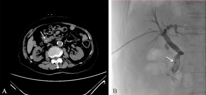
Figure 1: Preoperative imaging. (A) Upper abdominal CT scan revealing a common bile duct stone (arrow). (B) PTCD cholangiography demonstrating multiple stones in the common bile duct (arrow). Partial retrograde filling of the main pancreatic duct is also visible. Please click here to view a larger version of this figure.
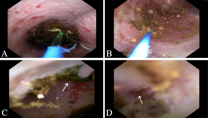
Figure 2: Intraoperative findings. (A) A stone is visualized in the mid common bile duct. (B) The mid-duct stone after pulverization into fine powder using laser lithotripsy. (C) An additional stone (arrow) is observed in the distal common bile duct near the ampullary orifice; notably, this stone was not detected on preoperative CT. (D) Complete stone clearance with a widely patent common bile duct orifice (arrow). Please click here to view a larger version of this figure.
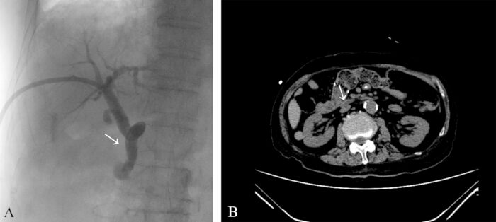
Figure 3: Postoperative recovery. (A) PTCD cholangiography showing a patent common bile duct without residual stones. (B) Upper abdominal CT scan confirming the absence of the previously identified stone (arrow indicates the original stone site). Please click here to view a larger version of this figure.
Tartışma
The management of impacted common bile duct stones in patients with surgically altered gastrointestinal anatomy, particularly those with prior Billroth II gastrectomy, remains a formidable clinical challenge. Because the papilla must be approached from a reversed direction, and in procedures such as Roux-en-Y reconstruction the afferent limb is markedly elongated, conventional ERCP frequently encounters substantial difficulty in this population5. In super-elderly patients presenting with acute cholangitis and multiple comorbidities, the treatment strategy must simultaneously address the urgent need for biliary decompression and avoid the substantial risks associated with open surgery. In the present approach, emergency PTCD is performed first to achieve rapid infection control, followed by peroral electronic cholangioscope-guided laser lithotripsy through a small-caliber sheath, thereby addressing both challenges in an integrated manner.
Several critical steps in the protocol warrant emphasis. The initial emergency PTCD is not merely a preparatory maneuver; it is a life-saving intervention that promptly controls cholangitis and creates a therapeutic window for subsequent definitive therapy. Chen et al.6 reported that, in patients with severe inflammation and acute cholangitis, PTCD is recommended before definitive stone extraction to achieve rapid infection control. After the acute infection has subsided, the establishment of a working sheath represents the pivotal technical transition. In this protocol, the transhepatic tract needs to be dilated to only 10 Fr, a striking contrast to the 16–18 Fr mature sinus tract required for conventional percutaneous transhepatic choledochoscopic lithotripsy4,7. By eliminating the weeks-long waiting period for tract maturation, this protocol shortens the overall treatment timeline and spares frail elderly patients from repeated interventions.
The significance of this method relative to existing alternatives is underscored by the well-recognized limitations of conventional PTCS. Although PTCS remains effective for complex biliary stones, it is inherently more invasive than retrograde endoscopic techniques, carries a higher complication rate, and entails a protracted course that compromises quality of life4,8. In patients with surgically altered anatomy, the SpyGlass direct visualization system has been demonstrated to offer clinical efficacy comparable to that of PTCS, while promoting faster return to baseline physiological status and superior short-term recovery9. Percutaneous stone extraction confers additional advantages: it facilitates identification of stone distribution, thereby enabling manipulation via the most accessible duct, which helps improve procedural success, reduce operative time and the need for multiple sessions, and shorten hospital stay10. Moreover, Dalal et al.11 have confirmed that SpyGlass cholangioscopy combined with percutaneous transhepatic biliary drainage (PTBD) is safe and effective for large bile duct stones, with good short-term outcomes and minimal complications. For the specific subgroup of super-elderly post-gastrectomy patients in whom ERCP is technically infeasible, PTCD represents the most commonly adopted initial intervention; yet definitive stone clearance has traditionally required either staged open surgery or a prolonged PTCS course5. The protocol presented here addresses this gap: it converts the PTCD tract into an immediate therapeutic conduit, thereby consolidating drainage and stone extraction into a single minimally invasive pathway. This integration is particularly critical for elderly patients with cardiovascular comorbidities, for whom any additional general anesthetic or surgical manipulation may further increase procedural risk.
Early intervention confers several advantages compared with conventional PTCSL after tract maturation: the present approach shortens hospital stay, decreases the number of procedures, and reduces medical costs; furthermore, it reduces the frequency of repeated tract dilation, thereby lowering the associated bleeding risk; and in high-risk elderly patients, it mitigates the risk of cardiovascular or cerebrovascular events and general deterioration during the waiting period. These benefits, however, must be balanced against the associated risks: since the tract is not fully mature, gentle manipulation is required during the procedure; otherwise, bleeding, bile leakage, and hepatic capsular tear may still occur; if infection remains incompletely controlled, the procedure may induce bacteremia or exacerbate sepsis; and incomplete stone clearance may necessitate a second procedure. Accordingly, early intervention should be predicated on infection control, correction of coagulopathy, multidisciplinary assessment, and adequate operator experience.
Several limitations of this method must be acknowledged. First, the protocol demands proficiency in both percutaneous biliary intervention and peroral cholangioscopy, and is best performed in centers equipped with a multidisciplinary team. The present experience is limited to a single case; therefore, definitive conclusions regarding safety and generalizability cannot be drawn. Patient selection is equally crucial, because the technique is currently reserved for patients who lack a safer alternative—ERCP-based stone extraction remains the preferred approach for those who are candidates for it. Furthermore, while the 10 Fr small-caliber working channel reduces invasiveness, it limits the size of retrievable fragments and obliges the operator to pulverize stones to a sufficiently small caliber, potentially prolonging procedural time; for stones that are too hard or large to fragment with holmium laser alone, electrohydraulic or mechanical lithotripsy may be required, although in the present case adequate pulverization was achieved by adjusting the laser fiber angle and energy settings. Bleeding from the transhepatic tract can usually be managed by prolonged balloon tamponade or coil embolization, and significant hemobilia may warrant temporary drainage and close monitoring. If sheath dislodgement occurs, re-advance the guidewire and re-establish the tract. Incomplete stone clearance should prompt second-look cholangioscopy or a staged procedure rather than forced extraction. These troubleshooting measures are essential to ensure safety and success in this fragile population.
Future refinements of this protocol may further enhance its safety and efficacy. For instance, the development of smaller-caliber cholangioscopes could allow further reduction in sheath size, thereby minimizing hepatic trauma. Intelligent laser systems that automatically adjust energy, frequency, and pulse width based on real-time stone composition analysis could improve fragmentation efficiency and reduce ductal injury. In addition, shortening the interval between PTCD and definitive lithotripsy may become feasible with improved infection control strategies, potentially allowing single-stage procedures in selected patients.
In conclusion, this strategy may provide a minimally invasive option for managing complex biliary stones in selected patients with surgically altered gastrointestinal anatomy. Emergency PTCD combined with peroral electronic cholangioscope-assisted laser lithotripsy enabled definitive management of impacted bile duct stones in this super-elderly, high-risk patient for whom conventional ERCP was technically infeasible. Future prospective studies are needed to refine the technical details, assess generalizability, and compare the long-term efficacy and safety of this strategy with staged PTCSL.
Açıklamalar
The authors have nothing to disclose.
Teşekkürler
Guangdong Province Health Services Appropriate Technology Promotion Project, Project Number: 202303182146326369; Guangdong Province Natural Science Foundation General Project, Project Number: 2020A1515010799; Guangzhou Science and Technology Plan–Joint Funding Project by University-Enterprise-Research Institute, Project Number: 2024A03J0670.
Malzemeler
| Ad | Şirket | Katalog numarası | Yorumlar |
|---|---|---|---|
| Drainage catheter and accessories | ARGON MEDICAL DEVICESINC. | 651506300 | |
| Drainage catheter and accessories | ARGON MEDICAL DEVICESINC. | 755308040 | |
| Catheter Sheath Sets | Zhejiang Barty Medical Technology Co.Ltd | QB100450A | |
| One-time use imaging catheter | Micro-Tech (Nanjing) Co., Ltd. | CDS22001 | |
| Pancreatic and Biliary System Imaging Controller | Micro-Tech (Nanjing) Co., Ltd. | BS-W-100 | |
| EMERALD Diagnostic Guidewire | Cordis Us Corp. | 502-731 | |
| Portable holmium laser | Guangzhou Potent Medical Science and Techology Co., Ltd. | HZ-40 | |
| Medical laser fiber | Guangzhou Potent Medical Science and Techology Co., Ltd. | 26072901 | 200μm |
Kaynaklar
- Fortunati F, et al. Endoscopic retrograde cholangiopancreatography in patients with different types of total and partial gastrectomy. Acta Gastroenterol Belg. 2025;88(1):23-30.
- Cheng Y, et al. Endoscopic retrograde cholangiopancreatography consultation after digestive tract reconstruction and risk factors for complications. Eur J Med Res. 2025;30(1):135.
- Li X, Ouyang J, Dai J. Current gallstone treatment methods, state of the art. Diseases. 2024;12(9):197.
- Ryu JK. Percutaneous approach for removal of difficult common bile duct stones. Clin Endosc. 2013;46(1):3-4.
- Wang D, et al. Diagnostic investigation, intervention, and outcome for post-subtotal gastrectomy patients who present with jaundice. Front Med (Lausanne). 2025;12:1485442.
- Chen L, et al. Evaluation of clinical indications of three treatments for choledocholithiasis with acute cholangitis. Int J Gen Med. 2023;16:4669-4680.
- Lee YS. Could the "SpyGlass Direct Visualization" system open new horizons for treating biliary tract diseases as a percutaneous cholangioscopy? Gut Liver. 2022;16(1):1-2.
- Wang S, Wu S. Percutaneous transhepatic choledochoscopy in the management of hepatolithiasis: a narrative review. Quant Imaging Med Surg. 2024;14(7):5164-5175.
- Shin KI, et al. SpyGlass Direct Visualization System: a cost-effective approach enhancing nutritional and immune recovery in difficult bile duct stone management. Yonsei Med J. 2026;67(6):466-476.
- Alabraba E, Travis S, Beckingham I. Percutaneous transhepatic cholangioscopy and lithotripsy in treating difficult biliary ductal stones: two case reports. World J Gastrointest Endosc. 2019;11:298-307.
- Dalal A, et al. Utility of cholangioscopy in patients with surgically altered anatomy after percutaneous transhepatic biliary drainage. Endosc Int Open. 2025;13:a24872890.
Yeniden basım ve izinler
Bu makale yayımlandı
Video yakında