We present an operative protocol with technical nuances of the TF approach utilized to perfor both a far-lateral and paracentral discectomy at L5-S1 to treat a 50-year-old male with two distinct and noncontiguous herniations with severe acute right-sided L and S1 radiculopathy.
A subscription to JoVE is required to view this content. Sign in or start your free trial.
Method Article
Transforaminal Endoscopic Approach for Combined Far Lateral and Paracentral Discectomies at L5-S1 Level
In This Article
Summary
Abstract
The transforaminal (TF) and interlaminar (IL) approaches compose the two endoscopic approaches to lumbar discectomy. There is a growing tendency to utilize the TF route for higher lumbar disc herniations and the IL approach related to anatomical factors for the caudal lumbar levels. The L5-S1 level produces a particular challenge due to anatomical barriers, including the height of the iliac crest and the orientation of the facet when confronted with a lateral or far-lateral disc herniation. Traditional approaches can lead to significant joint destabilization in the effort to resect the disc fragment
Here we present an operative video with technical nuances of the TF approach utilized to perform both a far-lateral and paracentral discectomy at L5-S1 to treat a 52-year-old male with two distinct and noncontiguous herniations with severe acute right-sided L5 and S1 radiculopathy. This would have otherwise required either two different endoscopic approaches or a more extensive open approach for successful discectomy
Introduction
Endoscopic spine surgery has evolved rapidly as an ultra-minimally invasive spine surgery solution over the last few decades. The work of Kambin describing a safe lateral approach to lumbar discectomy through Kambin's triangle1, allowed its wide utility in the transforaminal approach till today2. The posterolateral approach has been the main workhorse for spine endoscopic surgery till the beginning of this century, when the adoption of the interlaminar approach surged and expanded widely3,4. Nevertheless, the transforaminal approach remains a versatile approach with extended retraction-free access to the far lateral, foraminal, lateral recess, and ventral epidural compartments.
The interlaminar approach has been increasingly adopted as spine surgeons are usually comfortable with the related anatomy, as it is the natural corridor used in most open and minimally invasive surgeries. Similarly, the option of extending the same incision for conversion to open or tubular surgery to tackle technical difficulties is quite comforting to most early adopters. Although the transforaminal approach to learning might be initially demanding related to its unique anatomy and targeting principles, the flexibility and the advantages of using it are significant.
The interlaminar approach has been associated with good outcomes3. As a direct posterior access, it allows an unobstructed exposure of the posterior epidural compartment, allowing direct decompression of posterior pathologies, including thickened ligamentum flavus, facet hypertrophy, and sequestrated lateral posteriorly exophytic disc fragments. Ventral and ventrolateral epidural pathologies, including broad-based disc herniations, osteophytes, and sequestrated ventral fragments, would pose a challenge at times, and often significant retraction of the thecal and nerve root is unavoidable. Depending on how medial the pathology is and the ability to mobilize it, one could infer the corresponding retraction degree.
The interlaminar approach has been widely suggested for accessing the lower lumbar levels related to the corresponding wide interlaminar windows4,5,6,7. In addition, accessing the lumbosacral junction via the transforaminal route could well be hindered by a high iliac crest, steep angle of the access, and the smaller size of the foramen8. As higher lumbar levels and thoracic spine are contemplated, we start encountering the conus medullaris and spinal cord, which are structures sensitive to excessive retraction. Similarly, the corresponding intervertebral foramina are larger rostrally in the lumbar spine and more favorable. Hence, the transforaminal approach is generally more widely used at those levels.
Other factors that could help in decision-making between the two common endoscopic approaches are the location of the herniation in relation to the spinal canal, as extraforaminal "aka far lateral disc herniations" would require a significant bony resection of the facet, which would likely destabilize it when performed from a direct posterior approach. This could otherwise be treated with a posterolateral approach with no or very minimal bony resection, if needed, and hence no destabilizing effect.
The transforaminal approach is extremely helpful to access the extraforaminal compartment, the foramen, and the lateral recess. Traditionally, the initial access to the foramen during targeting is aimed at the inferior foraminal compartment near the junction of the superior endplate and upper pedicular margin of the vertebra below. It involves sequential dilation along with progressive reaming of the ventral superior articular process (SAP). In the setting of a tight foramen, a shallow docking technique is favored, which lands the access on the ventral (SAP) near the pedicle junction, and then drilling under direct visualization is utilized for foraminoplasty9. This provides a significant room for access, which could further be enhanced with a degree of safe partial pedicle resection and a ventral decompression through the disc space and shaving the adjacent osteophytes. Its safety and effectiveness have been well reported in literature10,11,12.
The L5-S1 transforaminal access has been increasingly deemed as less favorable for the aforementioned factors. Still, far lateral extraforaminal disc herniations would best be resected using a posterolateral approach when feasible without extensive bony resection. Selection of the best surgical approach for L5-S1 disc herniations remains an area of debate. The authors recommend an interlaminar approach for central and paracentral disc herniations at the L5-S1 level. For disc herniations with previous laminectomy at the L5-S1 level, which would present a challenge for a direct posterior endoscopic approach, we suggest the transforaminal or transpedicular approaches, especially in the setting of highly downward migrated disc herniations, be considered. For combined far-lateral L5-S1 disc herniations, the transforaminal approach is particularly helpful and presents a direct access and least invasive approach. For combined far-lateral and paracentral disc herniations at L5-S1, the authors recommend the transforaminal approach as illustrated in this case technical video.
CASE PRESENTATION:
The case presented involves a 52-year-old previously healthy, active male, who presented to the office with a history of right gluteal pain that developed acutely while he was jogging 1 month prior to presentation. The patient recalls a fall from a ladder where he landed on his back 2 months before the onset of his presenting pain. The pain was moderately severe, requiring a combination of pain medications (non-steroidal anti-inflammatory drugs (NSAIDs) and opioids), muscle relaxants, activity restriction, and lifestyle modifications. The patient tried physical therapy at this stage; however, he stopped after a few sessions as he perceived no improvement.
A week before his office visit, the patient had a sudden, severe exacerbation of pain with radiation down his right leg and foot, along with numbness. The patient describes his pain as sharp, electric-like, and shooting down his leg and foot. At this point, he was unable to walk because of the pain severity and limping for a few steps before turning to wheelchair use for mobility and significant restriction on his activities of daily living (ADL).
His physical exam was significant for 4 out of 5 motor power on the right extensor hallucis longus muscle, with otherwise normal strength of all other areas. Sensations, deep tendon reflexes, and the rest of the exam were found to be normal.
Diagnosis, Assessment, and Plan:
An MRI of the lumbar spine showed both a right-sided L5-S1 paracentral disc herniation with slight caudal migration and a second and distinct far-lateral disc herniation causing severe compression and posterior displacement of the right traversing S1 nerve root, and significant compression of the right exiting L5 nerve root at the extraforaminal compartment (Figure 1, Figure 2, and Figure 3).
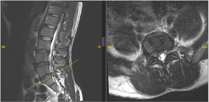
Figure 1: MRI images showing right paracentral L5-S1 disc herniation. MRI T2 sagittal on the left showing the right L5-S1 paracentral slightly caudally migrated fragment, with T2 axial images on the right showing the significant compression of the right lateral recess at the same level. Please click here to view a larger version of this figure.
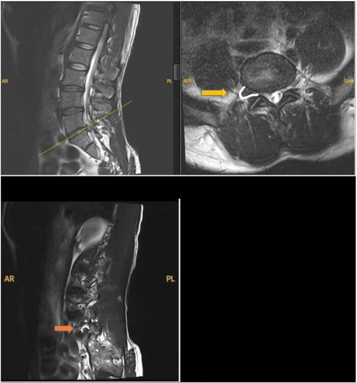
Figure 2: MRI images showing right far-lateral L5-S1 disc herniation. MRI T2 sagittal on the top left with T2 axial images on the top right showing the significant right L5-S1 far-lateral (extra-foraminal) herniated disc fragment (outlined in white) severely compressing the extra-foraminal compartment, as also shown on the bottom left MRI T2 right para-sagittal image (outlined in white). Please click here to view a larger version of this figure.
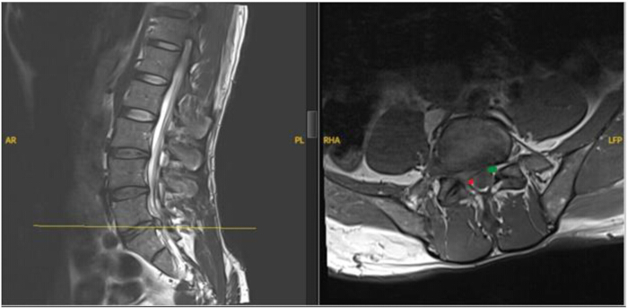
Figure 3: MRI images showing the extent of right S1 nerve root compression. MRI T2 sagittal on the left with T1 axial images on the right showing the displacement of the right L5-S1 traversing S1 nerve root (red dot) compressed and posteriorly displaced by the paracentral herniated disc fragment, where the contralateral S1 root relaxed and is in position (green dot) Please click here to view a larger version of this figure.
The patient was counselled for options, including conservative management with pain management, epidural steroid injections, and physical therapy versus surgical management, including open and minimally invasive options. As the pain was severely impacting the patient's ADL, he elected to proceed with minimally invasive discectomy. The technical difficulties related to the level and distributions of his disc herniations were discussed, and he agreed to the possibility of conversion to an open approach.
Access restricted. Please log in or start a trial to view this content.
Protocol
This research protocol was reviewed and approved by the Houston Methodist Human Research Ethics Committee, and all procedures were conducted in accordance with the ethical standards of the institutional research committee. Informed consent was obtained from the individual presented in the study.
1. Anesthesia induction and positioning
- Induce the patient with general endotracheal anesthesia, and place intraoperative neuromonitoring needles for somatosensory evoked potentials (SSEP), motor evoked potentials (MEP), and free-running electromyography (EMG).
- Position the patient prone on the Jackson Pro-Axis table.
- Prep and drape in standard fashion.
2. Localization
- Bring the C-arm into the field for fluoroscopic localization (Figure 4).
- Use a 6 inch needle to target the right L5-S1 foramen at Kambin's triangle in lateral and anteroposterior (AP) views (Figure 5, Figure 6, Figure 7).
- Make a ~1 cm skin incision using a 15-blade.
- Introduce serial dilators in the trajectory of the needle and confirm on fluoroscopy (Figure 8).
- Advance the working channel over the dilators and remove the dilators (Figure 8 and Figure 9).
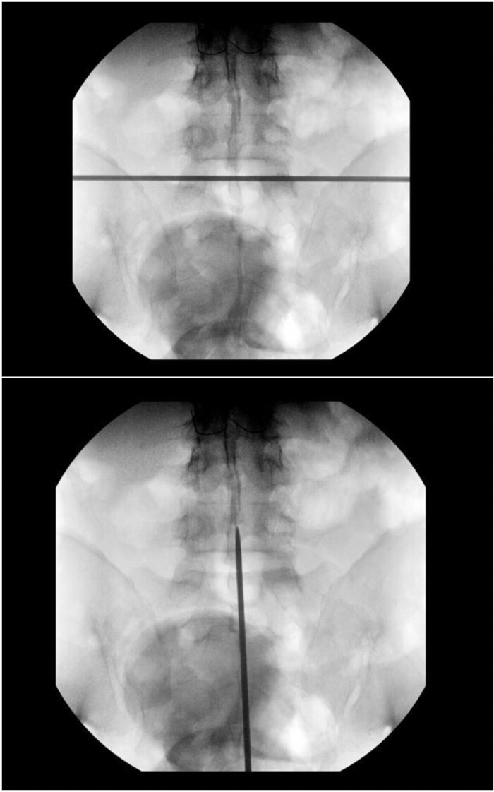
Figure 4: Skin marking of the level and the midline using fluoroscopy. The initial targeting with C-arm fluoroscopy starts with an AP view, squaring the upper endplate of the vertebra below the desired disc space. A transverse line is drawn here as a reference for the C-arm Wig-wag (swivel). Also, a vertical line is drawn along the midline for reference to the center of the field on AP. Please click here to view a larger version of this figure.
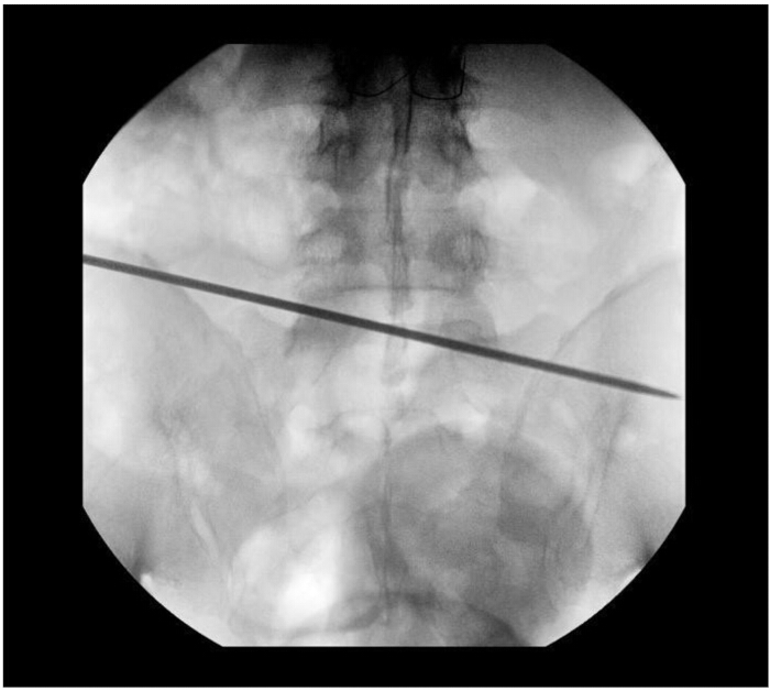
Figure 5: Marking the access line on the skin using fluoroscopy. AP image is obtained to outline the AP desired targeting trajectory line (for transforaminal approach, a slightly craniocaudal line crossing just above the ipsilateral pedicle and just below the contralateral pedicle of the vertebra below the disc space of interest is usually adequate; the line is usually extended on the approach side as far as 16-18 cm off midline as needed). It is recommended to stay at or just above the iliac crest line to avoid it during the approach. Please click here to view a larger version of this figure.
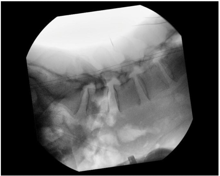
Figure 6: Marking the skin incision site using fluoroscopy. Lateral images are obtained along the Wig-wag line and centered on the disc level of interest. Using a protected spinal needle along the targeting line, a point is marked where the tip of the needle is opposite to the tip of the spinous process corresponding to lateral recess access. Please click here to view a larger version of this figure.
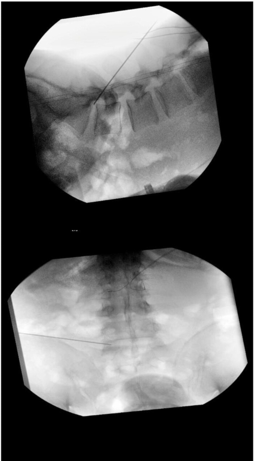
Figure 7: Initial needle access using fluoroscopy. The spinal needle is advanced from the marked point along the targeting line, with lateral fluoroscopy images to the inferior foraminal compartment. This is confirmed on AP images, and a K-wire is inserted to hold the path, and the needle is removed. Please click here to view a larger version of this figure.

Figure 8: Sequential dilation using fluoroscopy. A 7 mm skin incision is made at this point in the needle access site, and serial dilators are then introduced sequentially under fluoroscopic images. The scope cannula is advanced with the bevel turned dorsally, and then the dilators are removed. Please click here to view a larger version of this figure.

Figure 9: Confirmation of final cannula position using fluoroscopy. The final position of the scope cannula is confirmed in both lateral and AP images. A shallow dock was performed to avoid injuring the dorsally displaced exiting nerve. Please click here to view a larger version of this figure.
3. Exposure
- Remove superficial fat and muscle with the use of radiofrequency cautery and grasper instruments.
- Identify the landmarks of Kambin's triangle, including the superior articular process (SAP), exiting root, and cranial pedicle.
4. Far lateral microdiscectomy
- Identify the far lateral herniated fragments immediately in the foramen and remove sequentially with various graspers.
- Confirm full decompression of the swollen and congested exiting right L5 nerve root.
5. Paracentral microdiscectomy
- Rotate the bevel of the scope cannula to protect the exiting L5 nerve root.
- Perform a foraminoplasty by drilling the ventromedial SAP to enhance access to the lateral recess.
- Perform additional ventral foraminal widening by undermining the disc annulus and resecting disc herniated fragments from the subligamentous compartment.
- Release the ligamentum flavum with an endo-Kerrison punch for visualization of the lateral thecal sac.
- Interrogate the thecal sac to confirm it is floating freely with the fluid pressure denoting adequate decompression.
- Directly visualize the exiting L5 nerve and ensure integrity is maintained.
6. Closure
- Achieve adequate hemostasis with thrombin and radiofrequency ablation.
- Remove the working channel.
- Close the wound in layers with 3-0 PDS and 3-0 monocryl with skin adhesive (Figure 10).
- Ensure no neuromonitoring events were encountered and signals remained stable throughout the procedure.
Access restricted. Please log in or start a trial to view this content.
Results
This is an operative surgical video demonstrating the technical nuances of performing noncontiguous right-sided far lateral and paracentral discectomies at the L5-S1 level utilizing an endoscopic transforaminal approach to effectively decompress L5 and S1 nerve roots, respectively.
The patient had a smooth recovery from anesthesia and reported resolution of pain upon awakening from anesthesia with full strength on exam in all areas. He was discharged home in a few hours after surgery and repor...
Access restricted. Please log in or start a trial to view this content.
Discussion
The key to a successful L5-S1 transforaminal endoscopic lumbar discectomy (TELD) involves both patient selection and technical skill. Here, we have demonstrated a safe and effective way to treat both far lateral and paracentral disc herniations at L5/S1 from a TF approach. When selecting the TF approach for L5/S1 it is imperative to observe the angle of the iliac crest on lateral x-rays. An iliac crest that is superior to the rostral pedicle of the disc space of interest was previously considered a relative contraindica...
Access restricted. Please log in or start a trial to view this content.
Disclosures
Meng Huang is a consultant for Joimax, Arthrex, Amplify, Orthofix, MiRus, TrackX; Clinical Advisory Board/Consulting: XO Biologix. Paul Holman is a consultant for SeaSpine and XO Biologics.
Acknowledgements
The authors acknowledge the support of the Department of Neurosurgery, Houston Methodist Neurological Institute, for funding this project.
Access restricted. Please log in or start a trial to view this content.
Materials
| Name | Company | Catalog Number | Comments |
|---|---|---|---|
| Basics Instruments | Joimax | TESBASIC | TESBASIC set includes grasping forceps, cutting and punching forceps, guiding robs and tubes, and endo-Kerrison punches |
| Bone Drill | Joimax | DCC241121 | Bone drill |
| Camsource Duo | Joimax | CSFHD01CC | Endoscopic camera |
| Dermabond | Ethicon | https://www.jnjmedtech.com/en-US/product/dermabond-advanced-mini-topical-skin-adhesives | Skin adhesive |
| Disposable access kit | Joimax | TDAK0060 | Disposable access kit with 4 reamers |
References
- Kambin, P., Sampson, S. Posterolateral percutaneous suction excision of herniated lumbar intervertebral discs: Report of interim results. Clin Orthop Relat Res. 207, 37-43 (1986).
- Fanous, A. A., Tumialán, L. M., Wang, M. Y. Kambin's triangle: Definition and new classification schema. J Neurosurg Spine. 32 (3), 390-398 (2020).
- Ruetten, S., Komp, M., Godolias, G. Full-endoscopic interlaminar operation of lumbar disc herniations using new endoscopes and instruments. Orthop Praxis. 10, 527-532 (2005).
- Ruetten, S. The Full Endoscopic Interlaminar Approach for Lumbar Disk Herniations. Minimally Invasive Spine Surgery: A Surgical Manual. , Springer. New York. (2005).
- Choi, G., et al. Percutaneous endoscopic interlaminar discectomy for intracanalicular disc herniations at L5-S1 using a rigid working channel endoscope. Neurosurgery. 58 (1 Suppl), ONS59-ONS68 (2006).
- Ebraheim, N. A., et al. The location of the intervertebral lumbar disc on the posterior aspect of the spine. Surg Neurol. 48, 232-236 (1997).
- Mirkovic, S. R., Schwartz, D. G., Glazier, K. D. Anatomic considerations in lumbar posterolateral percutaneous procedures. Spine (Phila Pa 1976). 20 (18), 1965-1971 (1995).
- Kim, C. H., Chung, C. K. Endoscopic interlaminar lumbar discectomy with splitting of the ligament flavum under visual control. J Spinal Disord Tech. 25, 210-217 (2012).
- Hasan, S., White-Dzuro, B., Barber, J., Wagner, R., Hofstetter, C. The endoscopic trans-superior articular process approach: A novel minimally invasive surgical corridor to the lateral recess. Oper Neurosurg (Hagerstown). 19 (1), E1-E10 (2020).
- Hoogland, T., Schubert, M., Miklitz, B., Ramirez, A. Transforaminal posterolateral endoscopic discectomy with or without the combination of a low-dose chymopapain: A prospective randomized study in 280 consecutive cases. Spine (Phila Pa 1976). 31 (24), E890-E897 (2006).
- Lewandrowski, K. U. "Outside-in" technique, clinical results, and indications with transforaminal lumbar endoscopic surgery: A retrospective study on 220 patients on applied radiographic classification of foraminal spinal stenosis. Int J Spine Surg. 8, 1-17 (2014).
- Ahn, Y., Oh, H. K., Kim, H., Lee, S. H., Lee, H. N. Percutaneous endoscopic lumbar foraminotomy: An advanced surgical technique and clinical outcomes. Neurosurgery. 75 (2), 124-133 (2014).
- Song, Q. C., et al. Percutaneous endoscopic transforaminal discectomy for the treatment of L5-S1 lumbar disc herniation and the influence of iliac crest height on its clinical effects. Exp Ther Med. 22 (2), 866(2021).
Access restricted. Please log in or start a trial to view this content.