We outline a highly adaptable approach to using CRISPR-Cas9 ribonucleoprotein complex-mediated gene ablation in murine naïve CD4 T cells to investigate gene function in CD4 T cell differentiation.
Method Article
Investigating Murine CD4 T Cell Differentiation Using CRISPR-Cas9 Ribonucleoprotein Complex-mediated Gene Ablation
In This Article
Summary
Abstract
The widespread accessibility of clustered regularly interspaced short palindromic repeat (CRISPR)-Cas9 technology has made gene targeting in primary cells a routine method for evaluating gene function in T cells. Given the cost and limited availability of knockout (KO) mouse strains, testing preliminary hypotheses involving gene function in T cells can be prohibitive using gene-targeted animal models. However, using commercially available resources, including predesigned guide RNAs (gRNAs), researchers can conveniently generate gene-targeted naïve T cells that can be used for T cell activation and differentiation studies.
Here we outline a protocol for using nucleofection-delivered CRISPR-Cas9 ribonucleoprotein complexes (RNPs) to efficiently generate gene KO murine naïve CD4 T cells that can be used to evaluate gene function in CD4 T cell differentiation, in vitro. Isolation of naïve CD4 T cells from mouse secondary lymphoid organs, followed by nucleofection with Cas9-gRNA complexes ensures gene KO is initiated before downstream T cell activation, offering a strategic advantage over retroviral-mediated gRNA delivery, which typically requires preactivation of T cells, preventing the evaluation of effects in naïve T cells. Furthermore, this nucleofection-based method bypasses potential developmental issues associated with gene KO animals.
Following Cas9-gRNA delivery, we describe protocols for studying CD4 T cell differentiation into Th1, Th2, Th17, and Treg lineages using in vitro polarization. In addition, this protocol is adaptable to using gene-targeted CD4 or CD8 T cells for numerous downstream applications, including other T cell activation studies in vitro and adoptive transfer studies in vivo. The use of CRISPR-Cas9 methods has streamlined our ability to evaluate gene function in T cells and allows for the routine KO of many genes of interest, freeing researchers from limitations associated with studying gene KO animals.
Introduction
The use of clustered regularly interspaced short palindromic repeat (CRISPR)-based technologies has transformed our ability to manipulate genomic DNA sequences, greatly enhancing our ability to study gene function in countless biological systems. With respect to CD4 T cells, methods utilizing CRISPR-Cas9 ribonucleoprotein (RNP) complexes have emerged, facilitating efficient gene knock-out (KO) in primary naïve T cells that can be used for in vitro and in vivo studies1,2,3,4. Identification of new putative genes regulating CD4 T cell differentiation is often driven by transcriptomic and epigenomic analyses, generating novel targets for which there can be few or difficult-to-obtain resources to investigate, particularly if the gene affects multiple tissues and may require evaluation using conditional gene-targeted mice. Gene-targeting via retroviral transduction of gRNAs5,6 has been used to evaluate gene function in T cells, yet requires T cell activation for retroviral infection, and cannot be used to target genes in naïve T cells. Nucleofection-delivered CRISPR-Cas9 in naïve T cells offers a relatively inexpensive and fast alternative tool to validate and interrogate new genes of interest before investing in time-consuming and costly mouse models.
In our protocol described here, CRISPR-Cas9-mediated gene editing is carried out using recombinant Cas9 protein complexed with guide RNA (gRNA) molecules that are delivered into naïve CD4 T cells via nucleofection. The gRNA is made up of two distinct components, a trans-activating CRISPR RNA (tracrRNA) and a CRISPR RNA (crRNA). Functionally, tracrRNA-derived sequences facilitate association of the gRNA with Cas9, while the crRNA sequences contain specificity for target DNA regions of interest. Cas9-gRNA complexes mediate targeted double-stranded DNA (dsDNA) breaks, with gRNAs mediating the sequence specificity of Cas9 endonuclease activity7,8. The Cas9-mediated cleavage of dsDNA leads to gene inactivation, through the generation of indel mutations following non-homologous end joining in target cells9. In this protocol, we recommend using multiple gRNAs targeting genes of interest to ensure robust KO in naïve CD4 T cells.
Helper CD4 T cells are key players in the immune system, guiding immune responses through the production of a variety of cytokines that modulate the function of many immune cell types. Upon stimulation through the T cell receptor (TCR) in the presence of specific cytokines, naïve CD4 T cells can differentiate into distinct lineages of T helper (Th) CD4 T cells, including Th1, Th2, Th17, and regulatory T cells (Treg)10,11,12. Defining these distinct CD4 T cell lineages is the expression of specific lineage-defining transcription factors (TFs) and cytokines (Figure 1). CD4 T cell differentiation can be modeled using in vitro polarization13,14,15, using naïve CD4 T cells activated through the TCR in the presence of lineage-promoting cytokines and blocking antibodies that prevent inappropriate lineage adoption.
Combining CRISPR-Cas9 gene targeting in naïve CD4 T cells with in vitro CD4 T cell polarization offers a robust system to evaluate gene function in these cells (Figure 2). Given the widespread availability of reagents to assess CD4 differentiation by flow cytometry, key aspects of CD4 T cell differentiation, including TF expression and cytokine production, can easily be interrogated using the protocols described here. The identification of novel genes regulating CD4 T cell function enhances our understanding of these cells, and CRISPR-Cas9 methods paired with in vitro polarization offer a robust modality for assessing gene function before committing to gene KO mouse models.
Protocol
For all procedures described here, we used wild-type (WT) C57/BL6J mice. Mice were maintained and treated under specific pathogen-free (SPF) conditions in accordance with the guidelines of NIAID (protocol LISB-22E) and the Animal Care and Use committees at the NIH (Animal Welfare Assurance #A-4149-01).
1. Considerations before beginning
- Selecting gRNA reagents
NOTE: See the Table of Materials for obtaining predesigned gRNA reagents targeting the mouse genome used in this protocol.- Search the gene of interest (Table of Materials, Desired crRNAs) and select the top three ranked predesigned crRNAs (2 nmol format). In addition, obtain negative control crRNA to use in parallel (Table of Materials).
NOTE: The formation of functional gRNAs requires duplexing crRNA with tracrRNA; obtain a tracrRNA reagent and follow the steps in section 3. - Design custom targeted gRNA sequences.
NOTE: The design of custom targeted gRNA sequences is outside the scope of this protocol but can be performed using publicly available tools such as CRISPick (Table of Materials).
- Search the gene of interest (Table of Materials, Desired crRNAs) and select the top three ranked predesigned crRNAs (2 nmol format). In addition, obtain negative control crRNA to use in parallel (Table of Materials).
- Buffers, media, and equipment
NOTE: Unless otherwise noted, use cold (4 °C) buffers and reagents.- Prepare MACS buffer, FACS buffer, 0.5% Triton X-100 buffer, 1x permeabilization buffer, complete RPMI media, and complete IMDM media (Table of Materials) before beginning the procedure and store at 4 °C.
- Obtain naïve CD4 T cell isolation and anti-FITC microbead kits (Table of Materials) before beginning this procedure.
- Prior to beginning nucleofection (section 4), prewarm (37 °C) 5 mL of complete IMDM media and prepare a minimum of 10 mL of complete IMDM supplemented with IL-7 at 5 ng/µL warmed to 37 °C (Table of Materials). Adjust the volume of prewarmed media as needed to account for adding 125 µL per sample after nucleofection. Adjust the volume of IL-7-containing complete IMDM as needed, based on the numbers of cells nucleofected (section 4).
- Before starting antigen-presenting cell (APC) isolation, prepare the mitomycin C reagent by adding 4 mL of sterile H2O to the 2 mg vial of mitomycin C (0.5 mg/mL stock concentration).
NOTE: Perform all steps in a tissue culture hood and use sterile aseptic techniques.
2. Naïve CD4 T cell isolation
NOTE: Naïve CD4 T cells may be isolated using magnetic isolation or through cell sorting. Purity obtained from magnetic isolation is typically 97%, and cell viability is very high, whereas electronic sorting can improve cell purity at the expense of cell viability. The protocol described here will use magnetic isolation but can be adapted to either scenario. Ensure the LS columns are prepared appropriately (Table of Materials).
- Isolate the spleen and lymph nodes (inguinal, brachial, axial, and cervical) from the mice (WT C57BL/6J mice used here).
- Place a 70 µm screen in a 60 mm dish with 5 mL of complete RPMI (Table of Materials) for each tissue processed. Process the lymph nodes and spleen in different dishes to increase the yield. Gently grind the tissues through the filter using a plunger from a 3 mL syringe. Rinse the filter with 1-3 mL of media and transfer the single-cell suspensions to 15 mL conical tubes.
- Repeat step 2.2 for additional lymph nodes and splenic tissues, as needed.
- Centrifuge the single-cell suspensions at 500 × g for 5 min at 4 °C.
- Prepare to count the cells as follows.
- For splenic samples, aspirate the supernatant, gently resuspend the cell pellet in 1 mL of ACK lysis buffer, incubate for 2-5 min at RT, then add 2 mL of complete RPMI, spin down, resuspend in 5 mL of complete RPMI, and count the cells.
- For lymph node cells, aspirate the supernatant, resuspend the cell pellet in 5 mL of fresh complete RPMI, and count the cells.
- For input into the naïve CD4 isolation kit, mix the splenocytes with lymph node cells to reach the desired cell number for isolation.
NOTE: Typically, 1 × 10⁸ lymph node and spleen cells from WT-B6 mice yields 2-8 × 10⁶ naïve CD4 T cells. - Centrifuge the pooled spleen and lymph node cells at 500 × g for 5 min at 4 °C, remove the supernatant, and resuspend the cell pellet in the appropriate volume of MACS buffer (Table of Materials) as indicated in the manufacturer's protocol (naïve CD4 T cell isolation kit).
- Precisely follow the manufacturer's protocol to isolate naïve CD4 T cells by negative selection (Table of Materials) using Naïve CD4+ T Cell Biotin Antibody Cocktail, Anti-Biotin Microbeads and CD44 Microbeads; use LS columns for this isolation and collect the cells in 15 mL conical tubes.
- Following isolation, centrifuge the cells at 500 × g for 5 min, 4 °C, remove the supernatant, resuspend the cell pellet in 2 mL of complete RPMI media, and count the cells.
- Store the cells at 4 °C until ready to begin with nucleofection.
3. Cas9-gRNA complex formation
NOTE: Prepare negative control and gene-specific crRNA stocks. We outline using one unique crRNA for negative control conditions and three unique crRNAs for gene-targeting conditions. Using three distinct crRNAs simultaneously ensures KO of the genes of interest.
- Resuspend the crRNAs and tracrRNA to 100 µM stock concentrations using duplex buffer (Table of Materials). This amounts to 10 µL of buffer per 1 nmol of RNA.
- Combine individual crRNAs with tracrRNA at a 1:1 ratio in sterile PCR tubes and mix by pipetting. Heat to 95 °C for 5 min in a thermocycler. Then, let the complexes cool to RT on a benchtop for at least 10 min.
NOTE: crRNA/tracrRNA duplexes are referred to as gRNAs from this point onwards. Complexes are stable for at least 6 months if stored at -20 °C. We typically make 6 µL of gRNA (3 µL of crRNA + 3 µL of tracrRNA) per run, providing enough material for roughly five samples (1 µL/sample). - For gene-targeting gRNAs: In sterile PCR tubes, add 3 µL of gRNAs (1 µL each of three unique gRNAs per target), 1 µL of Cas9 protein, and 1 µL of Duplex buffer (5 µL total per sample). Pipet gently to mix and ensure all material is in the bottom of the tube. Incubate at RT for 10 min. Synchronize the timing of this step with nucleofection (steps 4.1-4.2).
- For negative control gRNA: In sterile PCR tubes, add 1 µL of gRNA, 1 µL of Cas9 protein, and 3 µL of Duplex buffer (5 µL total per sample). Pipet gently to mix and ensure all material is in the bottom of the tube. Incubate at RT for 10 min. Again, synchronize the timing of this step with nucleofection (steps 4.1-4.2).
NOTE: Using 50 pmol of gRNA for negative control (versus 3 x 50 pmol for test) is more cost-effective and has no impact on the results.
4. Nucleofection - Cas9-gRNA-mediated gene ablation
- Centrifuge 1-8 × 10⁶ purified T cells at 500 × g for 5 min, 4 °C per nucleofection sample. Carefully remove as much supernatant as possible.
- Resuspend the cells in 20 µL of P3 buffer (16.4 µL of P3 + 3.6 µL of supplement per sample; Nucleofector Kit, Table of Materials) and transfer the suspension to PCR tubes containing the Cas9-gRNA complexes. Mix and transfer to independent wells of a 16-well nucleocuvette (Nucleofector Kit, Table of Materials). Ensure bubbles have not formed when transferring cells to the cuvette, as this can negatively impact nucleofection efficiency. Tap the cuvette against a work surface to ensure all material is in the bottom of each well and free of bubbles.
- Perform nucleofection using the DN100 program of the referenced nucleofector (Table of Materials). Immediately add 125 µL of prewarmed complete IMDM media to the nucleocuvette wells and transfer the cells to 1.5 mL microcentrifuge tubes. Rinse each well one additional time with 125 µL of prewarmed media to ensure the collection of all cells.
- Centrifuge the cells at 500 × g for 5 min, 4 °C, remove all the media, and resuspend in 200 µL of warmed complete IMDM media + 5 ng/mL IL-7 (Table of Materials).
- Transfer 200 µL of the cells to the culture plate and add warmed complete IMDM media + 5 ng/mL IL-7 to make the final desired volume according to the plate size as follows: 1-2 × 10⁶ cells per well on a 48-well plate (1 mL of media per well), 2-4 × 10⁶ cells per well on a 24-well plate (1.5 mL of media per well) volume, 4-6 × 10⁶ cells per well on a 12-well plate (2 mL of media per well), and 6-10 × 106 cells per well on a 6-well plate (4 mL of media per well). Culture the cells in an incubator at 37 °C, 5% CO2 for 3 days.
NOTE: Depending on the turnover of protein and the downstream assay, optimize this culture period. We have found an optimal rest period of 3 days, but this may be adjusted based on target gene to between 1 and 7 days using these conditions. - Following culture in complete IMDM media + 5 ng/mL IL-7, resuspend the cells by pipetting and harvest them into 15mL conical tubes. Centrifuge the cells at 500 × g for 5 min, 4 °C, remove the supernatant, and resuspend the cell pellet in 1 mL of complete RPMI media.
- Count the cells to determine the number of live naïve CD4 T cells corresponding to the gRNA-nucleofected cells of interest.
NOTE: In the process of nucleofection, expect to lose ~ 50% of input cells over the course of this protocol and adjust the number of naïve CD4 T cells used accordingly. Also, at this point, you may test the efficiency of the gene targeting in naïve CD4 T cells using a variety of techniques (flow cytometry, western blot, qPCR, or T7EI assay).
5. Antigen-presenting cell (APC) isolation
NOTE: Ensure that the mitomycin reagent has been prepared, which will be used to treat APCs following isolation (Table of Materials). Read the manufacturer's anti-FITC microbead protocol before starting and ensure that the LS columns are prepared appropriately (Table of Materials).
- Isolate spleens from WT mice designated for APC isolation.
- Make single-cell suspensions as described above (2.2, 2.5.1)-lyse erythrocytes with ACK lysis buffer, resuspend the cell pellet in 5 mL of complete RPMI (4 °C), and count the cells.
NOTE: Following APC isolation, expect to obtain approximately half the number of input cells, for example, 1 × 10⁸ input splenocytes will yield ~40-50 × 106 APCs. Prepare the desired number of splenocytes for isolation. - Centrifuge the cells at 500 × g for 5 min, 4 °C, and resuspend the cell pellet in 800 µL of MACS buffer (Table of Materials).
- Add 4 µL each of FITC-conjugated anti-CD4 and anti-CD8 antibodies (Table of Materials) to the splenocytes. Mix well and incubate at 4 °C for 15 min.
- Centrifuge cells at 500 × g for 5 min, 4 °C and remove the supernatant. Following the manufacturer's instructions, resuspend the cells in the recommended volume of MACS buffer and add the appropriate amount of anti-FITC microbeads (Table of Materials).
- Incubate the cells at 4 °C for 15 min. Centrifuge the cells at 500 × g for 5 min, 4 °C. Resuspend the cells in 500 µL of MACS buffer and pass the cells through the prepared LS column as described in the manufacturer's protocol, collecting the cells in a 15 mL conical tube.
- Centrifuge the cells at 600 × g for 5 min, 4 °C, remove the supernatant, and resuspend the cells in 2.7 mL of sterile 1x PBS.
NOTE: APCs may pellet poorly; use an increased speed for centrifugation. - Add 300 µL of mitomycin C solution to the cells to make a final concentration of 50 µg/mL. Mix well and incubate at 37 °C in a water bath for 30 min or for 45 min at 37 °C, 5% CO2 in an incubator.
- Following incubation, centrifuge the cells at 600 × g for 5 min, 4 °C, and wash them with 10 mL of sterile 1x PBS. Use vacuum to remove as much supernatant as possible. Repeat the wash 2x.
- Centrifuge the cells again at 600 × g for 5 min, 4 °C, remove the supernatant, and resuspend the cell pellet in 5 mL of complete RPMI media (4 °C). Count the cells. Store APCs at 4 °C until ready to perform CD4 T cell differentiation.
6. CD4 T cell differentiation
NOTE: This protocol is set to culture cells in a 48-well plate format-2 × 10⁵ naïve CD4 + 1 × 10⁶ APC per well-a 1:5 ratio. Cells are cultured in complete IMDM media. Here, we will provide conditions for the differentiation of Th1, Th2, Th17, and Treg CD4 T cells; however, individual lineage conditions may be chosen depending on the focus of the user.
- Determine the number of cells in a 48-well plate to be used and calculate the number of cells needed for each cell type.
NOTE: For example, 10 wells would require 2 × 10⁶ naïve CD4 and 10 × 106 APCs to maintain the 1:5 ratio. If cell numbers permit, calculate enough cells for one additional well to account for any pipetting error when plating. - Mix together the desired number of naïve CD4 and APCs at 1:5, centrifuge the cells at 600 × g for 5 min, 4 °C, remove the supernatant, and resuspend the cell pellet in 500 µL of complete IMDM media per well (e.g., cells corresponding to 10 wells in 5 mL of media).
- Plate 500 µL increments of cells into wells of a 48-well plate, ensuring that each well contains 2 × 10⁵ naïve CD4 + 1 × 10⁶ APCs.
- For the desired CD4 T cell differentiation conditions, prepare 2x cytokine + antibody mixes in complete IMDM as described in Table 1. Add 500 µL of the 2x mix to the previously plated 500 µL increments of cells to make the final concentrations of reagents 1x.
NOTE: These examples are based on making 2.5 mL of 2x preparations, enough for five wells for each condition. Again, if the reagent amounts permit, make one additional increment of 2x mix to account for the pipetting error. - Add 500 µL of 2x cytokine + antibody media to 500 µL of CD4/APC cells-containing wells to make a final concentration of reagents at 1x.
- Place the 48-well plate in an incubator at 37 °C with 5% CO2. Culture the cells for 72 h to allow completion of CD4 polarization.
NOTE: Examine the plate after the culture period to ensure robust growth. The media should be an orange color after incubation, with yellow media indicating potential cell overgrowth.
7. Assessing the impact of gene ablation in CD4 T cell differentiation
NOTE: For all staining steps, prepare a master mix of antibodies added at the appropriate dilution to the required staining buffer. A 50 µL volume of the antibody/buffer cocktail is used per sample. Adjust specific antibodies used based on the experiment. Users should choose antibody panels relevant to their specific experiments; here we provide a template with which to approach this in the context of the genes targeted here.
- Following CD4 differentiation, carefully remove 600 µL of the supernatant, then resuspend the cells in each well by pipetting.
- Remove 200 µL of the cells from each well and add them to a labeled 96-well plate for transcription factor staining. Pellet the samples by spinning at 500 × g for 5 min at 4 °C, flick off the supernatant, and proceed to surface staining (step 7.5).
- For cytokine analysis, restimulate the remaining 200 µL of cells with PMA (50 ng/mL), Ionomycin (500 ng/mL), and Golgi Stop (1/2,000 dilution) in complete IMDM. Add 200 µL of the 2x restimulation mix to each well and incubate at 37 °C, 5% CO2 for 4 h.
NOTE: There should be 200 µL remaining in the 48-well plate; hence, adding 200 µL of the 2x mix of PMA (2x, 100 ng/mL), Ionomycin (2x, 1 µg/mL), and Golgi Stop (2x, 1/1,000) will create a final concentration of 1x (Table of Materials). - In the meantime, begin transcription factor staining. Perform surface antigen staining by resuspending the cells in 50 µL of FACS buffer containing the desired fluorochrome-conjugated antibodies per sample (Table 2). Incubate in the dark at 4 °C for 30 min.
- Add 150 µL of FACS buffer to each well to wash off the staining buffer. Centrifuge the plate at 500 × g for 5 min at 4 °C. Flick off the supernatant. Repeat for a total of two washes.
- Fix the cells with 100 µL of Foxp3 fixation solution (Table of Materials) in the dark at RT for 25 min.
- After 25 min, add 100 µL of FACS buffer to each well to wash off the fixative. Centrifuge the plate at 500 × g for 5 min at 4 °C and flick off the supernatant. Repeat for a total of two washes.
- Perform intracellular staining by resuspending the cells in 50 µL of 1x Foxp3 permeabilization buffer containing directly conjugated transcription factor-specific antibodies per sample (Table 2) for a minimum of 1 h at 4 °C in the dark.
- After intracellular staining is complete, add 100 µL of FACS buffer to each well. Centrifuge the plate at 500 × g for 5 min at 4 °C and flick off the supernatant. Repeat for a total of two washes. Resuspend cells in a final volume of 200 mL for flow cytometry.
- After PMA/ionomycin/Golgi Stop restimulation, resuspend the cells by pipetting and transfer 400 µL of each cytokine sample to 1.5 mL microcentrifuge tubes. Pellet the samples at 500 × g for 5 min at 4 °C and remove the supernatant. Resuspend the cell samples in 100 µL of FACS buffer and transfer them to a 96-well plate for surface staining. Pellet the samples again by spinning at 500 × g for 5 min at 4 °C, flick off the supernatant.
- Prepare a surface staining cocktail with FACS buffer containing fluorescent antibodies directed against surface antigens (Table 2). Resuspend the cells in 50 µL of staining buffer per sample and incubate for 30 min at 4 °C in the dark.
- Add 150 µL of FACS buffer to each well to wash off the staining buffer. Centrifuge the plate at 500 × g for 5 min at 4 °C. Flick off the supernatant. Repeat for a total of two washes.
- Fix the cells in 100 µL of 4% PFA (Table of Materials) in the dark at RT for 25 min.
- Add 100 µL of FACS buffer to each well to wash off the fixative. Centrifuge the plate at 500 × g for 5 min at 4 °C. Flick off the supernatant. Repeat for a total of two washes.
- Perform intracellular cytokine staining by resuspending the cells in 50 µL of anti-cytokine antibody containing 0.5% Triton X-100 staining buffer per sample (Table 2) for a minimum of 1 h at 4 °C in the dark.
- After intracellular staining is complete, add 100 µL of FACS buffer to each well. Centrifuge the plate at 500 × g for 5 min at 4 °C and flick off the supernatant. Repeat for a total of two washes. Resuspend cells in a final volume of 200 mL for flow cytometry.
- Analyze the transcription factor and cytokine-stained samples by flow cytometry.
NOTE: Expected flow cytometry profiles from the outlined staining panels will be shown in the representative results section.
Results
To validate that a pure population of naïve CD4 T cells was obtained using our protocol (Section 2), we used flow cytometry to identify these cells before and after magnetic isolation. Using our approach, we obtained a highly pure population of live CD4+TCRb+CD25-CD44-CD62L+ naïve CD4 T cells following isolation (Figure 3A). Furthermore, to confirm that Cas9-gRNA complexes were successfully nucleofected into naïve CD4 T cells (Section 4), we performed flow cytometry on control and Cas9-gRNA-treated cells and examined fluorescence of Atto550, a dye attached to tracrRNAs used in this protocol (Figure 3B). We observed robust detection of Cas9-gRNA complexes in nucleofected cells relative to control, consistent with robust entry of RNP complexes into naïve CD4 T cells. We also show our gating strategy for identifying live CD4 T cells following CD4 T cell differentiation (Section 7), which is critical for identifying activated CD4 T cells producing lineage-specific cytokines (Figure 3C).
Two broad classes of genes targeted using CRISPR-Cas9 in T cells are those encoding cell surface proteins and those encoding transcription factors. Here, we used gene-specific gRNAs to target the T cell marker Thy1, encoded by the Thy1 gene, as a representative cell surface protein and the transcription factor Tbet, encoded by the Tbx21 gene, as a representative transcription factor (Figure 4). As an important control, we include cells treated with a negative control gRNA that have gone through the same nucleofection process as Thy1 and Tbx21 gRNA-treated cells. Following CD4 T cell differentiation, cells were stained and analyzed by flow cytometry for TF and cytokine expression (Table 2), with live CD4 T cells identified by the gating strategy outlined in Figure 3C.
To evaluate CD4 T cell differentiation and KO efficiency of the gRNA constructs, we examined lineage-defining TF expression, along with surface expression of Thy1 in CD4 T cells following Th1, Th2, Th17, and Treg differentiation. As expected, negative control gRNA-treated CD4 T cells showed a classic pattern of lineage-defining TF expression, with Th1 cells showing robust induction of Tbet, Th2 cells showing high expression of GATA3, Th17 cells showing potent Rort expression, and Treg cells inducing expression of Foxp3 (Figure 4A). Confirming ablation of Thy1 expression, Thy1 gRNA-treated cells in all CD4 differentiation conditions showed strong reductions in surface Thy1 expression compared to negative control and Tbx21 gRNA-treated cells (Figure 4A). Thus, Thy1 gRNA-treated cells showed robust and specific ablation of Thy1 surface expression on in vitro differentiated CD4 T cells. In addition to Thy1, we also targeted intracellular, Th1-defining TF Tbet using Tbx21-targeting gRNAs. We evaluated the expression of Tbet in Th1 cells and observed markedly reduced Tbet expression in Tbx21 gRNA-treated cells compared to either negative control or Thy1 gRNA-treated cells (Figure 4A). Therefore, using the nucleofection-driven approach described here, we were able to achieve robust and specific gene ablation in the context of in vitro CD4 T cell differentiation.
The production of lineage-specific cytokines is a critical aspect of CD4 T cell differentiation. We evaluated cytokine production in Th1, Th2, Th17, and Treg cells using an intracellular cytokine staining protocol that relies on restimulating differentiated CD4 T cells with PMA and Ionomycin in the presence of the Golgi inhibitor monensin (Golgi Stop). This allows cytokines to be retained in the cytosol, facilitating intracellular detection of these normally secreted inflammatory mediators by intracellular staining and flow cytometry (Figure 4B). Focusing on Th1 polarized cells, we observed robust detection of IFNγ, with approximately 70% of negative control gRNA-treated cells being IFNγ+. Expression of Tbet is an important driver of Th1 cell differentiation and ablation of Tbet in Tbx21 gRNA-treated cells revealed a large reduction in IFNγ+ cells compared to negative control or Thy1 counterparts, with approximately 40% of Tbx21 gRNA-treated cells being IFNγ+ following KO. Differentiation in Th2 polarizing conditions supports the production of the cytokines IL-4 and IL-13. We detected robust populations of IL-4+, IL-13+, and IL-4+IL-13+ cells following Th2 polarization in all conditions. Following in vitro polarization, Th17 differentiated cells primarily produce the cytokine IL-17A. Using this protocol, we routinely observe between 30% and 60% IL-17A+ cells following Th17 polarizing conditions, with approximately 50% of CD4 cells being IL-17A+ in the experiment shown here. Under Treg polarizing conditions, we typically do not measure cytokine production and instead quantify frequencies of Foxp3+ cells as a readout of Treg differentiation. Here we observed approximately 60% of cells being Foxp3+, with a normal range being between 50% and 80% Foxp3+. Gating of cytokine and TF expressing cells from all CD4 polarizing conditions was determined according to negative control samples (Figure 4C). Altogether, with the protocol presented here, we are able to faithfully preserve the differentiation characteristics of CD4 T cells while targeting genes of interest using CRISPR-Cas9, allowing interrogation of a range of genes potentially involved in regulating this important process.

Figure 1: Overview of CD4 T cell differentiation. The activation of naïve CD4 T cells through the T cell receptor (TCR), as well co-stimulatory molecules like CD28, in the presence of lineage-defining cytokines, facilitates the differentiation of CD4 T cells into distinct lineages. The cytokine IL-12 drives the expression of Tbet, a transcription factor (TF) specifying Th1 cell differentiation16,17; Th1 cells are major producers of inflammatory cytokines IFNγ and TNFα. Differentiation of Th2 cells is driven by IL-4, leading to the induction of GATA318,19. Th2 cells are characterized by the production of the cytokines IL-4, IL-5, and IL-13. Specification of Th17 lineage differentiation is driven by the cytokines IL-6 and TGFβ, resulting in upregulation of Th17-specific TF Rorγt20. Differentiated Th17 cells are primary producers of the cytokines IL-17 and IL-22. Driving the differentiation of Treg cells, the cytokines TGFβ and IL-2 skew CD4 differentiation toward the Foxp3-expressing Treg lineage21,22. Treg cells produce the cytokines IL-10 and TGFβ and express inhibitory molecules like CTLA4. Please click here to view a larger version of this figure.
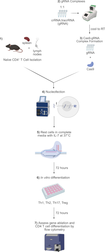
Figure 2: Overview of CRISPR-Cas9-mediated gene targeting in CD4 T cell differentiation. 1) Dissect spleen and lymph nodes from mice. Grind the organs and filter the cells to obtain single-cell suspensions. Isolate naive CD4+ T cells using magnetic isolation kits or cell sorting. 2) Combine crRNA and tracrRNA at a 1:1 ratio and heat in a thermocycler to form functional gRNA complexes. 3) Mix isolated naive CD4+ T cells with Cas9-gRNA complexes. 4) Performnucleofection of naïve CD4 T cells using a nucleofector device to deliver Cas9-gRNA complexes into target cells. 5) Culture naïve CD4 T cells in IL-7-containing media for 3 days to facilitate Cas-gRNA-mediated gene targeting prior to initiating T cell activation and differentiation. 6) Culture Cas9-gRNA-treated naïve CD4 T cells in CD4 T cell polarizing media for 3 days. 7) Analyze CD4 T cell differentiation by flow cytometry. Please click here to view a larger version of this figure.
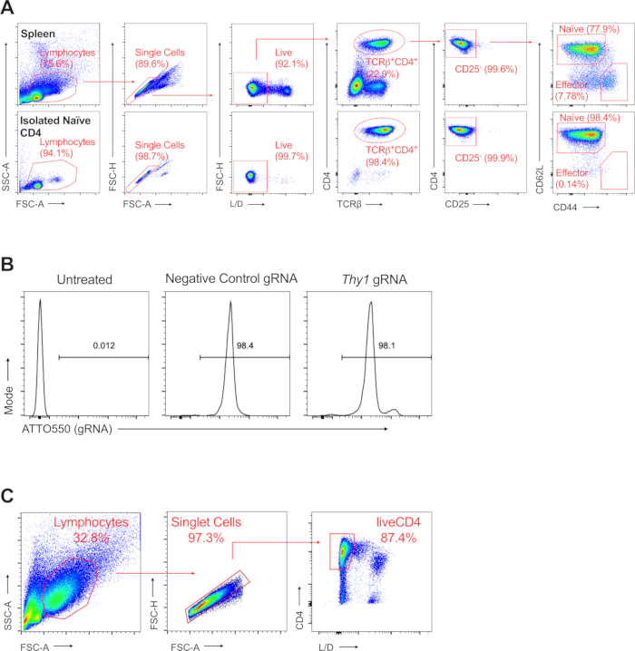
Figure 3: Validation of naïve CD4 T cell isolation, Cas9-gRNA nucleofection, and CD4 T cell differentiation by flow cytometry. (A) Representative data showing naïve CD4 T cell purity following magnetic isolation. Naïve CD4 T cells are gated as live CD4+TCRb+CD25-CD44-CD62L+ lymphocytes. Upper panel, naïve CD4 T cell gating in total splenocytes. Bottom panel, naïve CD4 T cell gating following magnetic isolation described in this protocol. (B) Control untreated cells, Negative control gRNA and Thy1 gRNA-nucleofected cells were examined by flow cytometry for Atto550 fluorescence. We used Atto550-labeled tracrRNA to generate gRNAs, allowing us to detect Cas9-gRNA entry into cells by flow cytometry. Cas9-gRNA-treated cells show a high efficiency of gRNA uptake, while control cells show minimal signal. (C) Representative gating of activated CD4 T cells following differentiation. Lymphocytes are first gated by size using FSC-A and SSC-A parameters. Singlet cells are then discriminated from doublet cells using FSC-A and FSC-H. To identify live CD4 T cells, we used L/D and CD4 staining to identify L/D-CD4+ T cells to specifically gate on CD4 T cells for downstream analysis. Abbreviations: FSC-A = forward scatter area; SSC-A = side scatter area; FSC-H = FSC-height; L/D = live/dead. Please click here to view a larger version of this figure.
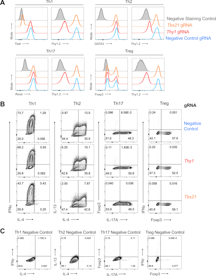
Figure 4: Representative results highlighting Cas9-gRNA-mediated gene ablation and CD4 T cell differentiation. A) Cell surface Thy1 and intracellular transcription factor staining from negative control (red), Thy1 (blue) and Tbx21 (orange) gRNA-treated liveCD4+ cells from Th1, Th2, Th17, and Treg differentiation conditions. Additionally, negative staining controls for each antibody are included in grey. Each CD4 T cell lineage panel shows lineage-defining transcription factor expression on the left and Thy1.2 surface expression on the right. B) Intracellular cytokine staining from negative control, Thy1 and Tbx21 gRNA-treated liveCD4+ cells from Th1, Th2, Th17, and Treg differentiation conditions. For Th1 conditions, we examined IFNγ and IL-4 staining, with IFNγ+IL-4- classified as cytokine-producing Th1 cells. For Th2 conditions, we examined IL-4 and IL-13 staining, with IL-4+ and IL-13+ cells characterizing Th2 cells. Under Th17 conditions, we evaluated the staining of IL-17A and IFNγ, with IL-17A+IFNγ- cells representing differentiated Th17 cells. To characterize Treg cells, we assessed Foxp3 and IFNγ staining, focusing on Foxp3+IFNγ- as a readout of Treg differentiation. C) Negative control gating for Th1, Th2, Th17, and Treg staining. Please click here to view a larger version of this figure.
Table 1: CD4 T cell differentiation media. This table includes stock concentrations for each cytokine and antibody reagent used for preparing CD4 T cell differentiation media, as well as 2x and final reagent concentrations needed to perform CD4 T cell differentiation studies. Here, we outline volumes of reagents needed to make 2.5 mL of 2x differentiation media, providing enough reagent for up to five conditions. Prepare the required volume of media for each CD4 T cell lineage condition based on the needs of the experiment. Please click here to download this Table.
Table 2: Flow cytometry antibodies used in representative data. This table includes the manufacturer, catalog number, fluorophore, and recommended dilution information for all antibodies used in the staining panels described in this protocol. Prepare staining cocktails in the indicated staining buffer, producing the volume of staining cocktail required to stain all conditions for the specific experiment. Please click here to download this Table.
Discussion
Integrating protocols for delivering CRISPR-Cas9 complexes into naïve CD4 T cells with methods for studying CD4 T cell differentiation provides a robust tool to explore novel genes that regulate CD4 T cell biology. Here, we provide a comprehensive guide for utilizing commercially available Cas9 and gRNA reagents that are straightforward to work with. Nucleofection-mediated delivery of Cas9-gRNA complexes into naïve CD4 T cells provides highly efficient gene editing, facilitating near-total knockout of genes of interest. In addition, flow cytometry analysis of in vitro polarized CD4 T cells allows comprehensive examination of lineage differentiation, as well a convenient tool to validate CRISPR-mediated gene ablation.
Here, we describe using Cas9-gRNA-nucleofected naïve CD4 T cells in the context of in vitro CD4 T cell polarization; however, this method can be adapted to numerous other applications. In particular, gene-targeted naïve CD4 T cells may also be used in adoptive transfer studies in vivo. Adoptive transfer of Cas9-gRNA-treated TCR transgenic CD4 T cells may be particularly useful in studying antigen-specific CD4 T cell responses to infection. In addition to CD4 T cells, the Cas9-gRNA nucleofection method outlined here also works well in naïve CD8 T cells. Nucleofected naïve CD8 T cells can similarly be used for a variety of downstream in vitro and in vivo applications. It should be noted that CRISPR-Cas9-nucleofected cells should always be validated for gene ablation before performing downstream studies.
In contrast to the recombinant Cas9-gRNA nucleofection approach described here, another common method of CRISPR-Cas9 gene ablation is using single guide RNA (sgRNA)-encoding retroviral vectors5. One advantage to this technique is the ability to follow the expression of reporters, such as GFP or Thy1.1, in these constructs, allowing the labeling of cells that have been successfully transduced. This is particularly useful for 1) following in vivo transfers, 2) in cases where gene-targeting gives a selective disadvantage to cells, and 3) in applications where sorting gene-ablated cells is desired, such as in many CRISPR screening protocols. This approach typically involves isolating T cells from Cas9-expressing mice for the delivery of sgRNA constructs into cells using retroviral vectors, which requires maintaining dedicated Cas9-expressing mouse lines. Furthermore, another major limitation of this method is that retroviral transduction of T cells requires preactivation via TCR stimulation. Therefore, T cell activation occurs before gene ablation in the T cell differentiation process, limiting the window of gene KO to later stages of activation. While this can be partially overcome by resting transduced cells in IL-7 prior to restimulation, this method still does not allow evaluation of true naïve cells. In contrast, Cas9-gRNA nucleofection can be performed directly in naïve T cells, allowing initiation of gene ablation prior to downstream activation, permitting both early and late activation phenotypes to more seamlessly be examined.
The use of CRISPR-Cas9 technology provides a highly adaptable tool to interrogate genes of interest in primary T cells, without committing to time- and cost-prohibitive mouse models. Researchers are often faced with multiple genes of interest in projects focused on CD4 T cell differentiation and CRISPR-Cas9 can provide a useful tool for deciding on which genes to prioritize before starting studies in mouse models that require a large time investment. Furthermore, many genes of interest lack accessible mutant mouse models altogether, making CRISPR-Cas9 editing in primary cells an attractive option for generating gene-targeted cells. CRISPR-Cas9-based approaches have revolutionized modern T cell research, providing researchers with new tools to understand fundamental biological process occurring in these important cells.
Disclosures
The authors have no conflicts of interest to declare.
Acknowledgements
This research was supported by the Intramural Research Program of NIAID, NIH.
Materials
| Name | Company | Catalog Number | Comments |
|---|---|---|---|
| 0.5 M EDTA, pH = 8.0 | IPM Scientific | 11005-016 | |
| 16% Paraformaldehyde, PFA | Electron Microscopy Sciences | 15710 | Dilute with PBS to make 4% PFA fixative solution |
| 1x eBioscience Buffer (1x Permeabilization buffer) | Thermo Fisher Scientific | 00-5523-00 | Use permeabilization buffer from kit to make 1x solution |
| 1x PBS, pH = 7.4 | Quality Biological | 114-058-101 | |
| 2-mercaptoethanol (1000x) | Gibco | 21985-023 | |
| 4D Nucleofector Core Unit | Lonza | AAF-1003B | |
| 4D Nucleofector X Unit | Lonza | AAF-1003X | |
| 60 mm dish | Falcon | 353002 | |
| 70 µm nylon mesh strainer | Fisherbrand | 22363548 | |
| ACK Lysing Buffer | Gibco | A10492-01 | |
| Amaxa P3 Primary Cell 4D-Nucleofector X Kit | Lonza | V4XP-3032 | This kit includes: P3 Primary Cell Solution, Supplement 1, and 16-well nucleocuvette strips |
| anti-FITC microbeads | Miltenyi Biotec | 130-048-701 | Follow Miltenyi Anti-FITC Microbeads Protocol |
| anti-mouse CD28 (37.51) | bioXcell | BE0015-1 | see Table 1 for stock concentration |
| anti-mouse CD3e (145-2C11) | bioXcell | BE0001-1 | see Table 1 for stock concentration |
| anti-mouse IFNγ (XMG1.2) | bioXcell | BE0055 | see Table 1 for stock concentration |
| anti-mouse IL-12p40 (C17.8) | bioXcell | BE0051 | see Table 1 for stock concentration |
| anti-mouse IL-4 (11B11) | bioXcell | BE0045 | see Table 1 for stock concentration |
| BioLite 48-well Mutlidish | Thermo Fisher Scientific | 130187 | |
| Bovine Serum Albumin, BSA | Sigma Life Science | A3059 | |
| Cas9 enzyme | IDT | 1081059 | Alt-R S.p. Cas9 Nuclease V3 (10mg/mL) - contains nuclear localization sequence |
| Complete IMDM + IL-7 Media | Complete IMDM with 5 ng/mL IL-7 | ||
| Complete IMDM Media | IMDM+GlutMAX, 10% FBS, 1% L-Glutamine, 1% Pen/Strep, 0.1% BME | ||
| Complete RPMI Media | RPMI-1640, 10% FBS, 1% L-Glutamine, 1% Pen/Strep, 0.1% BME | ||
| CRISPick | https://portals.broadinstitute.org/gppx/crispick/public | ||
| Desired crRNAs | IDT | Used predesigned from IDT website: https://www.idtdna.com/site/order/designtool/index/CRISPR_PREDESIGN | |
| eBioscience FOXP3/Transcription Factor Staining Buffer Set | Thermo Fisher Scientific | 00-5523-00 | The kit contains three reagents: Fixation/Permeabilization Concentrate (4x), Fixation/Permeabilization Diluent, and Permeabilization Buffer (10x) |
| FACS Buffer | 1x PBS, 1% FBS, 1 mM EDTA | ||
| Fetal Bovine Serum (FBS) | VWR Seradigm Life Science | 97068-085 | Heat inactivate prior to use (warm to 56°C for 45 minutes) |
| FITC anti-mouse CD4 (RM4-5) | Biolegend | 100510 | 1/200 dilution (0.5 mg/mL stock concentration) |
| FITC anti-mouse CD8α (53-6.7) | Biolegend | 100706 | 1/200 dilution (0.5 mg/mL stock concentration) |
| Golgi Stop | BD Biosciences | 51-2092KZ | 1/2000 dilution |
| IMDM (1x) + GlutMAX-1 Media | Gibco | 31980-030 | |
| Ionomycin calcium salt from Streptomyces conglobatus (1 mg/mL) | Sigma Aldrich | 10634 | Recommended final concentration of 500 ng/mL |
| L-Glutamine 200 mM (100x) | Gibco | 25030-081 | |
| LS columns | Miltenyi Biotec | 130-042-401 | |
| MACS Buffer | 1x PBS, 0.5% BSA, 1 mM EDTA, filter sterilized | ||
| Mitomycin C (0.5 mg/mL) | Millipore Sigma | M4287-2MG | |
| Mouse Naïve CD4 T Cell Isolation Kit | Miltenyi Biotec | 130-104-453 | Follow Miltenyi Naïve CD4 T Cell Isolation Protocol. This kit includes Naïve CD4+ T Cell Biotin Antibody Cocktail, Anti-Biotin Microbeads and CD44 Microbeads |
| Negative control crRNA | IDT | 1072544 | alternative to designing own negative control |
| Nuclease Free Duplex Buffer | IDT | 1072570 | |
| PCR tube strips | USA Scientific | 1402-2700 | |
| Penicillin Streptomycin | Gibco | 15140-023 | |
| Phorbol 12-myristate 13-acetate, PMA (100 µg/mL) | Sigma Aldrich | P8139 | Recommended final concentration of 50 ng/mL |
| ProSeries High Performance 15mL Centrifuge Tubes | Alkali Scientific | PS5600 | 15 mL conical tubes |
| recombinant human (h) IL-2 | Peprotech | 200-02 | see Table 1 for stock concentration |
| recombinant human TGF-b1 (HEK293 derived) | Peprotech | 100-21 | see Table 1 for stock concentration |
| recombinant murine IL-12p70 | Peprotech | 210-12 | see Table 1 for stock concentration |
| recombinant murine IL-4 | Peprotech | 214-14 | see Table 1 for stock concentration |
| recombinant murine IL-6 | Peprotech | 216-16 | see Table 1 for stock concentration |
| recombinant murine IL-7 | Peprotech | 217-17 | Prepare at 100 ng/mL |
| RPMI 1640 Media | Gibco | 21870-076 | |
| Thermal Cycler | Applied Biosystems | 4375786 | We use this model of thermocycler, however any similar equipment will work well in this protocol |
| tracrRNA Atto550 labeled | IDT | 1075928 | Allows detection of Cas9-gRNA complexes after nuceloefection using Atto550 fluoresence as a readout. We recommend this reagent if feasible. |
| Triton-X Buffer | 1x PBS, 0.5% TritonX-100, 0.1% BSA | ||
| TritonX-100 | BioRad | 161-0407 | |
| unlabeled tracrRNA | IDT | 1072534 | A more cost effective tracrRNA option, but does not permit evaluation of nucelofection efficiency of Cas9-gRNA complexes |
| Veriti Thermal Cycler, 96-well Fast | Thermo Fisher Scientific | 4375305 | We use this model of thermocycler, however any similar equipment will work well in this protocol |
References
- Seki, A., Rutz, S. Optimized RNP transfection for highly efficient CRISPR/Cas9-mediated gene knockout in primary T cells. J Exp Med. 215 (3), 985-997 (2018).
- Nussing, S., et al. Efficient CRISPR/Cas9 gene editing in uncultured naive mouse T cells for in vivo studies. J Immunol. 204 (8), 2308-2315 (2020).
- Albanese, M., et al. efficient and activation-neutral gene editing of polyclonal primary human resting CD4+ T cells allows complex functional analyses. Nat Methods. 19 (1), 81-89 (2022).
- Oh, S. A., Seki, A., Rutz, S. Ribonucleoprotein transfection for CRISPR/Cas9-mediated gene knockout in primary T cells. Curr Protoc Immunol. 124 (1), e69(2019).
- Huang, B., Johansen, K. H., Schwartzberg, P. L. Efficient CRISPR/Cas9-mediated mutagenesis in primary murine T lymphocytes. Curr Protoc Immunol. 124 (1), e62(2019).
- Huang, B., et al. In vivo CRISPR screens reveal a HIF-1α-mTOR network regulates T follicular helper versus Th1 cells. Nat Commun. 13 (1), 805(2022).
- Asmamaw, M., Zawdie, B. Mechanism and applications of CRISPR/Cas-9-mediated genome editing. Biologics. 15, 353-361 (2021).
- Li, T., et al. CRISPR/Cas9 therapeutics: progress and prospects. Signal Transduct Target Ther. 8 (1), 36(2023).
- Yang, H., et al. Methods favoring homology-directed repair choice in response to CRISPR/Cas9-induced double-strand breaks. Int J Mol Sci. 21 (18), (2020).
- Zhu, J., Yamane, H., Paul, W. E. Differentiation of effector CD4 T cell populations. Annu Rev Immunol. 28, 445-489 (2010).
- Luckheeram, R. V., Zhou, R., Verma, A. D., Xia, B. CD4+ T cells: differentiation and functions. Clin Dev Immunol. 2012, 925135(2012).
- Sun, L., Su, Y., Jiao, A., Wang, X., Zhang, B. T cells in health and disease. Signal Transduct Target Ther. 8 (1), 235(2023).
- Rumble, J., Segal, B. M. In vitro polarization of T-helper cells. Methods Mol Biol. 1193, 105-113 (2014).
- Yang, W., Chen, X., Hu, H. CD4(+) T-cell differentiation in vitro. Methods Mol Biol. 2111, 91-99 (2020).
- Wang, W., Ai, X. Primary culture of immature, naive mouse CD4+ T cells. STAR Protoc. 2 (3), 100756(2021).
- Hsieh, C. S., et al. Development of TH1 CD4+ T cells through IL-12 produced by Listeria-induced macrophages. Science. 260 (5107), 547-549 (1993).
- Szabo, S. J., et al. A novel transcription factor, T-bet, directs Th1 lineage commitment. Cell. 100 (6), 655-669 (2000).
- Zhang, D. H., Cohn, L., Ray, P., Bottomly, K., Ray, A. Transcription factor GATA-3 is differentially expressed in murine Th1 and Th2 cells and controls Th2-specific expression of the interleukin-5 gene. J Biol Chem. 272 (34), 21597-21603 (1997).
- Zheng, W., Flavell, R. A. The transcription factor GATA-3 is necessary and sufficient for Th2 cytokine gene expression in CD4 T cells. Cell. 89 (4), 587-596 (1997).
- Ivanov, I. I., et al. The orphan nuclear receptor RORγt directs the differentiation program of proinflammatory IL-17+ T helper cells. Cell. 126 (6), 1121-1133 (2006).
- Wan, Y. Y., Flavell, R. A. Regulatory T-cell functions are subverted and converted owing to attenuated Foxp3 expression. Nature. 445 (7129), 766-770 (2007).
- Williams, L. M., Rudensky, A. Y. Maintenance of the Foxp3-dependent developmental program in mature regulatory T cells requires continued expression of Foxp3. Nat Immunol. 8 (3), 277-284 (2007).
Reprints and Permissions
Request permission to reuse the text or figures of this JoVE article
Request Permission