Here, we present a protocol to visualize and standardize resazurin microtiter assay (REMA) and its checkerboard variation for evaluating the drug susceptibility and interaction profiles of Mycobacterium tuberculosis. This method provides insights, in vitro, for optimizing drug combinations to be used in treatment regimens and combating multidrug-resistant tuberculosis.
Method Article
Detection of Drug Susceptibility and Drug Interactions in Mycobacterium tuberculosis using Resazurin Microtiter and Checkerboard Assay
In This Article
Summary
Abstract
The emergence of multidrug-resistant tuberculosis (MDR-TB) necessitates rapid and accurate drug susceptibility testing (DST) methods to guide effective treatment. This study introduces a methodology combining the resazurin microtiter assay (REMA) and checkerboard assay to determine minimum inhibitory concentration (MIC) and evaluate drug-drug interactions of anti-tuberculosis drugs against Mycobacterium tuberculosis. The REMA, adapted to a 96-well format, leverages the reduction of resazurin dye by metabolically active M. tuberculosis as a visual indicator of drug susceptibility. Varying concentrations of anti-TB drugs are tested against M. tuberculosis isolates, and color changes are observed to determine the MIC. Subsequently, a checkerboard assay is employed to assess potential synergistic, additive, or antagonistic effects between drug combinations. This simple and inexpensive method yields results within seven days, offering a significant advantage over traditional DST methods. This method provides valuable insights into the DST of M. tuberculosis isolates and facilitates the identification of promising drug combinations for improved treatment outcomes against MDR-TB.
Introduction
Tuberculosis (TB), caused by Mycobacterium tuberculosis, remains a formidable global health challenge, exacerbated by the emergence of drug-resistant strains. According to the World Health Organization (WHO), in 2023, there were an estimated 10.6 million new TB cases and 1.3 million deaths, highlighting the pressing need for more effective diagnostic and therapeutic strategies1. Multidrug-resistant TB (MDR-TB) and extensively drug-resistant TB (XDR-TB) pose major challenges to disease control due to the complexity, toxicity, and prolonged duration of current treatment regimens2,3.
The recent introduction of novel anti-TB agents, such as bedaquiline and delamanid, targeting unique biochemical pathways in M. tuberculosis, represents a breakthrough in TB management4,5. These drugs have been approved primarily for use in fluoroquinolone- and rifampicin-resistant TB cases as part of WHO-endorsed longer and shorter MDR-TB regimens6,7. However, the determination of optimal drug combinations and dosage regimens, particularly when these agents are used in conjunction with established therapies, remains a critical area of investigation8,9. Current methods for drug susceptibility testing (DST), while reliable, are often time-intensive, resource-intensive, and impractical for evaluating complex drug combinations in a clinical or research setting10. This underscores the need for simple, scalable phenotypic assays that can evaluate both drug efficacy and interaction profiles in M. tuberculosis.
Among such phenotypic approaches, the resazurin microtiter assay (REMA) has emerged as a widely accepted and conventional method for assessing drug susceptibility. The assay is based on the reduction of the blue resazurin dye to pink resorufin by metabolically active bacteria, serving as a visual indicator of cell viability. Although popularized by Palomino et al. in 200211, the principle of using resazurin to assess bacterial growth dates back to the work of Pital et al.12. Over the past two decades, the REMA method has been extensively validated and incorporated into standard operating procedures for mycobacterial research and diagnostic testing13,14,15.
Complementing the REMA, the checkerboard assay, as introduced by Caleffi-Ferracioli et al., extends the utility of REMA by enabling two-drug interaction studies in a microdilution format using the same resazurin-based readout16. While promising in principle, this method remains primarily a research tool useful for screening potential synergistic or antagonistic drug combinations during early preclinical evaluation rather than a routine diagnostic assay.
This manuscript does not present REMA or its checkerboard variant as novel methods. Rather, the goal of this study is to provide a visual representation of a useful and validated methodology for MIC determination and drug interaction testing, which has been described and successfully applied in earlier studies. We provide a step-by-step protocol with detailed guidance on inoculum preparation, drug titration, plate layout, MIC interpretation, and checkerboard interaction analysis. This manuscript aims to support broader, more consistent use of REMA and checkerboard assays in TB drug development and experimental pharmacology.
Protocol
NOTE: Chemicals and materials utilized in the example experiments are available in the Table of Materials file.
1. Preparation of 7H9-S Media
- For 100 mL of 7H9-S media, weigh 0.47 g of 7H9 powder and 0.1 g of casitone, then dissolve in 90 mL of distilled water; stir until fully dissolved.
- Sterilize the broth in a 250 mL flask using an autoclave at 121 °C for 15 min.
- Once autoclaved and cooled, add 10 mL of Oleic Albumin Dextrose Catalase (OADC) supplement and 0.5 mL of glycerol.
- Incubate overnight at 37 °C to check for sterility.
NOTE: The sterility of the prepared 7H9-S medium is verified by incubating it overnight at 37 °C. The medium should remain clear the next day, with no turbidity observed. - Store the 7H9-S medium at 4 °C, away from direct light.
2. M.tuberculosis Inoculum preparation
- Preparation of inoculum from M.tuberculosis culture grown in Lowenstein Jensen (LJ) solid medium
- Using a sterile inoculation loop, collect a full loop of M. tuberculosis culture from a freshly grown (21-28 days old) LJ medium.
NOTE: Carefully avoid scraping too deep into the medium, which may disturb older colonies. - Place the collected bacteria into a sterile bijou bottle containing sterile glass beads and 2.5 mL of 7H9-S broth. Ensure that the glass beads help in the mechanical disruption of clumps during vortexing.
NOTE: Fresh cultures between 21-28 days are essential to ensure reliable susceptibility test results. Avoid using older cultures, as they may lead to inconsistent outcomes. - Secure the bijou bottle and vortex vigorously for at least 1 min. Ensure continuous mixing to break up bacterial clumps, resulting in a relatively turbid suspension. The goal is a homogenous distribution of bacteria within the medium.
- Transfer the culture suspension into a fresh bijou bottle and allow it to stand undisturbed for 15 min. This step allows the larger bacterial clumps to settle at the bottom.
- Using the McFarland 1.0 Standard as a reference, check the turbidity of the culture suspension and adjust with 7H9-S broth until the desired turbidity is achieved.
NOTE: A McFarland Standard, typically 1.0, is used as the reference for turbidity equivalent to approximately 1.97 × 10⁶ CFU/mL17. The standard is prepared by combining 0.1 mL of 1% barium chloride and 9.9 1 mL of 1% sulfuric acid, resulting in a fine precipitate of barium sulfate, which provides the turbidity. The bacterial suspension is visually compared against the McFarland Standard by holding both tubes against a white background with black lines. The turbidity of the bacterial suspension is adjusted to match the standard.
- Using a sterile inoculation loop, collect a full loop of M. tuberculosis culture from a freshly grown (21-28 days old) LJ medium.
- Preparation of inoculum from M.tuberculosis culture grown in 7H9 liquid medium
- Using a sterile pipette, transfer 2.5 mL of M. tuberculosis culture (2-3 weeks old) to a fresh bijou bottle with glass beads.
- Vortex the mixture for at least 1 min to ensure a homogenous suspension. The vortexing action helps to break any clumps.
- Transfer the 2.5 mL of culture suspension to a fresh bijou bottle and leave it for 15 min to allow clumps to settle at the bottom.
- Compare the turbidity against the McFarland 1.0 Standard and adjust using 7H9-S broth to achieve the required concentration.
- Dilution of inoculum
- Prepare a 1:10 dilution: To dilute the inoculum, add 1 mL of the bacterial suspension to 9 mL of 7H9-S broth in a sterile container (This will give approximately 1.97 × 10⁵ CFU/mL).
3. Preparation of antibiotics and storage
- Preparation of stock and working solutions: Prepare the drugs used in the example experiment, along with their stock and working solution preparation methods, as summarized in Table 1.
- Isoniazid (INH or I): To prepare a 1 mg/mL stock solution of isoniazid, dissolve 2 mg of isoniazid powder in 2.0 mL of distilled water. Sterilize the solution using a 0.22 µm syringe filter.
- Moxifloxacin (MOX or M): To prepare a 1 mg/mL stock solution of Moxifloxacin, dissolve 2 mg of moxifloxacin powder in 2.0 mL of distilled water. Sterilize the solution using a 0.22 µm syringe filter.
- Bedaquiline (BDQ or J): Bedaquiline is poorly soluble in water. To prepare a 1 mg/mL stock solution, dissolve 2 mg of bedaquiline powder in 2.0 mL of DMSO. Sterilize the solution using a 0.22 µm syringe filter.
- Delamanid (DEL or D): Similar to bedaquiline, delamanid has poor water solubility. To prepare a 1 mg/mL stock solution, dissolve 2 mg of delamanid powder in 2.0 mL of DMSO. Sterilize the solution using a 0.22 µm syringe filter.
- Storage: Aliquot 100 µL of each drug stock solution into sterile cryovials and store them at -20 °C for up to 3 months. Once thawed, discard any remaining solution. Do not refreeze.
4. Preparation of Resazurin solution and storage
- Dissolve resazurin to a final concentration of 0.01% or 0.02% in distilled water. Filter the solution through a 0.2 µm filter for sterilization.
- Store the sterilized resazurin solution at 4 °C for up to 1-2 weeks, protected from light to prevent degradation.
5. Preparation of the REMA plate
NOTE: Refer to Figure 1 for a visual guide on the REMA 96-well microtiter plate setup. The 96-well plate provides sufficient space to run drug assays in duplicate for each drug in six two-fold dilutions.
- Add 7H9-S broth: Using a multichannel pipette, add 100 µL of 7H9-S broth to columns 2-11, rows B-G. Ensure the distribution is even across all wells.
- Add drug solutions: Add 100 µL of the working INH solution to wells B2 and B6. Similarly, add the MOX, DEL, and BDQ working solutions to their respective wells (B3 and B7, B4 and B8, B5 and B9).
- Dilute the drug concentrations: Using a multichannel pipette, perform two-fold serial dilutions from rows B to G (columns 2-9), ensuring to discard the final 100 µL after mixing in row G.
- Growth control wells: Add 100 µL of 7H9-S broth to wells B10 and C10 for the growth control.
- Negative and sterility controls: Add 200 µL of 7H9-S broth to wells B11 and C11 to serve as negative and sterility controls.
- Prevent evaporation: Add 200 µL of sterile distilled water to all outer wells to minimize evaporation during incubation.
- Inoculate plates: Inoculate each well with 100 µL of the 1:10 diluted culture suspension, except for the negative control well. Ensure uniform inoculation across all wells.
- Seal and incubate: Seal the plates in plastic bags to maintain sterility and incubate at 37 °C for 7 days.
6. REMA checkerboard titration assay
NOTE: Refer to Figure 2A, B for a visual guide on the REMA checkerboard titration assay template for drug combinations (e.g., BDQ and DEL).
- Dilute drugs vertically and horizontally: Dilute the first drug (BDQ) vertically (rows B-H) and the second drug (DEL) horizontally (columns 2-8) to generate various two-drug combinations.
NOTE: Prepare the serially diluted working solution in a sterile vial (from 4 µg/mL- 0.06 µg/mL) for a drug that is diluted horizontally. - Add BDQ working solution for vertical dilution: Add 100 µL of the working BDQ solution to well B2 and B8.
- Add 7H9-S broth: Using a multichannel pipette, add 50 µL of 7H9-S broth to columns C2-H8. Ensure the distribution is even across all wells.
- Dilute the drug concentrations: Using a multichannel pipette, perform two-fold dilutions from rows B-H (columns 2-8), ensuring to discard the final 50 µL after mixing in row H.
- Add DEL working solution for horizontal dilution: Add serially diluted working solutions of the drugs from 4 µg/mL to 0.06 µg/mL to the corresponding wells.
- Growth control wells: Add 100 µL of 7H9-S broth to wells B9 and C9 for the growth control.
- Negative and sterility controls: Add 200 µL of 7H9-S broth to wells B10 and C10 to serve as negative and sterility controls.
- Inoculate plates: Inoculate each well with 100 µL of the 1:10 diluted culture suspension, except for the negative control well. Ensure uniform inoculation across all wells.
- Seal and incubate: Seal the plates in plastic bags to maintain sterility and incubate at 37 °C for 7 days.
7. Incubation and interpretation of results
- Resazurin staining: After 7 days of incubation, add 30 µL of resazurin (0.01%-0.02%) to each well. Seal and incubate the plate overnight for color development.
- Interpret color change.
- Examine the wells visually under consistent white light or daylight-balanced illumination.
NOTE: The growth control (no drug) should turn blue to pink, indicating active bacterial metabolism. A blue color indicates complete inhibition of growth. Pink denotes active growth, while purple (a mixed hue) represents partial inhibition. - Compare the intensity of the color to both the negative control (media only) and the positive control (bacterial growth without drug) for accurate interpretation. To minimize observer bias, use dual independent readings or plate photography.
NOTE: Visual judgment can vary with ambient lighting conditions, so consistent illumination is essential.
- Examine the wells visually under consistent white light or daylight-balanced illumination.
- Minimal inhibitory concentration (MIC)
- Define MIC as the lowest concentration of drug at which the well remains completely blue, indicating ≥99% inhibition of metabolic activity. Consider wells showing purple or pink color indicative of partial or complete growth.
NOTE: The MIC determination by REMA approximates the principles of the classical proportion method, in which the MIC is considered the lowest concentration that inhibits ≥99% of the inoculum relative to the drug-free control. While REMA does not use CFU counts, the visual endpoint represents a phenotypic correlate of this proportion-based threshold. This interpretation aligns with previously established colorimetric protocols11,14.
- Define MIC as the lowest concentration of drug at which the well remains completely blue, indicating ≥99% inhibition of metabolic activity. Consider wells showing purple or pink color indicative of partial or complete growth.
8. Calculation of fractional inhibitory concentration (FIC) Index
NOTE: The fractional inhibitory concentration (FIC) index helps determine the interaction between two drugs (synergy, antagonism, or indifference). It is calculated based on the MIC values obtained from the checkerboard assay.
- Identify the MIC for each drug alone: From the REMA assay, determine the MIC for each drug when used individually.
- Identify the MIC for each drug in combination: From the checkerboard titration assay, identify the MIC of each drug when used in combination with the second drug.
- Calculate the FIC for each drug.
NOTE: Refer to Table 2 for the formulas used to calculate the FIC and FIC index. Table 3 shows consolidated MIC based on REMA assay. Representative MIC and FIC index values are shown in Table 4.- Interpret the FIC Index as follows:
FIC ≤ 0.5: Indicate synergy - the drugs work together to inhibit growth more effectively than either drug alone.
FIC > 0.5 to < 4.0: Indicate additivity - the combined effect is equal to the sum of the individual effects.
FIC ≥ 4.0: Indicate antagonism - the drugs interfere with each other's activity.
- Interpret the FIC Index as follows:
- Example: Calculate the FIC Index for BDQ and DEL in H37Rv
- Determine the MIC of BDQ for H37Rv (e.g., 0.125 µg/mL). In combination with DEL, record the MIC of BDQ as 0.03 µg/mL.
- Determine the MIC of DEL for H37Rv (e.g., 0.063 µg/mL). In combination with BDQ, record the MIC of DEL as 0.125 µg/mL.
- Calculate FIC for BDQ:
FIC(BDQ) = MIC of BDQ in combination/MIC of BDQ alone = 0.03 / 0.125 = 0.24 - Calculate FIC for DEL:FIC(DEL) = MIC of DEL in combination / MIC of DEL alone = 0.125 / 0.063 = 1.98
- Add both FICs to obtain the FIC Index:FIC Index = FIC(BDQ) + FIC(DEL) = 0.24 + 1.98 = 2.26
- Interpret the result.
NOTE: An FIC index of 2.26 falls within the additive interaction range (>0.5 to <4.0), indicating that the combination of BDQ and DEL has an additive effect on M. tuberculosis growth inhibition.
Results
Determination of MIC of anti-TB drugs by REMA assay
The activities of Bedaquiline, Delamanid, Moxifloxacin, and Isoniazid were evaluated using REMA against drug-sensitive H37RV and drug-resistant clinical isolate of M. tuberculosis. Bedaquiline, Delamanid, and Isoniazid were used in the concentration range of (1 µg/mL to 0.03 µg/mL) and Moxifloxacin was in the range of (2 µg/mL to 0.06 µg/mL). The bacterial suspensions of drug-resistant clinical isolates of M. tuberculosis and H37Rv were then inoculated and incubated for 7 days in the presence of various drug concentrations.
The viabilities of the bacterial growth were markedly inhibited in a concentration-dependent manner and were 0% at 0.25 to 0.125 µg/mL range of concentrations for Bedaquiline, 0.063 µg/mL concentration for Delamanid and 0.063 µg/mL for Moxifloxacin against drug-sensitive H37RV and drug-resistant clinical isolate of M. tuberculosis (Table 3). Bedaquiline, Delamanid, and Moxifloxacin demonstrated their MTB inhibitory effects, causing activity through color change observed in REMA (Figure 3A,B).
In addition, isoniazid against drug-resistant clinical isolate of M.tuberculosis showed MIC of > 1 µg/mL, which indicates clinical isolates are intrinsically resistant to this particular drug, which was evident by the viability of growth in REMA with the change of color to pink. These results demonstrate that all the new anti-TB drugs tested against clinical isolates of M. tuberculosis strongly inhibit growth and proliferation.
Anti-TB drug interactions assessed by REMA checkerboard titration assay
To evaluate the efficacy and activity of new anti-TB drugs in combinations with H37Rv and against drug-resistant clinical isolate of M. tuberculosis, REMA checkerboard titration assay was performed. First, the MIC of the individual compounds was evaluated by REMA, and it was found to be consistent with previously reported MIC values. Second, using the REMA checkerboard titration assay (as illustrated in Figure 4A,B) compound interactions were assessed by considering sub-MIC fractions of Bedaquiline in combinations with sub-MIC fractions of Delamanid by growing drug-sensitive and drug-resistant clinical isolate of M. tuberculosis. The ΣFIC of each combination tested against drug-sensitive and drug-resistant clinical isolates of M.tuberculosis were calculated and summarised in (Table 4).
The checkerboard experiments were performed twice, and the results obtained between the results were consistent with each other. The data obtained from the titration assay indicates that the new anti-TB drug tested in combination with Bedaquiline exhibits additive effects, meaning that each compound exerts its activity without affecting the activity of other compounds (ΣFIC between 0.5 and 4).
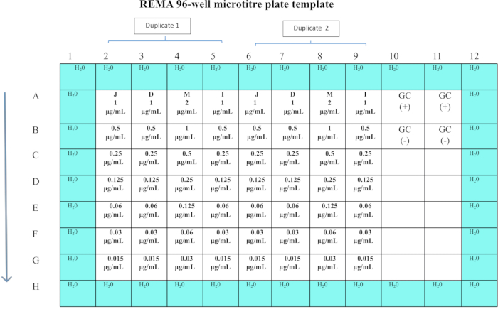
Figure 1: Template diagram of REMA 96 well plate assay for determination of MIC of anti-TB drugs. 4x concentration of drugs (BDQ, DEL, MOX, and INH) were added to row B respectively and were serially diluted in a two-fold dilution down the plate with the exception of the control and perimeter wells. Please click here to view a larger version of this figure.
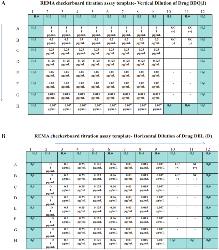
Figure 2: Template diagram of the Resazurin microtiter checkerboard titration assay for evaluating drug interactions. The REMA checkerboard titration assay evaluates the interaction between (A) Bedaquiline (BDQ) and (B) Delamanid (DEL) against M. tuberculosis. The vertical dilution series represents varying concentrations of BDQ (2a), while the horizontal dilution series represents DEL (2b). Please click here to view a larger version of this figure.
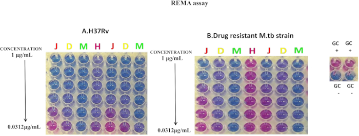
Figure 3: Individual MIC determination trials by REMA assay. MIC determination for laboratory strain (A) H37Rv (drug-sensitive) and (B) drug-resistant clinical isolate of M. tuberculosis. The initial concentrations of drugs, Bedaquiline(J), Delamanid(D), Moxifloxacin (M), and isoniazid (H), are also shown individually. These drugs were serially diluted in a two-fold dilution down the plate with the exception of the control and perimeter wells. Viability is determined by the change in color of Resazurin (blue) to Resorufin (pink). This figure has been modified and reused with permission from Chandramohan et al.18. Please click here to view a larger version of this figure.
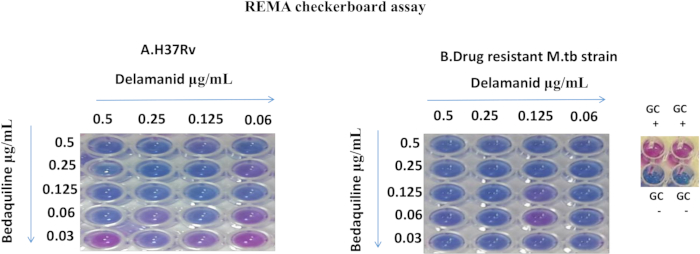
Figure 4: REMA checkerboard assay results. The results from the REMA checkerboard assay for laboratory strain (A) H37Rv (drug-sensitive) and (B) drug-resistant clinical isolate of M. tuberculosis. Depicted are the wells containing different strains growing in the sub-MIC fractions of Bedaquiline that are diluted vertically in combinations with sub-MIC fractions of Delamanid. Viability is determined by the change in color of Resazurin (blue) to Resorufin (pink). This figure has been modified and reused with permission from Chandramohan et al.18. Please click here to view a larger version of this figure.
| S.No | Drugs | Weight | Solvent / Volume | Drug Stock(μg/mL) | Drug Dilution | Working Solution (4x) | |
| Drug stock (μL) | 7H9-S (μL) | ||||||
| 1 | INH | 2 mg | distilled water / 2 mL | 1000 | 10 | 2490 | 4 μg/mL |
| 2 | MOX | 2 mg | distilled water / 2 mL | 1000 | 20 | 2480 | 8μg/mL |
| 3 | BDQ | 2 mg | DMSO / 2 mL | 1000 | 10 | 2490 | 4 μg/mL |
| 4 | DEL | 2 mg | DMSO / 2 mL | 1000 | 10 | 2490 | 4 μg/mL |
Table 1: The drugs used in the example experiment, preparation of their stock, and working solutions.
| 1. Calculation of FIC for each drug | |
| For Drug A | FIC (A) = (MIC of drug A in combination/MIC of drug A alone) |
| For drug B | FIC (B) = (MIC of drug B in combination/MIC of drug Balone) |
| 2. Calculation of FIC Index | |
| FIC Index = FIC (A)+ FIC (B) |
Table 2: Calculation of fractional inhibitory concentrations (FICs) and FIC index for Drug A and Drug B in combination.
| Stain Name | BDQ | DEL | MOX | INH |
| H37Rv | 0.125 | 0.063 | 0.063 | 0.063 |
| H-Mono | 0.25 | 0.063 | 0.063 | >1 |
Table 3: Consolidated MIC based on REMA assay against laboratory drug-sensitive H37RV and drug-resistant clinical isolate of M. tuberculosis. This table has been modified with permission from Chandramohan et al.18.
| STRAIN | DRUG COMBINATION | MIC (µg/mL) | ΣFIC | OUTCOME | |
| ALONE | COMBINATION | ||||
| BDQ | 0.125 | 0.03 | |||
| H37RV | DEL | 0.063 | 0.125 | 2.26 | ADDITIVE |
| BDQ | 0.25 | 0.03 | |||
| H-Mono | DEL | 0.063 | 0.06 | 1.12 | ADDITIVE |
Table 4: REMA-checkerboard assay: The MICs of selected anti-tuberculous compounds against drug-sensitive and drug-resistant clinical isolate of M. tuberculosis and corresponding interactions profiles with Bedaquiline assessed by REMA-checkerboard assay. This table has been modified with permission from Chandramohan et al.18.
Discussion
The global threat posed by drug-resistant tuberculosis (DR-TB) has become a pressing issue1,19. This has created an urgent need for developing improved, rapid methods for drug susceptibility testing (DST), particularly for second-line and newer anti-TB drugs. Such methods are essential to enhance treatment strategies, manage anti-TB drugs effectively, and reduce the risks of disease progression and further resistance20. A method that evaluates both drug susceptibility and interaction profiles would provide significant benefits in this context, improving treatment outcomes and supporting more effective TB control strategies.
Resazurin, a redox-sensitive dye, is extensively used in bacterial viability assays as an indicator of metabolic activity21. The Resazurin Microtiter Assay (REMA) and its checkerboard variation are cost-effective, reliable tools for assessing the efficacy of individual anti-TB drugs and their combinations11,16. Despite their utility, obtaining consistent results requires careful adherence to key experimental steps. The preparation of a uniform inoculum is vital; freshly cultured bacteria aged 21-28 days and adjusted to McFarland 1.0 turbidity are recommended22,23. Dispersing bacterial clumps is necessary to avoid skewed results, typically achieved by vigorous vortexing. Throughout the procedure, maintaining strict sterility is paramount, as well as using sterile equipment such as glass beads and pipettes to minimize contamination risks. Positive controls confirm bacterial growth, while negative controls ensure that contamination has not occurred. To prevent evaporation, which could alter drug concentrations and outcomes, sterile water is used to fill the outer wells of the microtiter plates11.
Another critical aspect is the precise preparation and dilution of drugs. Any inaccuracy during these steps can distort the determination of minimum inhibitory concentrations (MICs) or the fractional inhibitory concentration (FIC) indices used in evaluating drug interactions. In REMA assays, multichannel pipettes are preferred for accurate dilutions, while in checkerboard assays, one drug is diluted along rows and the other along columns to test interactions comprehensively. Mistakes at this stage could lead to incorrect conclusions about the synergistic or antagonistic effects of drug combinations24. Post-incubation handling is equally important; plates must be sealed during incubation at 37°C for seven days to prevent contamination or evaporation. After incubation, resazurin is carefully added at a concentration of 0.01-0.02%, with its color change from blue to pink serving as an indicator of bacterial growth. Deviations, such as immediate color change upon adding resazurin, may indicate contamination and should be excluded from analysis25.
Compared to other phenotypic methods like the BACTEC MGIT 960 or agar proportion tests, REMA is significantly less expensive and easier to use, making it especially suitable for resource-limited settings26,27. Unlike molecular methods such as GeneXpert, which focuses on detecting genetic mutations associated with drug resistance, REMA provides insights into the actual phenotypic susceptibility of bacteria and their interactions with drugs. However, REMA's lower sensitivity, with a detection limit of approximately 1 × 10⁵ CFU/mL, is a drawback when compared to colony-forming unit (CFU)-based evaluations28. This detection threshold is typically reached after the standard 7-day incubation period, after which the resazurin is added for final visualization. Nonetheless, the straightforward execution and low-cost setup of REMA assays make them highly practical in many research and resource-limited settings.
It is essential to emphasize that the colorimetric shift observed in REMA reflects cellular viability, i.e., metabolic activity sufficient to reduce resazurin, rather than active replication or growth. This distinction is critical in interpreting MIC values. As described by Mouton and Vinks, viable but non-dividing cells can maintain respiratory activity and convert resazurin from blue to pink, even in the presence of growth-inhibitory drug concentrations29. This phenomenon underscores the importance of aligning the timing of resazurin addition with the exponential growth phase of M. tuberculosis to ensure that observed color changes more accurately reflect drug effects on proliferation rather than mere viability. The MIC determined through REMA thus represents a phenotypic endpoint of metabolic inhibition, which may differ slightly from MIC values derived through growth-based methods. Moreover, the pharmacodynamic concept of the stationary concentration (SC), the concentration at which bacterial kill rate equals growth rate, provides a more nuanced understanding of antimicrobial action beyond fixed time-point MIC readings.
Despite their many advantages, REMA and checkerboard assays also have limitations. The reliance on visual interpretation of color changes introduces potential variability between observers, which could impact reproducibility. Color perception can be influenced by ambient lighting conditions and subjective bias. While this protocol emphasizes consistent lighting and reference controls, further improvements can be achieved by incorporating automated colorimetric or fluorescence-based detection systems. Fluorescent derivatives of resazurin and digital image analysis-such as those using smartphone cameras or spectrophotometers, have been shown to improve precision and reduce observer bias. For example, recent studies by Sychev et al. and Postnikov et al. have demonstrated that smartphone-based imaging can provide quantitative readouts that correlate well with spectrophotometric analysis30,31. Moreover, high-sensitivity fluorescence methods, as described by Mehlawat et al., can enhance early detection of viable organisms, although they require more advanced instrumentation32. Additionally, the use of liquid culture media raises biosafety concerns due to the potential for aerosol generation. The limited number of M. tuberculosis strains and drug combinations tested in the study described here further underscores the need for broader validation of these methods to establish their general applicability across diverse strains and combinations.
In this study, we used the REMA assay to determine MICs for INH, MOX, BDQ, and DEL in M. tuberculosis (Table 3). Additionally, the REMA checkerboard assay was used to evaluate drug interactions between BDQ and DEL. The purpose of the checkerboard assay is to evaluate the in vitro interactions of novel anti-TB drugs when combined with existing or preclinical TB drugs, which are urgently needed to shorten treatment duration33. In this study, we aimed to determine the interaction effects of bedaquiline combined with delamanid on drug-sensitive and drug-resistant clinical isolates of M. tuberculosis. The findings demonstrate that the BDQ+DEL drug combination exhibits an additive effect in its sterilizing activity against both drug-sensitive and drug-resistant M. tuberculosis clinical isolates (Table 4).
In conclusion, the REMA and REMA checkerboard assays represent accessible, cost-effective methods for studying anti-TB drugs. Their reliability depends on strict adherence to established protocols, especially regarding inoculum preparation, drug dilutions, incubation conditions, and resazurin application. Although these methods provide valuable in vitro data, they may not fully replicate the complexities of in vivo systems, and manual color change interpretation can introduce variability. Future improvements, such as integrating automated colorimetric readers, could address these challenges and enhance precision. Despite these limitations, these assays remain indispensable tools for studying multidrug-resistant and extensively drug-resistant TB, particularly for identifying synergistic drug combinations and advancing TB treatment strategies.
Disclosures
The authors have nothing to disclose.
Materials
| Name | Company | Catalog Number | Comments |
|---|---|---|---|
| 250 mL square bottle with cap | Schott Duran | 218203655 | Media preparation |
| 7H9 broth | Becton Dickinson | 271310 | Growth media for M. tuberculosis (Room temparature) |
| 96-well plate with lid | Corning | 3596 | 96-well Clear Flat Bottom Polystyrene TC-treated Microplates, Individually Wrapped, with Lid, Sterile (Room temparature) |
| ART-Tips (1000 μL) | TARSONS | 528106 | Hydrophobic polyethylene filters with Polypropylene Racked Filter Tip-Sterile (Room temparature) |
| ART-Tips (200 μL) | TARSONS | 528104 | Hydrophobic polyethylene filters with Polypropylene Racked Filter Tip-Sterile (Room temparature) |
| Barium chloride dihydrate | Merck | 217565 | McFarland Standard preparation (Room temparature) |
| Bedaquiline (BDQ) | Jansen and Jansen | Gift from Jansen and Jansen | Anti-TB drug (4 °C) |
| Casitone | Becton Dickinson | 225930 | Supplement for bacterial culture (Room temparature) |
| Delamanid (DEL) | Otsuka Pharmaceuticals | Gift from Otsuka Pharmaceuticals | Anti-TB drug (4 °C) |
| Dimethyl sulfoxide | Sigma-Aldrich | 67-68-5 | Drug stock preparation (Room temparature) |
| Filter 0.2 μm | Millipore | SLGVR33RS | 0.22 μm PVDF ,disposable , sterile syringe filter (Room temparature) |
| Glycerol | Merck | 356350 | Added to culture media (Room temparature) |
| Inoculating Loop | Sigma-Aldrich | I8263 | For culture suspension preparation (Room temparature) |
| Isoniazid (INH) | Sigma-Aldrich | I3377-5G | Anti-TB drug (4 °C) |
| LJ medium Slant | BD Mycobactosel | 221414 | Growth media for M. tuberculosis (4 °C) |
| McCartney Bijou Narrow Mouth bottle, 14 mL | Supertek | 12.124.0014 | For culture suspension preparation (Room temparature) |
| McCartney Bijou Narrow Mouth bottle, 7 mL | Supertek | 12.124.0007 | For culture suspension preparation (Room temparature) |
| Micropipettes (1000 μL) | Gilson | F144059M | For precise liquid handling (Room temparature) |
| Micropipettes (200 μL) | Gilson | FA10005M | For precise liquid handling (Room temparature) |
| Moxifloxacin (MOX) | Sigma-Aldrich | SML1581-50MG | Anti-TB drug (4 °C) |
| Multichannel pipette(20–200 μL) | VWR | 849070024 | For simultaneous pipetting across wells |
| OADC | Becton Dickinson | 211886 | Supplement for bacterial culture (4 °C) |
| Pipette fillers | Merck | Z333980 | For precise liquid handling (Room temparature) |
| Resazurin sodium salt | Sigma-Aldrich | 62758-13-8 | Redox indicator for REMA assays (Room temparature) |
| Serological pipettes,10 mL | Corning | CLS4100 | Sterile, polystyrene disposable pipettes were used (Room temparature) |
| Solid-glass beads, diam. 6 mm | Sigma-Aldrich | Z265950 | For culture suspension preparation (Room temparature) |
| Sulfuric acid 95–97% | Merck | 100731 | McFarland Standard preparation (Room temparature) |
| Syringes for filter | Dispo Van | 100032 | For use with syringe filters |
| Vortex Mixer | REMI | CM-101 PLUS | For culture suspension preparation |
| Zipper-top poly bag | Sigma-Aldrich | Z162949 |
References
- Global tuberculosis report 2023. , World Health Organization. https://www.who.int/publications/i/item/9789240083851 (2023).
- Khawbung, J. L., Nath, D., Chakraborty, S. Drug-resistant tuberculosis: A review. Comp Immunol Microbiol Infect Dis. 74, 101574(2021).
- Alame Emane, A. K., Guo, X., Takiff, H. E., Liu, S. Highly transmitted M. tuberculosis strains are more likely to evolve MDR/XDR and cause outbreaks, but what makes them highly transmitted. Tuberculosis. 129, 102092(2021).
- Koul, A., et al. Delayed bactericidal response of Mycobacterium tuberculosis to bedaquiline involves remodelling of bacterial metabolism. Nat Commun. 5, 3369(2014).
- Matsumoto, M., et al. OPC-67683, a nitro-dihydro-imidazooxazole derivative with promising action against tuberculosis in vitro and in mice. PLoS Med. 3 (11), e466(2006).
- WHO consolidated guidelines on tuberculosis: Module 4. treatment drug-resistant tuberculosis treatment. , World Health Organization. At https://www.who.int/publications/i/item/9789240063129 (2020).
- Conradie, F., et al. Treatment of highly drug-resistant pulmonary tuberculosis. N Engl J Med. 382 (10), 893-902 (2020).
- Lechartier, B., Hartkoorn, R. C., Cole, S. T. In vitro combination studies of benzothiazinone lead compound BTZ043 against Mycobacterium tuberculosis. Antimicrob Agents Chemother. 56 (11), 5790-5793 (2012).
- Mallikaarjun, S., et al. Delamanid coadministered with antiretroviral drugs or anti-tuberculosis drugs shows no clinically relevant drug-drug interactions in healthy subjects. Antimicrob Agents Chemother. 60 (10), 5976-5985 (2016).
- Pai, M., Nicol, M. P., Boehme, C. C. Tuberculosis diagnostics: State of the art and future directions. Microbiol Spectr. 4 (5), (2016).
- Palomino, J. -C., et al. Resazurin microtiter assay plate: Simple and inexpensive method for detection of drug resistance in Mycobacterium tuberculosis. Antimicrob Agents Chemother. 46 (8), 2720-2722 (2002).
- Pital, A., Pital, R. C., Leise, J. M. A rapid method for determining the drug susceptibility of Mycobacterium tuberculosis. Am Rev Tuberc. 78 (1), 111-116 (1958).
- Präbst, K., Engelhardt, H., Ringgeler, S., Hübner, H. Basic colorimetric proliferation assays: MTT, WST, and Resazurin. Methods Mol Biol. 1601, 1-17 (2017).
- Schön, T., et al. Antimicrobial susceptibility testing of Mycobacterium tuberculosis complex isolates - the EUCAST broth microdilution reference method for MIC determination. Clin. Microbiol. Infect. 26 (11), 1488-1492 (2020).
- Singh, S., et al. Rapid identification and drug susceptibility Testing of Mycobacterium tuberculosis: Standard operating procedure for non-commercial assays: Part 3: Colorimetric Redox Indicator Assay v1.3.12. J Lab Physicians. 4 (2), 120-126 (2012).
- Caleffi-Ferracioli, K. R., Maltempe, F. G., Siqueira, V. L. D., Cardoso, R. F. Fast detection of drug interaction in Mycobacterium tuberculosis by a checkerboard resazurin method. Tuberculosis. 93 (6), 660-663 (2013).
- Peñuelas-Urquides, K., et al. Measuring of Mycobacterium tuberculosis growth: a correlation of the optical measurements with colony forming units. Braz J Microbiol. 44 (1), 287-290 (2013).
- Chandramohan, Y., et al. In vitro interaction profiles of the new antitubercular drugs bedaquiline and delamanid with Moxifloxacin against clinical Mycobacterium tuberculosis isolates. J Glob Antimicrob Resist. 19, 348-353 (2019).
- Hameed, H. A., et al. Characterization of genetic variants associated with rifampicin resistance level in Mycobacterium tuberculosis clinical isolates collected in Guangzhou Chest Hospital, China. Infect Drug Resist. 15, 5655-5666 (2022).
- Lienhardt, C., et al. New drugs for the treatment of tuberculosis: Needs, challenges, promise, and prospects for the future. J Infect Dis. 205, S241-S249 (2012).
- Anoopkumar-Dukie, S., et al. Resazurin assay of radiation response in cultured cells. Br J Radiol. 78 (934), 945-947 (2005).
- Baker, C. N., Thornsberry, C., Hawkinson, R. W. Inoculum standardization in antimicrobial susceptibility testing: evaluation of overnight agar cultures and the rapid inoculum standardization system. J Clin Microbiol. 17 (3), 450-457 (1983).
- McFarland, J. The nephelometer: an instrument for estimating the number of bacteria in suspensions used for calculating the opsonic index and for vaccines. J Am Med Assoc. XLIX (14), 1176(1907).
- Bellio, P., Fagnani, L., Nazzicone, L., Celenza, G. New and simplified method for drug combination studies by checkerboard assay. MethodsX. 8, 101543(2021).
- Martin, A., Camacho, M., Portaels, F., Palomino, J. C. Resazurin microtiter assay plate testing of Mycobacterium tuberculosis susceptibilities to second-line drugs: Rapid, simple, and inexpensive method. Antimicrob Agents Chemother. 47 (11), 3616-3619 (2003).
- Zhao, L., et al. Evaluation of BACTEC MGIT 960 system for the second-line drugs susceptibility testing of Mycobacterium tuberculosis in China. J Microbiol Methods. 91, 212-214 (2012).
- Said, H. M., et al. Comparison between the BACTEC MGIT 960 system and the agar proportion method for susceptibility testing of multidrug resistant tuberculosis strains in a high burden setting of South Africa. BMC Infect Dis. 12, 369(2012).
- Caviedes, L., Delgado, J., Gilman, R. H. Tetrazolium microplate assay as a rapid and inexpensive colorimetric method for determination of antibiotic susceptibility of Mycobacterium tuberculosis. J Clin Microbiol. 40 (5), 1873-1874 (2002).
- Mouton, J. W., Vinks, A. A. Pharmacokinetic/pharmacodynamic modelling of antibacterials in vitro and in vivo using bacterial growth and kill kinetics. Clin Pharmacokinet. 44 (2), 201-210 (2005).
- Sychev, A. V., Lavrova, A. I., Dogonadze, M. Z., Postnikov, E. B. Establishing compliance between spectral, colourimetric and photometric indicators in resazurin reduction test. Bioengineering. 10 (8), 962(2023).
- Postnikov, E. B., Sychev, A. V., Lavrova, A. I. Dose-response curve in REMA test: Determination from smartphone-based pictures. Analytica. 5 (4), 619-631 (2024).
- Mehlawat, N., Chakkumpulakkal Puthan Veettil, T., Sharpin, R., Wood, B. R., Alan, T. Ultrafast and ultrasensitive bacterial detection in biofluids: Leveraging resazurin as a visible and fluorescent spectroscopic marker. Anal Chem. 96 (45), 18002-18010 (2024).
- Grosset, J., Truffot-Pernot, C., Lacroix, C., Ji, B. Antagonism between isoniazid and the combination pyrazinamide-rifampin against tuberculosis infection in mice. Antimicrob Agents Chemother. 36 (3), 548-551 (1992).
Reprints and Permissions
This article has been published
Video Coming Soon