Constipation is a common prodromal feature of Parkinson's disease (PD), which often manifests years before the diagnosis. This study validated a proposed method to measure constipation in Drosophila melanogaster, and deficits in different fly models of monogenic PD were confirmed.
Method Article
Gut Transit Defects in Drosophila Models of Monogenic Parkinson's Disease Using a Constipation Assay
Opens in a new tab
In This Article
Summary
Loading...
$$\rightleftharpoonup{xx}$$
$$\longleftharp{xx}$$,
$$\longrightharp{xx}$$,
Abstract
Loading...
$$\rightleftharpoonup{xx}$$
$$\longleftharp{xx}$$,
$$\longrightharp{xx}$$,
Parkinson's disease (PD) is a common neurodegenerative motor disorder and is frequently accompanied by several non-motor symptoms. Among the latter, gastrointestinal problems, including constipation, often emerge years before the manifestation of locomotion difficulties and before a diagnosis of PD is established. A recent study proposed a method to measure constipation in a PD fly model that expresses wild-type human alpha-synuclein. In this study, we examined the feasibility of this method and tested whether other genetic fly PD models equally result in constipation. Available fly lines were used to measure the constipation level in autosomal dominant and recessive PD models, including three different mutant human alpha-synuclein-expressing Drosophila lines and Pink1- and Parkin-deficient flies. These flies and their corresponding controls were placed on blue-colored food one day before the experiment to induce blue fecal spots. On the day of the experiment, these flies were transferred to standard cornmeal. The percentage of blue fecal spots to total fecal spots was recorded and calculated every 90 min. The different fly PD models show slower or incomplete expulsion of blue-colored food compared to their respective control flies. Hence, a previously proposed method to measure constipation was validated and applied to various fly models of different forms of genetic PD.
Introduction
Loading...
$$\rightleftharpoonup{xx}$$
$$\longleftharp{xx}$$,
$$\longrightharp{xx}$$,
Parkinson's disease (PD) is a neurodegenerative disorder primarily characterized by motor dysfunction, including the cardinal diagnostic signs of bradykinesia, rigidity, and tremor. Although PD is best known for its motor signs, non-motor symptoms are common, including depression, pain, sleep disturbances, and constipation1,2. Often, these non-motor symptoms emerge early in the disease course and may precede motor symptoms by several years3. Recognizing these non-motor features in the initial stage of the disease may be critical in the early diagnosis of PD, potentially facilitating more timely intervention and disease management.
Constipation and alterations in gut motility are commonly reported non-motor symptoms in PD and are often present long before the onset of motor symptoms3,4. Furthermore, their severity and frequency increase upon PD progression5. Nonetheless, our understanding of constipation patterns and mechanisms remains limited. Hence, the early identification of constipation linked to PD could aid in diagnosing PD at a stage when therapeutic interventions may be more effective in slowing disease progression. Despite its significance, understanding the underlying mechanisms driving constipation in PD remains limited, partially due to the lack of appropriate experimental models and design. Recent advances in studying constipation in Drosophila melanogaster using overexpression of human wild-type alpha-synuclein provide a valuable tool for investigating the mechanisms underlying this early non-motor symptom6.
SNCA, encoding alpha-synuclein, represents the first identified cause of PD7. Both gene multiplication (duplication and triplication) and point mutations in SNCA lead to autosomal dominant inheritance of PD8. Notably, constipation affects up to 90% of patients carrying SNCA mutations5. In addition, several other genes have been identified to cause PD when mutated, including VPS35, which also causes autosomal dominant forms of PD8. Other genes, such as PINK1 and Parkin, cause autosomal recessive PD when mutated9. Non-motor symptoms also occur in these other genetic forms of PD; however, reported data is sparse and, hence, challenging to conclude or investigate the underlying mechanisms10,11.
Several animal models have been created for PD, including in Drosophila melanogaster12. Although Drosophila does not contain the SNCA gene, overexpression of wild-type and mutant forms of the gene induces Parkinsonian phenotypes, including motor symptoms13. In addition, PD-linked mutant vps35, pink1, and parkin flies are well-established models for studying the effect of the respective mutant genes in relation to PD12. Furthermore, the ability to measure constipation in different Drosophila models of PD, including dominant and recessive forms, allows a more in-depth analysis of gastrointestinal dysfunction and, hence, a better understanding of the pathways involved in PD-related constipation.
Here, the feasibility of the recently proposed method to measure constipation was assessed and validated6 using different genetic PD models. By expanding the application of this method to other genetic PD models and making adjustments, we confirm the feasibility of this method as a general assay to measure constipation or altered gut motility and endorse the possibility of performing modifier screens to gain insights into the molecular mechanisms underlying PD's constipation.
Protocol
Loading...
$$\rightleftharpoonup{xx}$$
$$\longleftharp{xx}$$,
$$\longrightharp{xx}$$,
Ten-day-old male flies were used in this assay. For overexpression of SNCA, the ubiquitous driver DaughterlessGal4 was used.
Flies used in this assay:
w1118;;SNCA p.A53T/DaGal4 and its control w1118;;SNCA p.A53T
w1118;;SNCA p.A30P/DaGal4 and its control w1118;;SNCA p.A30P
w1118;;SNCA p.V15A/DaGal4 and its control w1118;;SNCA p.V15A
w1118;;Vps35D650N/DaGal4 and its control w1118;;vps35D650N
w pink1B9;; and their control w pink1RV;;
w1118;;parkD1/D21 and their control w1118;;parkRV
SNCA p.A53T, SNCA p.A30P, w*;;P(EP)park1, w*;;parkΔ21, Vps35D650N, and DaGal4 were commercially obtained (see Table of Materials). w pink1B9 null mutants and controls (w pink1RV) were kindly provided by Jeehye Park and Jongkyeong Chung (Korea Advanced Institute of Science and Technology)14. SNCA p.V15A flies were previously created15. The reagents and the equipment used in this study are listed in the Table of Materials.
1. Preparation of the blue food
- Prepare fresh standard cornmeal fly medium and keep it warm and liquid.
NOTE: A pre-prepared fly medium can be used, but it needs to be heated to make the medium liquid. Standard cornmeal consists of 50 g of cornmeal, 7 g of yeast, 7 g of agar, 13.5 g of dextrose, 10 mL of ethanol (100%), 1.1 g of Methol-4-hydroxybenzoate, 55 g of molasses, and 4.8 mL of propionic acid for 1 L of fly medium. - Mix blue food dye with distilled water in a 1:1 ratio (v/v).
- Add the blue food mix to the standard cornmeal medium to saturate it with the blue food and obtain a uniform blue color.
NOTE: In this study, a 1:100 ratio was used. - Dispense the blue food into large food vials using a Pasteur pipette so the bottom is covered, and let them air dry to solidify.
- Make the same number of food vials with standard cornmeal medium.
- Cover the food vials with a thin paper towel whilst drying to prevent the accumulation of moisture and contamination.
2. Preparation of the assay
- Place a wet Whatman paper in empty food vials.
NOTE: Ensure to soak the Whatman paper to stick to the bottom when the flies are transferred to the vials with the blue medium. - Add 15-20 flies to each vial and leave them for 1 h.
NOTE: Anesthetizing flies enabled the selection of flies with the correct genotype. Subsequently, the flies were swiped in a vial with a brush and aged for ten days. Transfer of the flies is performed by cautiously flipping the flies from one vial to the other and using stoppers to prevent the flies from escaping. - Transfer the flies to the vials containing the blue medium and leave them overnight in an incubator at 25 °C.
3. Execution of the constipation assay
- The following morning, transfer the flies to the vials with standard cornmeal medium and place them back in the incubator for 90 min.
NOTE: One can check if the bellies of all the flies in the vials are blue by placing them under a microscope. - After 90 min, count the number of blue and colorless, opaque dots, mark them with a colored marker, log them, and place the flies back in the incubator (Figure 1).
NOTE: Marking the already counted dots avoids repeated counting. Furthermore, the dots on the food or the vial plug were ignored. - Repeat step 4.2 four more times with the same flies to obtain a time course of the fecal dots. Each time, a different-colored marker was used to avoid repeated counting of previous rounds.
4. Analysis
- Calculate the percentages of blue dots to the total number of dots (blue and opaque dots) for each time point.
- Plot the data in a graph using the percentage of blue dots over the different time points, and via the 'area under curve' analysis in GraphPad, the area under the curve was calculated and plotted.
- Analyze the data in GraphPad Prism to perform an unpaired t-test with Welch's correction16.
NOTE: Welch's correction was included as equal standard deviations could not be tested in all populations.
Results
Loading...
$$\rightleftharpoonup{xx}$$
$$\longleftharp{xx}$$,
$$\longrightharp{xx}$$,
Minor adjustments were made to a previously presented method to measure constipation in flies7 and to test the intestinal function in different genetic PD-related fly models. Overnight incubation of the flies on the blue-colored medium resulted in fly bellies that appeared blue (Figure 2) and fecal matter that left blue spots on the fly food vial (Figure 2). Over time, the blue bellies returned back to the normal yellowish-colored bellies (Figure 2). In addition, the blue fecal spots diminished, and increasing yellow, opaque spots were observed (Figure 2). This transition from blue to yellow spots gives an idea of the level of gut motility or constipation. The transition was complete in control flies after 7 h 30 min, while in PD-related flies, this removal of the blue-colored medium occurred at a slower pace (Figure 2).
Previously, delayed clearance of blue medium was described in 10-day-old flies expressing wild-type alpha-synuclein7, suggesting a level of constipation or delayed clearance due to alterations in gut motility being present in these flies compared to control flies. In addition to duplications or triplications of SNCA, several pathogenic missense variants have been identified in SNCA linked to PD, and we tested three common pathogenic missense variants in SNCA. Expression of mutant alpha-synuclein (p.A30P, p.A53T, and p.V15A) resulted in an incomplete clearance of blue medium within 7 h 30 min, suggesting the presence of constipation in these flies compared to control flies (Figure 3).
Alpha-synuclein is linked to autosomal dominant forms of PD. Interestingly, expression of alpha-synuclein reduces VPS35 protein levels17. VPS35 is a subunit of the retromer and, when mutated, causes PD9,18,19. Increased constipation was observed in the mutant vps35-expressing flies compared to control flies (Figure 4).
Flies deficient for Pink1 and Parkin display delayed clearance of the blue medium; however, after 7 h 30 min, no blue fecal spots were detected anymore in these mutant flies (Figure 5).
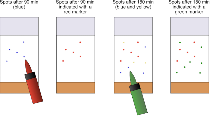
Figure 1: Schematic representation of the counting process with colored markers. After 90 min, the fecal spots were counted and indicated with a colored marker to prevent them from being recounted in the next round. Another color was used to indicate the additional fecal spots during the next counting round. Please click here to view a larger version of this figure.
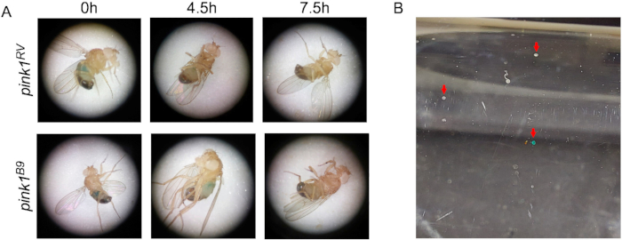
Figure 2: Blue medium results in blue belly and blue fecal spots. (A) Images of the bellies of control flies (pink1RV) and pink1-mutant flies at time points 0 h, 4 h 30 min , and 7 h 30 min show fading of the blue color. (B) Blue and yellow, opaque fecal spots on the fly food vial are indicated with red arrows. Please click here to view a larger version of this figure.
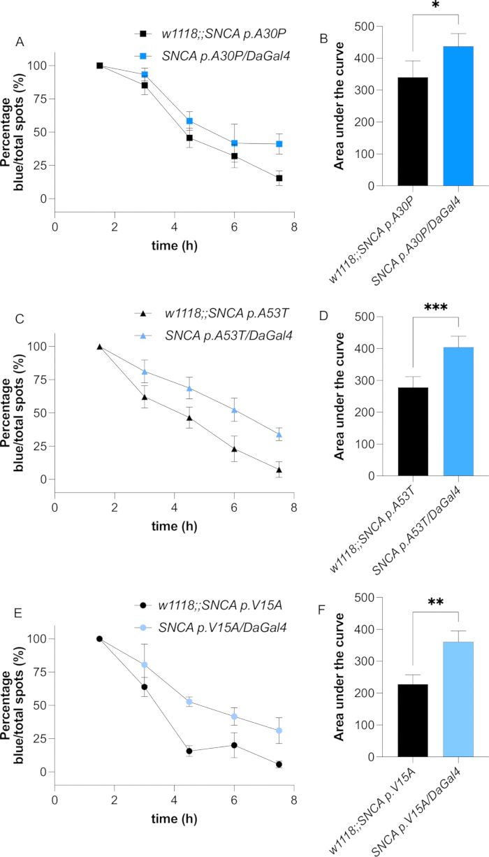
Figure 3: Expression of mutant alpha-synuclein results in constipation. Percentage of blue fecal spots over time in flies overexpressing SNCA p.A30P (A), SNCA p.A53T (C), and SNCA p.V15A (E), and their respective controls. The associated area under the curve (B,D,F) is presented. Data are the mean and standard deviation. n ≥ 3 vials with 15-20 flies per vial. Welch's T-test was performed; *: p < 0.05; **: p < 0.01; ***: p < 0.001. Please click here to view a larger version of this figure.
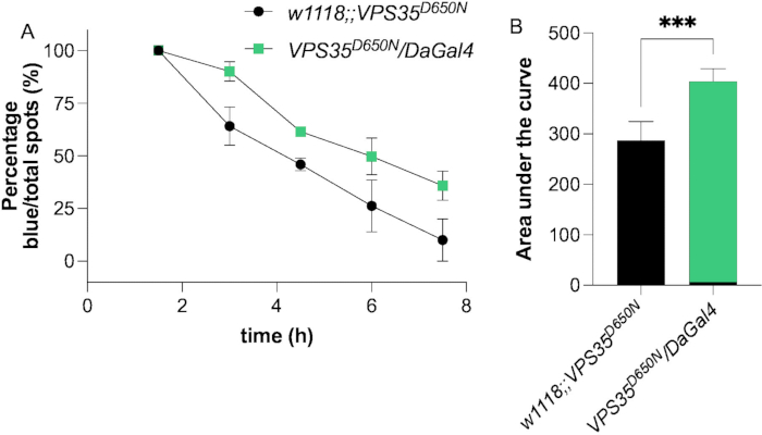
Figure 4: Expression of mutant vps35 results in constipation. (A) Percentage of blue fecal spots over time in flies overexpressing the PD-linked mutant vps35D650N and its control. (B) The area under the curve is presented. The data are the mean and standard deviation. n = 3 vials with 15-20 flies per vial. Welch's T-test was performed; ***: p < 0.001. Please click here to view a larger version of this figure.
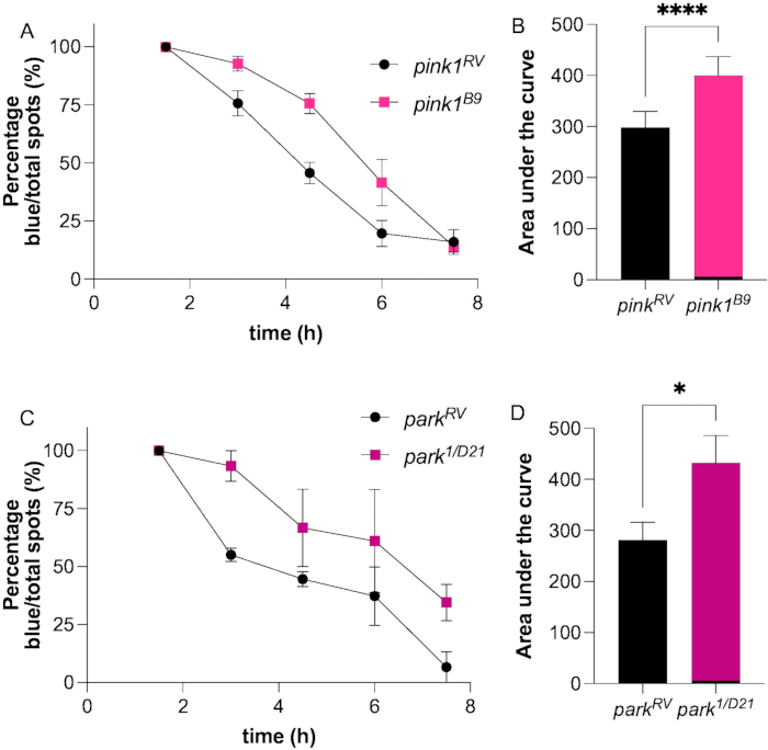
Figure 5: Pink1 and Parkin deficiency induce delayed fecal clearance. Percentage of blue fecal spots over time in Pink1- (A) and Parkin-deficient (C) and their respective control flies. The associated area under the curve (B,D) is presented. Data is the mean with the standard deviation. n ≥ 3 vials with 15-20 flies per vial. Welch's T-test was performed; *: p < 0.05; ****: p < 0.0001. Please click here to view a larger version of this figure.
Discussion
Loading...
$$\rightleftharpoonup{xx}$$
$$\longleftharp{xx}$$,
$$\longrightharp{xx}$$,
Recently, a method to measure constipation in Drosophila melanogaster was proposed. With the application of this method, constipation or altered gut motility was observed in flies expressing wild-type alpha-synuclein7. This method was validated with several alterations, and constipation in all genetic PD models tested was identified.
Compared to the original description of the method, several changes were incorporated to increase the feasibility and robustness. To avoid alterations in blue medium saturation, one batch of blue medium was prepared and divided into the required number of food vials. Furthermore, before adding the flies to the blue medium, they were briefly deprived of food, only providing access to water. This step was added to stimulate the uptake of the blue medium. Without this additional step, not all flies showed a clear blue belly, a critical starting point for obtaining representative results. Finally, several PD fly models show a level of bang sensitivity13,14,20. Hence, the flies were not transferred during each counting step to avoid any influence of this sensitivity on the fecal output. However, different-colored markers were used to indicate the spots to avoid double-counting.
Similar to the original description, it was also observed that flies defecate less frequently in the afternoon. Nonetheless, as this is the case for both control and mutant flies, this did not lead to abnormalities in the recording of the spots. Furthermore, having enough fecal spots to count at each time point is key to obtaining robust results. Hence, a minimum of fifteen flies per vial is recommended7.
In the original manuscript, constipation was observed in 10-day-old flies but not in 1-day-old flies. Thus, in this assay, the constipation assay was performed in ten-day-old flies, and constipation or altered gut motility was observed in all tested PD fly models. Earlier locomotion studies revealed that constipation precedes motor dysfunction in alpha-synuclein-expressing flies, with flight impairments only becoming apparent at three weeks of age16. This temporal sequence of symptoms mirrors the progression observed in PD patients, where gastrointestinal issues often manifest before motor deficits. In contrast to alpha-synuclein models, pink1- or parkin-mutant flies exhibit impairments as early as day one after eclosion21. Therefore, investigating whether gut transit defects manifest at this early time point would provide valuable insights into the progression of non-motor symptoms in these genetic models. Hence, more in-depth experiments should be performed to understand the underlying biology further.
The assay described here facilitates quantitative assessment of gastrointestinal motility and constipation phenotypes but cannot evaluate parameters such as fecal consistency, content, coloration, or morphology. Previously, fecal output was measured, in which color and morphology were evaluated using established methodologies in Drosophila models22,23,24. However, this assay requires specialized equipment (e.g., transparency scanners) and dedicated analytical software, introducing distinct technical and resource considerations compared to the protocol described here. Nonetheless, this assay shows gastrointestinal defects in flies deficient in Pink122,23.
Gastrointestinal dysfunctions are recognized as comorbid manifestations across multiple neurological and neurodegenerative disorders, including autism spectrum disorder, Alzheimer's disease, and multiple sclerosis25,26,27,28, for which Drosophila melanogaster serves as a tractable model organism to investigate the mechanistic underpinnings. The methodological framework established for studying gut motility and constipation in Drosophila can be extrapolated to these diseases.
In conclusion, this study confirmed a feasible and robust method previously presented to measure constipation in flies. This assay shows that constipation is present in multiple genetic Drosophila PD models that are particularly well-suited to study constipation, as they offer the opportunity to study genetically defined at-risk flies before the manifestation of motor features appears. Furthermore, other disease models in Drosophila that manifest gut motility irregularities can be assessed with this as well as drug screening.
Disclosures
Loading...
$$\rightleftharpoonup{xx}$$
$$\longleftharp{xx}$$,
$$\longrightharp{xx}$$,
C. K has served as a consultant for Centogene, Takeda, Biogen, and the Lundbeck Foundation, received speakers' honoraria from Bial, and research funding from DFG, MJFF, and ASAP.
Materials
| Name | Company | Catalog Number | Comments |
|---|---|---|---|
| Bacto Agar | Becton, Dickinson and company | 214010 | |
| Ceapran plugs | Greiner Bio-One | 330070 | |
| Cornmeal | Dr. Oetker GmbH | ||
| Dextrose | Sigma-Aldrich | D9434-1KG | |
| Drosophila container | Greiner Bio-One | 217101 | |
| Ethanol (100%) | J.T. Baker | BAK8025 2500 | |
| Filterpapier Cellulose, Whatman (Grade 3) | Sigma-Aldrich | WHA1003185 | |
| GraphPad Prism | GraphPad | ||
| Methyl-4-hydroxybenzoat | Sigma-Aldrich | H5501-100G | |
| Molasses | Rapunzel Naturkost GmbH | ||
| permanenet colored marker | Stadtler | 11-348 | |
| Propionic acid | Thermo Fisher Scientific Inc. | 149300010 | |
| Royal Blue Azul real icing colour | Wilton Lebensmittelfarben | ||
| Stereo microscope | Nikon | ||
| Yeast | Algist Bruggeman | 1278 | |
| Drosophila stocks | |||
| DaGal4 | Bloomington Stock Center | 95282 | |
| parkD1/D21 | Jeehye Park and Jongkyeong Chung | ||
| parkRV | Jeehye Park and Jongkyeong Chung | ||
| SNCA p.A53T | Bloomington Stock Center | 8148 | |
| SNCA p.A30P | Bloomington Stock Center | 8147 | |
| SNCA p.V15A | previously created in the laboratory | ||
| Vps35D650N | Bloomington Stock Center | 91646 | |
| w pink1B9 | Jeehye Park and Jongkyeong Chung | ||
| w pink1RV | Jeehye Park and Jongkyeong Chung | ||
| w1118 | Bloomington Stock Center | 3605 |
References
Loading...
$$\rightleftharpoonup{xx}$$
$$\longleftharp{xx}$$,
$$\longrightharp{xx}$$,
- Fanciulli, A., et al. Diagnosis and treatment of autonomic failure, pain and sleep disturbances in Parkinson's disease: Guideline "Parkinson's disease" of the German Society of Neurology. J Neurol. 272 (1), 1-32 (2025).
- Kasten, M., Marras, C., Klein, C. Nonmotor signs in genetic forms of Parkinson's disease. Int Rev Neurobiol. 133, 129-178 (2017).
- Bloem, B. R., Okun, M. S., Klein, C. Parkinson's disease. Lancet. 397 (10291), 2284-2303 (2021).
- Higinbotham, A. S., Kilbane, C. W. The gastrointestinal tract and Parkinson's disease. Front Cell Infect Microbiol. 13, 1158986(2023).
- Yuan, X. Y., Chen, Y. S., Liu, Z. Relationship among Parkinson's disease, constipation, microbes, and microbiological therapy. World J Gastroenterol. 30 (3), 225-237 (2024).
- Sodders, M. J., Shen, M., Olsen, A. L. Measuring constipation in a Drosophila model of Parkinson's disease. J Vis Exp. (199), e65966(2023).
- Spillantini, M. G., et al. Alpha-synuclein in Lewy bodies. Nature. 388 (6645), 839-840 (1997).
- Trinh, J., et al. Genotype-phenotype relations for the Parkinson's disease genes SNCA, LRRK2, VPS35: MDSGene systematic review. Mov Disord. 33 (12), 1857-1870 (2018).
- Kasten, M., et al. Genotype-phenotype relations for the Parkinson's disease genes Parkin, PINK1, DJ1: MDSGene systematic review. Mov Disord. 33 (5), 733-741 (2018).
- Deutschländer, A., Ross, O. A., Wszolek, Z. K. VPS35-related Parkinson disease. GeneReviews. , https://www.ncbi.nlm.nih.gov/books/NBK447258/ (2023).
- Wasner, K., Grünewald, A., Klein, C. Parkin-linked Parkinson's disease: From clinical insights to pathogenic mechanisms and novel therapeutic approaches. Neurosci Res. 159, 34-39 (2020).
- Vos, M., Klein, C. The importance of Drosophila melanogaster research to uncover cellular pathways underlying Parkinson's disease. Cells. 10 (3), 579(2021).
- Feany, M. B., Bender, W. W. A Drosophila model of Parkinson's disease. Nature. 404 (6776), 394-398 (2000).
- Park, J., et al. Mitochondrial dysfunction in Drosophila PINK1 mutants is complemented by parkin. Nature. 441 (7097), 1157-1161 (2006).
- Diaw, S. H., et al. Characterization of the pathogenic α-synuclein variant V15A in Parkinson's disease. NPJ Parkinsons Dis. 9 (1), 148(2023).
- Ruxton, G. D. The unequal variance t-test is an underused alternative to Student's t-test and the Mann-Whitney U test. Behav Ecol. 17 (4), 688-690 (2006).
- Lin, G., et al. Phospholipase PLA2G6, a Parkinsonism-associated gene, affects Vps26 and Vps35, retromer function, and ceramide levels, similar to α-synuclein gain. Cell Metab. 28 (4), 605-618.e6 (2018).
- Mandik, F., Vos, M. Neurodegenerative disorders: Spotlight on sphingolipids. Int J Mol Sci. 5 (21), ijms222111998(2021).
- Vos, M., Klein, C., Hicks, A. A. Role of ceramides and sphingolipids in Parkinson's disease. J Mol Biol. 435 (12), 168000(2023).
- Clark, I. E., et al. Drosophila pink1 is required for mitochondrial function and interacts genetically with parkin. Nature. 441 (7097), 1162-1166 (2006).
- Vos, M., et al. Vitamin K2 is a mitochondrial electron carrier that rescues pink1 deficiency. Science. 336, 1306-1310 (2012).
- Fedele, G., et al. Suppression of intestinal dysfunction in a Drosophila model of Parkinson's disease is neuroprotective. Nat Aging. 2 (4), 317-331 (2022).
- Popovic, R., et al. Blocking dPerk in the intestine suppresses neurodegeneration in a Drosophila model of Parkinson's disease. Cell Death Dis. 14 (3), 1-9 (2023).
- Wayland, M. T., et al. Spotting the differences: Probing host/microbiota interactions with a dedicated software tool for the analysis of faecal outputs in Drosophila. J Insect Physiol. 69, 126-135 (2014).
- Heston, M. B., et al. Gut inflammation associated with age and Alzheimer's disease pathology: A human cohort study. Sci Rep. 13 (1), 1-12 (2023).
- Levinthal, D. J., et al. Adding to the burden: Gastrointestinal symptoms and syndromes in multiple sclerosis. Mult Scler Int. 2013, 319201(2013).
- Sorboni, S. G., et al. A comprehensive review on the role of the gut microbiome in human neurological disorders. Clin Microbiol Rev. 35 (1), e0033820(2022).
- Salim, S., et al. The gut-microbiota-brain axis in autism: What Drosophila models can offer. J Neurodev Disord. 13 (1), 37(2021).
Reprints and Permissions
Request permission to reuse the text or figures of this JoVE article
Request Permission