This study reveals that preliminary and backup Antarctic inland expedition team members demonstrated significantly elevated anxiety levels, prefrontal neuroplastic compensation (left anterior prefrontal cortex activation), and sleep fragmentation (Wake After Sleep Onset WASO) under high-altitude conditions. These findings provide an empirical basis for developing adaptive training protocols for extreme environments.
A subscription to JoVE is required to view this content. Sign in or start your free trial.
Method Article
Psychological and Sleep Changes in Pre-Selected Members of the Polar Inland Expedition Team During Rapid Ascent High-Altitude Training
* These authors contributed equally
In This Article
Summary
Abstract
This study established and implemented a multidimensional assessment protocol to systematically investigate neurobehavioral changes among 30 preliminary and backup members of the Antarctic inland expedition team undergoing rapid altitude training. The protocol integrates psychometric scales (BFI-44, DASS-21, PSQI), behavioral paradigms (N-back task), functional near-infrared spectroscopy (fNIRS), and wrist actigraphy to assess neurobehavioral adaptations across varying altitudes (0 m above sea level (a.s.l) / 3,700 m a.s.l / 4,300 m a.s.l) over an 8-day cycle. The core design comprises pre-ascent baseline testing; synchronized cognitive-fNIRS measurements at target altitudes; 24/8 sleep monitoring (including 8 h nocturnal actigraphy). This multimodal framework enables quantitative characterization of prefrontal compensatory activation, sleep fragmentation dynamics, and altitude-induced anxiety fluctuations. Findings reveal that, compared to Chinese normative data, participants exhibited significantly elevated agreeableness (t=3.940, P<0.001) and conscientiousness (t=9.736, P<0.001), alongside reduced neuroticism (t=-14.087, P<0.001). Acute hypoxia exacerbated anxiety (Z=-4.098, P<0.001), with greater severity in ethnic minority members (H=6.405, P=0.011). Actigraphy demonstrated altitude-dependent sleep fragmentation (WASO: 4,300 m a.s.l versus baseline, P=0.028). N-back tasks confirmed preserved working memory at 3,700 m a.s.l. (accuracy P=0.027) mediated by compensatory prefrontal activation (L-aPFC activation P=0.043). These data establish evidence-based selection criteria for polar science expeditions in high-altitude settings and delineate neuroplastic resilience thresholds under hypoxic stress.
Introduction
Polar expedition members must adapt to coexisting physiological and psychological challenges in extreme environments1. The high-altitude conditions of Antarctica's interior (elevation > 4000 m a.s.l) can induce hypoxia-related cognitive impairment and sleep disruption. Although only Dome A (>4000 m a.s.l) is a permanent high-altitude Antarctic research station, traverses to inland sites such as Dome A and Dome F involve prolonged exposure (1-2 weeks) to altitudes exceeding 3000-4000 m a.s.l2,3. Moreover, hypoxia is a key stressor encountered both during these traverses and at Dome A itself, reflecting the conditions simulated in this training protocol. Therefore, understanding neurobehavioral adaptation to hypoxia is essential for personnel selection and training optimization for inland traverse teams and Dome A operators, to ensure safety and performance in these logistically critical and scientifically valuable missions. The overall goal of this method is to establish a multimodal assessment protocol that systematically quantifies neurobehavioral adaptations during rapid altitude acclimatization, integrating psychological, cognitive, neurovascular, and sleep parameters into a unified framework. This technique was developed in response to critical limitations in existing research, which predominantly examines psychological or physiological parameters in isolation4,5,6,7. The advantages of this multimodal technique over conventional methods include:
Ecological validity: Simultaneous fNIRS-behavioral coupling captures real-time neurocognitive adaptations8,9, overcoming retrospective biases inherent in scale-only assessments7
Comprehensive profiling: Integrating actigraphy with validated scales (PSQI) provides objective verification of subjective sleep reports10,11
Dynamic tracking: Repeated measures across altitudes reveal temporal adaptation patterns unattainable through single-point assessments4
Within the wider literature, this protocol addresses the escalating operational demands of Antarctic research12 by providing the first standardized framework that: Quantifies prefrontal compensatory mechanisms by fNIRS during cognitive loading under hypoxia8,13; correlates personality traits (BFI-44) with altitude-induced anxiety dynamics (DASS-21)14,15; synchronizes objective sleep fragmentation (actigraphy) with neurobehavioral outcomes16,17.
This method is appropriate for researchers and practitioners who require personnel selection systems for high-altitude polar operations, validation of neuroplastic resilience thresholds under hypoxic stress (>3,700 m a.s.l.), and objective biomarkers (e.g., L-aPFC activation) to predict task performance degradation. Implementation considerations: Recommended for adults aged 25-45 without cardiopulmonary comorbidities, requires 3-day acclimatization per altitude tier (3,700 m a.s.l to 4,300 m a.s.l), technical constraints: fNIRS optode placement limits concurrent headgear use. With increasing Antarctic research stations and diversified scientific missions12, this protocol provides evidence-based solutions for optimizing adaptive training in extreme environments.
Access restricted. Please log in or start a trial to view this content.
Protocol
1. Study participants
- Participant selection
- Sampling method: A cluster sampling of 30 pre-selected and alternate team members from the Antarctic inland expedition team was taken as the study subjects.
- Study subjects: Include all pre-selected candidates and reserve members from the Chinese Antarctic Inland Expedition Team. Exclude individuals unable to complete the training regimen during the training period (e.g., those who failed to complete the training plan due to illness, physical discomfort, or other reasons).
- Data collection
- Demographic documentation: Record comprehensive sociodemographic data, including age, educational attainment, and previous polar expedition experience (Table 1).
- Ensure that strict anonymization measures are enforced for all psychometric data, including: assignment of unique anonymized IDs (e.g., EXP##) to replace personal identifiers, removal of direct identifiers (e.g., names, addresses) from datasets, storage of identifiable data (e.g., consent forms) in password-protected, encrypted files separate from research data.
| n=30 | N | % |
| Ethnicity | ||
| Han Chinese | 28 | 93.3 |
| Ethnic Minority(Tibetan and Manchu) | 2 | 6.67 |
| Highest educational attainment | ||
| High school and below | 1 | 3.3 |
| College or Bachelor's Degree | 11 | 36.7 |
| Master's Degree | 8 | 26.7 |
| Doctoral Degree | 10 | 33.3 |
| Only Child | ||
| Yes | 16 | 53.3 |
| No | 14 | 46.7 |
| Marital Status | ||
| Unmarried | 15 | 50 |
| Married | 15 | 50 |
| Number of Scientific Expeditions Participated | ||
| Not Participated | 24 | 80 |
| 1 Time | 2 | 6.7 |
| 2 or More Times | 4 | 13.3 |
| Age: 32.72±7.73 |
Table 1: Demographic data. (A) This table presents the ethnic composition, educational level, only-child status, marital status, and frequency of research participation for the sample, with the last row showing age data (Mean ± SD). (B) N: Sample count; %: Percentage relative to total sample size (n=30), data shows Mean ± SD.
2. Preparation of psychological and sleep assessment instruments
- Standardized psychometric scales: Carry out a researcher-administered questionnaire-based assessment in a standardized interview format to ensure comprehension and minimize self-reporting bias. Guide participants verbally through each item, with clarifications provided as needed.
- Big five inventory-44 (BFI-44)14
- Select the 44-item BFI-44 questionnaire to assess five personality dimensions. Instruct participants to rate statements using a 5-point Likert scale (1=Strongly disagree to 5=Strongly agree).
- Calculate domain scores for Extraversion, Agreeableness, Neuroticism, Openness, and Conscientiousness. Higher scores indicate stronger expression of corresponding traits. The scale includes 28 positively keyed items.
- Depression anxiety stress scales-21 (DASS-21)15
- Administer the 21-item DASS-21 with a 4-point Likert scale (1= Did not apply to me at all to 4=Applied to me very much). Record subscale scores for Depression, Anxiety, and Stress. Elevated scores reflect greater severity of emotional disturbances.
- Pittsburgh Sleep Quality Index (PSQI)10
- Distribute 18-item PSQI questionnaires to participants. Instruct completion within 10 min using 0-3 scoring per domain: Subjective sleep quality, Sleep latency, Sleep disturbances, hypnotic medication use, daytime dysfunction. Calculate global scores (0-21 scale). Higher total scores on the PSQI indicate poorer overall sleep quality.
- Big five inventory-44 (BFI-44)14
- Sleep monitoring
NOTE: The apparatus used here for wrist actigraphy was ActiGraph wGT3X-BT16.- Initialize devices using the software with standardized timestamps.
- Launch ActiLife v6.15.0. Click on Device > Initialize > Set. Set the sampling rate: 60 Hz, Start time: YYYY-MM-DD HH:MM:SS (synchronized with atomic clock), Subject ID: EXP## (anonymized format).
- Set a checkpoint to verify that the initialization log displays Initialization Successful.
- Calibrate the software using manufacturer protocols after placement on the non-dominant wrist. Insert the device into the USB Dock. Click Calibrate and then follow the on-screen gravity vector test protocol.
- Collect 24/8 activity data at a 60 Hz sampling rate. Perform a visual checkpoint and confirm the green status LED blinks every 15 s (normal operation).
- Carry out data acquisition. Post-deployment, insert the device into the dock. Select ActiLife. Click on Download > RAW .gt3x format > Process using Cole-Kripke algorithm. Use Cole-Kripke algorithm for sleep-wake classification. Validate device-derived sleep parameters via participant sleep diaries17.
- Perform sampling with continuous 24/8 recording at 60 Hz (ensure space between value/unit: 60 Hz)
3. Cognitive assessment and fNIRS acquisition
- Letter N-back task8,9
- Using E-Prime 2.0 software, uppercase letters are randomly presented in the center of a 16-inch LCD monitor. Participants are required to make judgments: press the F key if the current letter matches the one presented N positions earlier or press the J key if it does not match (N=1, 1-back; N=2, 2-back; N=3, 3-back).
- Record accuracy (ACC) and reaction time (RT) metrics for 50 trials per difficulty level.
- In the E-Prime output settings, configure the DataFile.Name as "EXP##Nback.edat2". For the stimulus onset time, select the variables: ACC (Correct = 1 / Incorrect = 0) and RT (in milliseconds). Click Export > Excel format.
- Click on Analyze > General Linear Model > Repeated MeasuresWithin-Subject Factors: N-level(1,2,3). Set checkpoints at Raw data: 50 trials/level = 150 trials total.
- For the practice phase, in E-Prime, click on BlockProc > Practice Properties. Set Max trials: 10, Feedback Screen: Correct! (green) / Incorrect (red), and Advancement: Press the 'Q' key to repeat the practice phase, press the 'P' key to proceed to the formal experiment. A checkpoint is set, limiting the practice phase to a maximum of 3 repetitions (Figure 1).
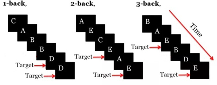
Figure 1: Letter N-back paradigm. This schematic illustrates the letter n-back task paradigm across three difficulty levels (1-back, 2-back, and 3-back). Please click here to view a larger version of this figure.
- fNIRS data acquisition
NOTE: The NirScan-500C, Huichuang, Danyang was used here.- Carry out optode configuration. Position a 48-channel fNIRS headpiece according to the 10-20 international system.
- Set spatial arrangement with source-detector distance at 30 mm, coverage: Prefrontal cortex (PFC) and bilateral temporal regions, and optode matrix: 15 source optodes + 16 detector optodes (Table 2, Table 3, Figure 2).
- Signal Acquisition Parameters
- Set signal acquisition parameters using the NirScan-500C software. Select Dual-wavelength near-infrared light: Set wavelengths to 730 nm ± 5 nm and 850 nm ± 5 nm.
- Configure Sampling rate: Set to 11 Hz by clicking Settings > Acquisition Rate. Enable Task-event synchronization: Activate hardware triggers by selecting Sync > External Trigger.
- Record raw optical intensity changes for subsequent conversion to oxygenated/deoxygenated hemoglobin concentrations using the modified Beer-Lambert law. Verify parameter settings in the software log (e.g., Wavelengths: 730/850 nm, Rate: 11 Hz).
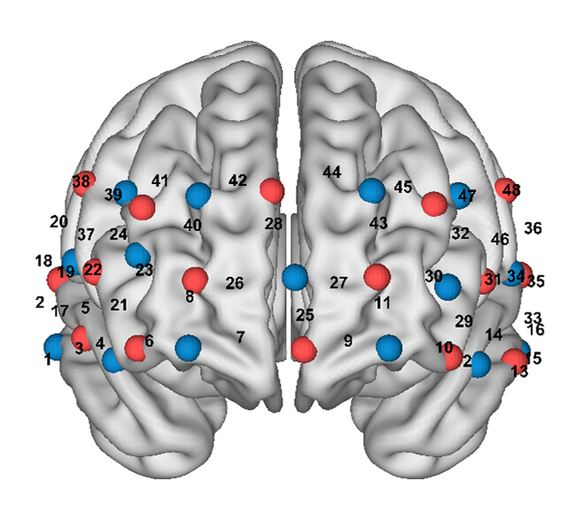
Figure 2: Channel arrangement, position, and configuration of the fNIRS probe. (A) Red circles represent light sources, blue squares represent detectors, and numbered areas represent the nearest source-detector pairs (channels) to measure the brain activities. (B) Coregistered positions of the optodes on a standard brain atlas. The anatomical position of each channel on the brain atlas is reported in detail in Table 1. Please click here to view a larger version of this figure.
| Channel | Brodmann area | Anatomical area | Coverage percentage |
| CH1 | 21 | Middle Temporal gyrus | 97.44% |
| CH2 | 22 | Superior Temporal Gyrus | 43.21% |
| CH3 | 38 | Temporopolar area | 51.84% |
| CH4 | 47 | Inferior prefrontal gyrus | 100.00% |
| CH5 | 47 | Inferior prefrontal gyrus | 56.35% |
| CH6 | 10 | Frontopolar area | 62.75% |
| CH7 | 10 | Frontopolar area | 51.96% |
| CH8 | 10 | Frontopolar area | 100.00% |
| CH9 | 11 | Orbitofrontal area | 59.66% |
| CH10 | 11 | Orbitofrontal area | 46.67% |
| CH11 | 10 | Frontopolar area | 98.66% |
| CH12 | 38 | Temporopolar area | 57.25% |
| CH13 | 21 | Middle Temporal gyrus | 98.63% |
| CH14 | 22 | Superior Temporal Gyrus | 43.46% |
| CH15 | 21 | Middle Temporal gyrus | 92.79% |
| CH16 | 21 | Middle Temporal gyrus | 75.85% |
| CH17 | 22 | Superior Temporal Gyrus | 62.67% |
| CH18 | 43 | Subcentral area | 36.79% |
| CH19 | 44 | pars opercularis_ part of Broca's area | 57.05% |
| CH20 | 6 | Pre-Motor and Supplementary Motor Cortex | 73.08% |
| CH21 | 10 | Frontopolar area | 49.10% |
| CH22 | 46 | Dorsolateral prefrontal cortex | 78.62% |
| CH23 | 10 | Frontopolar area | 93.20% |
| CH24 | 9,46 | Dorsolateral prefrontal cortex | 77.95% |
| CH25 | 10 | Frontopolar area | 87.63% |
| CH26 | 10 | Frontopolar area | 100.00% |
| CH27 | 10 | Frontopolar area | 100.00% |
| CH28 | 10 | Frontopolar area | 99.65% |
| CH29 | 47 | Inferior prefrontal gyrus | 58.95% |
| CH30 | 10 | Frontopolar area | 92.46% |
| CH31 | 45 | pars triangularis Broca's area | 64.19% |
| CH32 | 46 | Dorsolateral prefrontal cortex | 86.52% |
| CH33 | 21 | Middle Temporal gyrus | 56.73% |
| CH34 | 22 | Superior Temporal Gyrus | 53.70% |
| CH35 | 42 | Primary and Auditory Association Cortex | 64.69% |
| CH36 | 43 | Subcentral area | 46.23% |
| CH37 | 9,46 | Dorsolateral prefrontal cortex | 60.54% |
| CH38 | 9 | Dorsolateral prefrontal cortex | 52.09% |
| CH39 | 9,46 | Dorsolateral prefrontal cortex | 92.77% |
| CH40 | 10 | Frontopolar area | 89.88% |
| CH41 | 9,46 | Dorsolateral prefrontal cortex | 91.08% |
| CH42 | 9 | Dorsolateral prefrontal cortex | 84.34% |
| CH43 | 10 | Frontopolar area | 92.46% |
| CH44 | 9 | Dorsolateral prefrontal cortex | 83.67% |
| CH45 | 9,46 | Dorsolateral prefrontal cortex | 90.37% |
| CH46 | 45 | pars triangularis Broca's area | 39.27% |
| CH47 | 9,46 | Dorsolateral prefrontal cortex | 98.16% |
| CH48 | 6 | Pre-Motor and Supplementary Motor Cortex | 80.97% |
Table 2: Brain region distribution of fNIRS measurement channel after calibration by 3D locator. (A) Spatial distribution characteristics of 48 fNIRS channels calibrated by a 3D positioning system across Brodmann areas and corresponding anatomical brain regions. Coverage percentage indicates the effective signal acquisition proportion from target brain regions. (B) Channel: fNIRS optical measurement channel number (CH1-CH48); Brodmann area: Brodmann area number (e.g., 22 = Auditory Association Cortex); Anatomical area: Standard terminology per Terminologia Anatomica (1998); Coverage %: Effective coverage percentage of the optode pairs for the target brain region.
| Anatomical area | Channel |
| L-aPFC | 11,25,27,28,30,43 |
| R-aPFC | 6,7,8,21,23,26,40 |
| L-DLPFC | 32,44,45,47 |
| R-DLPFC | 22,24,37,38,39,41,42 |
Table 3: Areas of region of interest. (A) Display the channel configuration distribution for specific functional brain regions in the fNIRS measurement system, including the anterior Prefrontal Cortex (aPFC) and Dorsolateral Prefrontal Cortex (DLPFC) in both left and right hemispheres. (B) Abbreviations: L = Left hemisphere; R = Right hemisphere; aPFC = anterior Prefrontal Cortex; DLPFC = Dorsolateral Prefrontal Cortex.
4. Data acquisition
- Psychological assessment implementation
NOTE: The team members departed from sea level (Shanghai, 0 m above sea level) and took a commercial flight (approximately 8 h) to Lhasa, located at 3,700 m above sea level, where they conducted a 72 h (3-day) scientific research skills training. Subsequently, the team transferred y ground transportation (approximately 2 h) to Yangbajain at 4,300 m above sea level to continue another 72 h (3-day) scientific research skills training. Data collection was carried out immediately upon the team’s arrival at each of the three aforementioned altitudes (Figure 3).- Conduct standardized administration: Launch pre-assessment group briefing sessions. Administer questionnaires with a 30 min time constraint.
- Mitigate bias: Read standardized instructions from scripted protocols. Eliminate suggestive phrasing by following pre-approved text.
- Synchronize multimodal data: Enable synchronization between cognitive tasks and fNIRS acquisition. Time-lock stimuli to fNIRS triggers by selecting E-Prime > Trigger Settings > Sync to fNIRS.
- Ensure all sessions adhere to identical scripts and timing constraints.
- Sleep parameter monitoring
- Implement continuous actigraphy protocol: Collect 24 h/8 h wrist actigraphy data. Concurrently record sleep diary.
- Ensure data integrity: Perform daily battery checks at 07:00-08:00 by clicking Device > Status > Battery. Validate data continuity by clicking ActiLife > Tools > Data Integrity Check.
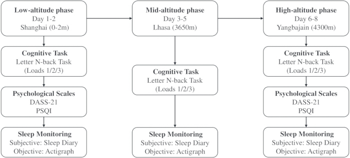
Figure 3: Psychological assessment flowchart. (A) This flowchart illustrates a three-stage psychometric assessment protocol across graded altitude exposures: Low altitude (0-2 m a.s.l.): Baseline measurements in Shanghai (Day 1-2), Mid altitude (3650 m a.s.l.): Acclimatization-phase measurements in Lhasa (Day 3-5), High altitude (4300 m a.s.l.): High-exposure measurements in Yangbajing (Day 6-8). (B) Identical assessment modules implemented at each stage: Cognitive Task: Letter N-back (3 difficulty levels); Psychological Scales: Depression Anxiety Stress Scale (DASS-21), Pittsburgh Sleep Quality Index (PSQI); Sleep Monitoring: Subjective (Sleep diary), Objective (Actigraphy). Please click here to view a larger version of this figure.
5. Data analysis and statistics
- For data preprocessing in a spreadsheet, apply exclusion criteria to remove incomplete datasets with >10% missing responses by selecting Data > Filter > Missing Values. Filter cognitive data to discard RT outliers (<150 ms or> mean ±2.5 SD) using Data > Sort & Filter > Advanced, and calculate the HitRT (in milliseconds) and rejRT (in milliseconds). Handle missing data to implement multiple imputation in SPSS 29.0 by clicking Transform > Multiple Imputation (5 iterations).
- fNIRS signal processing
NOTE: This was done using NirSpark v1.8.8, Huichuang, Danyang.- Convert raw signals to optical density (OD). Click File > Import Raw > Select .nirs file. Select Convert to Optical Density and apply algorithm: OD = -log10(I/I0). Verify OD range 0.1-1.2 AU in View > Channel Metrics. Reject channels with OD >1.5(poor contact) by selecting Tools > Reject Channels.
- Calculate hemodynamic changes. Navigate to Analysis > Hemodynamic > Parameters. Set Law as Modified Beer-Lambert, DPF as 66, and Image Mode as adult_head_template. Execute calculation as [HbO, HbR] = mBLL(OD, [730,850], [6,6]) and HbT = HbO + HbR. Confirm DPF value matches participant age (e.g., 6.0 for 25-45 years).
- Denoise signals by clicking on Preprocessing > Filter > Settings. Select Bandpass Butterworth 4th-order and set the range to 0-0.2 Hz. Perform motion correction via the automatic detection (std_thr=6, amp_thr=0.5) and spline interpolation method and set artifact rejection threshold to SD >±6. Verify SNR improvement >10 dBin by clicking View > Pre/Post Comparison.
- Quantify activation as follows. Define baseline as Mean HbO during the entire post-stimulus task period. Calculate task response as Mean HbO during 2-20 s post-stimulus. Generate ROI averages by selecting Analysis > ROI Masks > Anatomical Templates.
- Sleep efficiency calculation
- Import and preprocess data by clicking File > Import > Select .gt3x file. Set the Epoch to 60s and validate the wear time by selecting Tools > Wear Time Validation > Choi algorithm. To perform actigraphy and sleep diary synchronization, click on Tools > Sleep > Detect Sleep Periods. Then, calibrate the actigraphy data against the sleep diary. Ensure sleep diary timestamps are within ± 5 min of actigraphy data.
- Extract parameters as follows. Navigate to Reports > Sleep Report. Select parameters: Latency, TIB, TST, WASO. Compute Sleep Efficiency = (TST / TIB) x 100 by selecting Options > Formulas. Export results by clicking on Export > Excel .xlsx > Filename: EXP##_SleepEfficiency.xlsx. Confirm actogram displays ≥3 clear sleep/wake cycles.
- Statistical testing
- Perform descriptive statistics. Report normally distributed data as mean ± SD and non-normal data as median [IQR].
- Execute parametric tests. Run one-sample t-tests by clicking Analyze > Compare Means > One-Sample T Test. Conduct RM-ANOVA by clicking Analyze > General Linear Model > Repeated Measures. Apply Greenhouse-Geisser correction for sphericity violations.
- Apply nonparametric alternatives as needed. Use Nonparametric Tests on Related Samples for Wilcoxon signed-rank tests. Control covariates by selecting Analyze > General Linear Model > ANCOVA. Set significance threshold by applying α=0.05 (two-tailed) with Bonferroni correction for multiple comparisons.
Access restricted. Please log in or start a trial to view this content.
Results
Personality traits
Personality characteristics of the pre-selected polar inland expedition members were evaluated using the Big Five Inventory (BFI). Compared to Chinese undergraduate norms18, candidates demonstrated (Table 4) significantly higher scores in Agreeableness (t=3.940, P<0.001) and Conscientiousness (t=9.736, P<0.001); a markedly lower score in Neuroticism (t=−14.078, P<0.001)18. No ...
Access restricted. Please log in or start a trial to view this content.
Discussion
Personality traits of pre-selected polar inland expedition members differ significantly from Chinese norms
As measured by the Big Five Inventory-44 (BFI-44), candidates exhibited elevated agreeableness and conscientiousness alongside reduced neuroticism compared to undergraduate norms (Table 3). This triad-heightened social cooperativeness, task-oriented rigor, and emotional stability-aligns with the psychosocial demands of Antarctic expeditions.
Access restricted. Please log in or start a trial to view this content.
Disclosures
The authors declare no competing interests.
Acknowledgements
The authors extend their sincere gratitude to the Chinese Arctic and Antarctic Administration (Chinese Polar Research Center) for providing critical logistical support and participant access throughout this study. We specifically acknowledge their coordination of high-altitude training protocols and expedition team engagement. The data set is provided by National Arctic and Antarctic Data Center (https://datacenter.chinare.org.cn).
Access restricted. Please log in or start a trial to view this content.
Materials
| Name | Company | Catalog Number | Comments |
|---|---|---|---|
| Actigraph | Actigraph | wGT3X-BT | America |
| ActiLife | Actigraph | 6.15.0 | America |
| E-Prime | Psychology Software Tools | 2.0 | America |
| IBM SPSS Statistics | IBM | 29 | America |
| Microsoft Office | Microsoft Corporation | 2021 | America |
| NirScan | Huichuang Medical Equipment | 500C | Danyang, China |
| NirSpark | Huichuang Medical Equipment | 1.8.8 | Danyang, China |
References
- Wang, Z. J., et al. Applied research on low-altitude wind field using coherent Doppler wind lidar at Zhongshan Station, Antarctic. Chinese J Polar Res. 34 (1), 11-19 (2022).
- Wang, T., et al. Assessment of solar photovoltaic power potential at Kunlun Station, Antarctica. Chinese J Polar Res. 34 (3), 292-302 (2022).
- Liu, X. Y., Wang, Y. Y., Lu, Z. H. Effects of different altitudes on sleep and cognitive function. J Air Force Med Uni. , https://www.fmmu.edu.cn/en/Resources1.htm (2024).
- Guo, Z. M., Huang, F. M., Lu, H., Zu, S. Y., Xu, C. L. Effects of Different Altitudes on Cardiac Hemodynamics and Electrocardiogram in Healthy Adult Males. Chinese J Appl Physiol. 28 (1), 1-4 (2012).
- Lu, Y. H., Xiong, Y. L., Fang, L., Chen, N., Xu, C. L. Comparative Effects of Rapid versus Gradual Tibetan Acclimatization on Cardiovascular Function and Stress Biomarkers in Kunlun Station Pre-Selection Candidates. Chinese J Appl Physiol. 36 (5), 419-424 (2020).
- Huang, F. M., et al. Screening for Hypoxia Susceptibility in Dome A Antarctic Expedition Candidates Using Pulmonary Function Indices. Basic Clin Med. 31 (4), 400-403 (2011).
- Xiaopei, W., et al. Different physiopsychological changes between AMS-susceptible and AMS-resistant pre-selected Antarctic expeditioners in Tibet. Adv Polar Sci. 32 (3), 239-247 (2021).
- Owen, A. M., McMillan, K. M., Laird, A. R., Bullmore, E. N-back working memory paradigm: a meta-analysis of normative functional neuroimaging studies. Human Brain Map. 25 (1), 46-59 (2005).
- Owen, A. M., et al. Putting brain training to the test. Nature. 465 (7299), 775-778 (2010).
- Buysse, D. J., Reynolds, C. F., Monk, T. H., Berman, S. R., Kupfer, D. J. The Pittsburgh Sleep Quality Index: a new instrument for psychiatric practice and research. Psychiatry Res. 28 (2), 193-213 (1989).
- Carney, C. E., et al. The Consensus Sleep Diary: Standardizing Prospective Sleep Self-Monitoring. Sleep. 35 (2), 287-302 (2012).
- Liu, Y. Comprehensive Assessment of Antarctic Operational Capabilities Among Major Polar Expedition Nations. , (2024).
- Virués-Ortega, J., Buela-Casal, G., Garrido, E., Alcázar, B. Neuropsychological functioning associated with high-altitude exposure. Neuropsychol Rev. 14 (4), 197-224 (2004).
- John, O. P., Donahue, E. M., Kentle, R. L. The Big-Five Inventory. Uni California Berkeley. 18 (5), 367-385 (1991).
- Gong, X., Xie, X. Y., Xu, R., Luo, Y. J. Psychometric Validation of the Depression Anxiety Stress Scales-21 (DASS-21) Simplified Chinese Version in Chinese University Students. Chinese J Clin Psychol. 18 (4), 443-446 (2010).
- Dong, J., et al. Anxiety correlates with somatic symptoms and sleep status at high altitudes. Physiol Behav. 112, 23-31 (2013).
- Carney, C. E., et al. The Consensus Sleep Diary: Standardizing Prospective Sleep Self-Monitoring. Sleep. 35 (2), 287-302 (2012).
- Eng, Y., Yue, C. Z. Exploring the Relationship Between Big Five Personality Self-Assessment, Reflective Evaluation, and Mental Health in University Students. Health Med Res Pract. 14 (1), 28-32 (2017).
- Yan, G. G., Ye, Q. Psychological Research in Polar Environments. Adv Psychol Sci. 17 (1), 227-232 (2009).
- Kious, B. M., et al. Altitude and risk of depression and anxiety: findings from the intern health study. Int Rev Psychiat. 31 (7-8), 637-645 (2019).
- Chen, N., et al. Alterations in Circadian Rhythms, Sleep Patterns, and Psychological States Among Overwintering Crew Members During Prolonged Residence at Zhongshan Station, Antarctica. Adv Psychol Sci. , https://kns.cnki.net/KCMS/detail/detail.aspx?dbcode=IPFD&dbname=IPFDLAST2015&filename=OGSF201406001161 (2014).
- Jaeggi, S. M., Buschkuehl, M., Perrig, W. J., Meier, B. The concurrent validity of the N-back task as a working memory measure. Memory. 18 (4), 394-412 (2010).
- Greenberg, K., Zheng, R. Cognitive load theory and its measurement: a study of secondary tasks in relation to working memory. J Cognitive Psychol. 34 (4), 497-515 (2022).
- Jenkins, J. R., Salmon, O. F., Smith, C. M. Cognitive function is unaffected during acute hypoxic exposure but was improved following exercise. Int J Exerc Sci. 15 (5), 1481-1491 (2022).
- Yan, X., Zhang, J., Gong, Q., Weng, X. Adaptive influence of long-term high altitude residence on spatial working memory: an fMRI study. Brain Cognit. 77 (1), 53-59 (2011).
- Zhang, X., Zhang, J. The human brain in a high altitude natural environment: a review. Front Human Neurosci. 16, 915995(2022).
- Ji, X., et al. EEG and fNIRS datasets based on stroop task during two weeks of high-altitude exposure in new immigrants. Sci Data. 11 (1), 350(2024).
- Duncan, J., Owen, A. M. Common regions of the human frontal lobe recruited by diverse cognitive demands. Trends Neurosci. 23 (10), 475-483 (2000).
- Burgess, P. W., Dumontheil, I., Gilbert, S. J. The gateway hypothesis of rostral prefrontal cortex (area 10) function. Trends Cognitive Sci. 11 (7), 290-298 (2007).
- Curtis, C. E., D'Esposito, M. Persistent activity in the prefrontal cortex during working memory. Trends Cognitive Sci. 7 (9), 415-423 (2003).
- Minamoto, T., Kawakami, N., Tsujimoto, T. Robustness of the self-referential process under normobaric hypoxia: an fNIRS study using the GLM and homologous cortical functional connectivity analyses. Front Human Neurosci. 18, 1337798(2024).
- Wang, L., et al. Effects of acute high-altitude exposure on working memory: a functional near-infrared spectroscopy study. Brain Behav. 12 (12), e2776(2022).
- Deary, I. J., Strand, S., Smith, P., Fernandes, C. Intelligence and educational achievement. Intelligence. 35 (1), 13-21 (2007).
- Jaeggi, S. M., Buschkuehl, M., Jonides, J., Perrig, W. J. Improving fluid intelligence with training on working memory. Proc Natl Acad Sci. 105 (19), 6829-6833 (2008).
Access restricted. Please log in or start a trial to view this content.