The protocol describes methods to study micromorphological changes in neurons and the transition of mitochondrial cytochrome c by immuno-transmission electron microscopy.
Method Article
Highlighting the Transition of Mitochondrial Cytochrome C by Immuno-transmission Electron Microscopy
* These authors contributed equally
In This Article
Summary
Abstract
Protein location changes in the microstructure often present a precise regulation mechanism at the level of pathological organelles. How to accurately capture the location of this organelle protein transfer has positive significance and value for the diagnosis and treatment of diseases. Mitochondria play a central role in necrosis and the intrinsic pathway of apoptosis. Under normal conditions, cytochrome c (Cyt c) only exists between the inner and outer membranes of mitochondria and in the nucleus. When cells undergo apoptosis, the mitochondrial inner membrane permeability is altered, and Cyt c is released from mitochondria into the cytoplasm. In this study, a rat model of cerebral ischemia-reperfusion injury was established, and the micromorphological changes of neurons undergoing apoptosis were shown by immuno-transmission electron microscopy. Additionally, it shows the vision of immunogold labeling Cyt c transfer from the mitochondrial matrix to the cytoplasm after cerebral ischemia-reperfusion injury more precisely. The immuno-transmission electron microscopy technique presented in this protocol is suitable for the demonstration of any protein position transitions at the microscopic level.
Introduction
According to the 2021 global burden of disease study, stroke was the 3rd leading cause of death and the 4th leading cause of disability-adjusted life-years. There were 11.9 million new stroke events, in which ischemic stroke constituted 65.3% (7.8 million)1. Neuronal death occurs after cerebral ischemia, including necrosis and apoptosis. The most severely affected brain tissue subsequently undergoes necrotic cell death. Neurons in the ischemic penumbra or periinfarct zone may undergo apoptosis after several hours or days2. In addition to bioenergetic and metabolic functions, mitochondria also play a central role in the intrinsic pathway of apoptosis3,4,5,6.
Cyt C is a protein encoded by nuclear genes. Under normal conditions, Cyt C only exists between the inner and outer membranes of mitochondria and in the nucleus. The apoptotic pathway mediated by its release is associated with the regulatory control of Bcl-2 family members. After being stimulated by apoptosis, Bax/Bak oligomers are formed and inserted into the mitochondrial outer membrane pore, resulting in changes in mitochondrial osmotic pressure and loss of transmembrane potential. Cyt C is released from mitochondria to the cytoplasm, where it binds to the activator of apoptosis 1 to form the apoptotic complex and activate pro-caspase-9. In turn, caspase-3 and caspase-7 are activated, which initiates the Caspase cascade and induces cell apoptosis7.
Transmission electron microscopy (TEM) can clearly observe the changes in cell structure at different stages of apoptosis. Electron microscopy morphological observation is a reliable method for determining apoptosis8. However, it cannot observe changes in Cyt C in ultrastructure. There are several techniques for detecting specific proteins in ultrastructure: flow cytometry, enzyme-Linked Immunosorbent assay, enzyme-linked immunosorbent assay, spectrophotometry, and Western blotting. However, they cannot accurately capture the location changes of protein in the microstructure. Colloidal gold has particles of different sizes, and the electron density of colloidal gold is high. Immunogold technology is particularly suitable for single or multi-label localization studies of immuno-electron microscopy. Due to its inherent light-to-dark red coloration, colloidal gold is also amenable to observation under light microscopy. When specific proteins are Immunogold labeled, the location changes can be visualized by means of a TEM at a subcellular resolution9.
This protocol describes highlighting the transition of mitochondrial Cyt C by immuno-transmission electron microscopy (IEM). The overall goal of this method is to visualize the subcellular translocation of Cyt C during ischemia-induced apoptosis usingIEM. The rationale for IEM is to label Cyt C with colloidal gold and visualize its localization changes at subcellular resolution using TEM9. IEM can observe the dynamic changes of proteins at the spatial level, providing a powerful means for explaining physiological and pathological activities10.
Protocol
The animal program has been approved by the Ethical Committee of SLAS (IACUC Issue No: SYST-2024-018). Here, 6 pathogen-free male SD rats (200-220 g) were raised in independent ventilation cages, maintaining the laboratory environment at 22-25 °C and 45%-65% relative humidity. All rats underwent adaptive feeding for 1 week with free water and diet during the period.
1. Establishment of middle cerebral artery occlusion (MCAO) model
- Randomly divide the rats into the MCAO group and the sham operation group, with three rats in each group. See Figure 1 for the experimental flowchart.
- Verify the equipment involved in this protocol according to the list in Figure 2.
- To establish a model of MCAO of rat ischemia-reperfusion injury, use the intraluminal suture procedure.
- Fast the rats for 12 h before surgery with water accessible.
- Anesthetize SD rats with 2% isoflurane (see Table of Materials) in an air-oxygen mixture at 0.6 L/min. Confirm proper anesthetization by clipping the skin of the limbs with forceps; rats should have no retraction or leg reflex. Fasten the thoroughly unconscious rats on the operating table and maintain the body temperature at 37 °C using an animal temperature maintainer. Tape their eyes closed during the surgery to prevent dryness.
- Remove the fur around the neck with an electric shaver and disinfect the skin with betadine. Make a 1.5 cm midline neck incision with surgical scissors (see Table of Materials), perform blunt dissection on muscle and perivascular fascia, and expose the trachea to locate the right common carotid artery (CCA), external carotid artery (ECA), and internal carotid artery (ICA). Surgical tools should be sterilized to prevent infection.
- Ligate the distal end of the ECA with a 6-0surgicalsuture and place another surgical suture loosely around the proximal end of the ECA to the bifurcation point. Take care not to harm the vagal nerve.
- Clip the CCA and ICA with vascular clips and make a small incision in the ECA between two surgical sutures.
- Introduce the monofilament suture into the ECA lumen down toward the CCA, tighten the surgical suture around the ECA stump to secure the intraluminal suture and prevent bleeding. Ensure the monofilament suture matches the weight of the animal to minimize damage and achieve a target infarct volume of 40% in the ipsilateral hemisphere.
- Cut the remaining portion of the ECA to free the stump and position the ECA stump below the bifurcation point such that it is roughly in line with the ICA.
- Remove the microvascular clip from the ICA, continue advancing the suture until a mild resistance is felt, indicating the occlusion of the middle cerebral artery; this length is typically around 2 cm. Tie the suture around the monofilament tightly. Remove the other microvascular clip from the CCA.
- Close the incision with surgical suture and put the rat in a recovery cage, take care of the animal for up to 2 h post-ischemia.
2. Restoration of middle cerebral artery blood flow
- Before re-anesthesia, check the neuroscore with the Longa Score Scale to evaluate the success of the surgery as described in step 3.
- Anesthetize the rat as above and reopen the incision. Place a microvascular clip on the CCA, withdraw the occluding suture partway from the ICA until the suture end is visible through the ICA.
- Place another micro clip on the ICA above the location of the intraluminal suture, completely remove the occluding suture, and tie off the ECA stump. Reperfusion for 24h.
- Remove the clips from the CCA and ICA. Close the incision with a surgical suture. Administer buprenorphine (0.05 mg/kg; see Table of Materials) subcutaneously.
- Put the rat back into the cage, provide water and mashed food, and take care until it regains consciousness. Maintain the body temperature of the rats at 37± 0.5 °C during the whole experimental procedure.
3. Assessment of neuroscore
- Score neurological deficits after the operation and 24 h post-reperfusion using the Longa Score Scale11: (0) no deficit; (1) failure to extend left (paralyzed side) forepaw fully; (2) circling to the left (paralyzed side); (3) falling to the left (paralyzed side); (4) no spontaneous walking and decreased consciousness; (5) dead.
NOTE: Three operators were evaluated according to the above standards.
4. Brain tissue collection
- Collect brain tissues 24 h post-reperfusion after neurological deficits are scored and assessed.
- Anesthetize the rat as described above. Cut the skin to expose the head and neck and cut the cervical cord from the cervical spine with surgical scissors (see Table of Materials).
- Separate and remove the posterior neck muscles. Carefully remove the skull with curved forceps (see Table of Materials). Pay attention to the dura when separating the skull to avoid scratching the brain tissues. Remove the brain tissues completely.
- Place the brain tissues in TEM and IEM fixative precooled at 4 °C, and store in the refrigerator at 4 °C.
5. Sham operation
- For sham operations, perform all procedures as above except that the occlusion is not done.
6. Ultrathin section preparation and imaging
- TEM ultrathin section preparation
- Fixation: Determine the sampling site of fresh tissue to minimize mechanical damage such as traction, contusion, and extrusion, and ensure the tissue thickness does not exceed 2 mm. Ensure the tissue volume does not exceed 1 mm x 1 mm x 1 mm.
- Fix with TEM fixative at 4 °C for 24 h. Wash 3x for 15 min each in 0.1 M phosphate buffer PB (pH 7.4; see Table of Materials).
- Post fixation: Add 1% osmic acid·0.1 M PB (see Table of Materials) for 2 h at room temperature (25 °C).
- Dehydration: Dehydrate the tissue successively in 30% ethanol for 10 min, 50% ethanol for 10 min, 70% ethanol for 15 min, 90% ethanol for 15 min, absolute ethanol for 15 min, and 100% acetone for 15 min.
- Gum dipping: Immerse tissues successively in solution epoxy resin SPI-Pon 812: acetone=1:3 for 30 min, solution SPI-Pon 812: acetone = 1:1 for 30 min, and solution SPI-Pon 812: acetone = 3:1 for 1 h (see Table of Materials).
- Polymerization and embedding: Immerse the sample in pure SPI-Pon 812 overnight at 37 °C, change out to fresh SPI-Pon 812 for 8 h, and polymerize at 60°C for 48 h (see Table of Materials).
- Sectioning: Slice the embedded block semi-thinly at 0.8 µm thickness with a diamond knife, stain with aminotoluene blue at 25 °C for 5 min. Locate and select areas with densely packed neurons with a light microscope for high-magnification observation. Make ultrathin sections with a microtome at 90 nm thickness. Put the samples on 200-Mesh formvar-coated copper grids (see Table of Materials).
- Post-staining: Stain grids in uranyl acetate at 25 °C for 15 min, then stain grids in lead citrate at 25 °C for 5 min.
- IEM ultrathin section preparation
- Perform step 6.1.1-6.1.4.
- Gum dipping: Immerse tissues successively in solution L. R. White Resin: Ethanol = 1: 3 for 2 h, solution L. R. White Resin: Ethanol =1: 1 for 2 h, and solution L. R. White Resin: Ethanol = 3: 1 for 2 h.
- Polymerization and embedding: Immerse the sample in pure L. R. White Resin overnight at 4 °C, change out to fresh L. R. White Resin for 4h at 25 °C and polymerize at 55 °C for 78 h.
- Sectioning (as done in step 6.1.7): Slice the embedded blocks semi-thinly at 0.8 µmthickness, stain with aminotoluene blue at 25 °C for 5 min. locate and select areas with densely packed neurons with a light microscope. Make ultrathin sections at 90 nm thickness. Put the samples on copper grids.
7. Immunogold labelling
- Rinse 2x with PBS and 1x with glycine buffer at 25 °C for 5 min each, then add 2% phosphate buffer of bovine serum albumin (PBS-BSA) and incubate for 30 min.
- Incubate in the primary antibody (Rabbit polyclonal antibody to Cytochrome C) solution at 37 °C for 2 h. The primary antibody is diluted with PBS-BSA at a ratio of 1:50.
- Rinse with PBS at 25 °C for 5x for 5 min each. Incubate in the colloidal gold-labeled secondary antibody (Gold-conjugated Goat anti-rabbit IgG) solution at 25 °C for 1 h. The secondary antibody is diluted with PBS-BSA at a ratio of 1:20 (see Table of Materials).
- Rinse with PBS at 25 °C for 5x for 5 min each. Rinse with distilled water at 25 °C for 5x for 5 min each (see Table of Materials).
- Post-staining: Stain grids in uranyl acetate at 25 °Cfor 10 min, then stain grids in lead citrate at 25 °C for 2 min (see Table of Materials).
- Image the ultrathin section and take photos on a transmission electron microscope at 80 kV.
Results
The neuroscore evaluation confirms the success of MCAO, according to Longa Score Scale, rats in MCAO group presented at least circling to the left (neuroscore 2). Rats of the sham operation group showed no obvious neurological dysfunction (neuroscore 0). No mortality was observed during the surgery day.The animals' neuroscore are listed in Table 1.
IEM analysis of the MCAO group revealed neurons with shrunken nuclei and elevated cytoplasmic electron density, accompanied by partial mitochondrial structural disruption. Critically, the immunogold-labeled Cyt c exhibited a marked redistribution: compared to the sham group's predominant mitochondrial localization, the MCAO samples showed a significant decline in mitochondrial gold particles alongside abundant cytosolic and nuclear labeling. This spatial shift of Cyt c from mitochondria to the cytoplasm and nucleus directly visualizes mitochondrial outer membrane permeabilization and confirms the activation of the intrinsic apoptotic pathway following cerebral ischemia-reperfusion injury. The correlation between these ultrastructural changes and Cyt c translocation provides conclusive evidence of apoptosis initiation at the subcellular level.
TEM showed that the ultrastructure of hippocampal neurons in the sham operation group was normal, and no lesions were observed in mitochondria and endoplasmic reticulum. In the MCAO group, some neurons showed pyknosis and degeneration, showing the characteristics of pre-necrotic changes, mitochondrial swelling or destruction, and endoplasmic reticulum dilation. The results of IEM showed that the colloidal gold particles (mark Cyt C) in the sham-operated group were mainly distributed in the nucleus and mitochondria, and a few were scattered in the cytoplasm. The colloidal gold particles in the MCAO group were less distributed in the mitochondria and were abundant in the endoplasmic reticulum of the cytoplasm. This shows that after cerebral ischemia-reperfusion injury, brain tissues undergo necrotic cell death and apoptosis, and in those processes, Cyt C is released from the mitochondria into the cytoplasm.Mitochondria play a central role in bioenergetic and metabolic functions. Release of Cyt c from mitochondria is a critical checkpoint in cerebral I/R injury, bridging mitochondrial damage to cell death execution12 (Figure 3 and Figure 4).
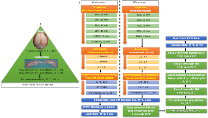
Figure 1: Experimental flowchart. (A) Brain tissue fixation process. (B)TEM process and IEM process workflow. Please click here to view a larger version of this figure.
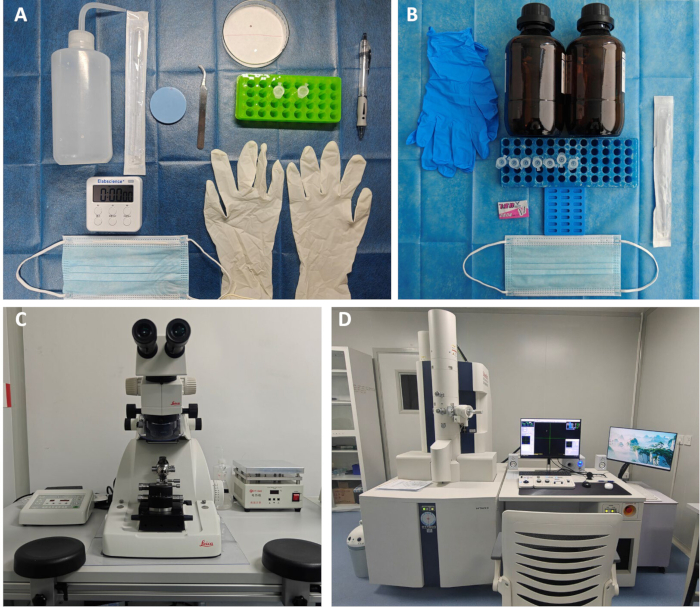
Figure 2: Equipment and apparatus used here. (A) Brain tissue collection. (B) Brain tissue fixation process. (C) Ultrathin section preparation. (D) Transmission electron microscopy. Please click here to view a larger version of this figure.
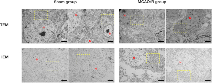
Figure 3: TEM analysis. TEM images of neurons in the sham operation group and MCAO group.In the sham group, neurons show normal ultrastructure: the nuclear membrane (N) is smooth, chromatin is evenly distributed, and the nucleolus is visible. Mitochondria (red arrows) have intact membranes and clear cristae. The rough endoplasmic reticulum (blue arrows) and Golgi apparatus (yellow arrows) are normal. In contrast, neurons in the MCAO group are pyknotic and degenerated, with wrinkled nuclear envelopes and increased electron density. Mitochondria show disrupted cristae, and the rough endoplasmic reticulum and Golgi apparatus are dilated. Abbreviations: N = nucleus; TEM = transmission electron microscopy; MCAO = middle cerebral artery occlusion; IEM = immuno-transmission electron microscopy. Please click here to view a larger version of this figure.
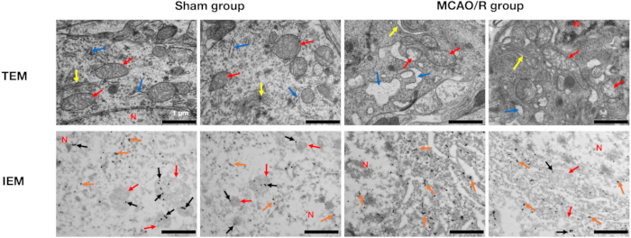
Figure 4: IEM analysis. IEM images showing subcellular localization of gold-labeled proteins. In the sham group, neurons display normal ultrastructure. Colloidal gold particles (black arrows) are found in mitochondria, and others (orange arrows) are distributed in nuclei and cytoplasm. In the MCAO group, neurons show slight shrinkage and increased electron density. Fewer gold particles are present in mitochondria, but more are found in nuclei and cytoplasm. Abbreviations: N = nucleus; TEM = transmission electron microscopy; MCAO = middle cerebral artery occlusion; IEM = immuno-transmission electron microscopy. Please click here to view a larger version of this figure.
| Animal | Neuroscore |
| Sham 1 | 0 |
| Sham 2 | 0 |
| Sham 3 | 0 |
| MCAO 1 | 2 |
| MCAO 2 | 3 |
| MCAO 3 | 2 |
Table 1: Comparison of neuroscore between sham and MCAO groups. p = 0.034. Abbreviations: MCAO = middle cerebral artery occlusion.
Discussion
The primary objective of reperfusion therapy in acute ischemic stroke is to restore blood perfusion to the ischemic region. Now we can achieve this goal through intravenous thrombolysis, endovascular treatment, and antithrombotic therapy. However, restoration of blood perfusion leads to ischemia-reperfusion injury, aggravates neuronal necrosis and apoptosis, leading to neurological impairment. During ischemia-reperfusion, abnormal leakage of electrons from the mitochondrial electron transport chain leads to the production of a large amount of mitochondrial reactive oxygen species, which damages the mitochondrial membrane and releases Cyt c and mitochondrial DNA13. Studies have demonstrated that Bax channel inhibitors and insulin can activate cell survival signaling, prevent apoptotic release of Cyt c, and exert neuroprotective and antiapoptotic effects14,15.
Immunohistochemistry allows us to visualize and localize specific biochemical components in cell compartments and tissues. Combined with TEM, we can accurately capture the location changes of protein in the microstructure. Immune colloidal gold technique has made great progress in successfully applying to various aspects of cell biology and the study of cellular ultrastructure level of immunology, histology, and pathology16,17,18. It has been used in renal diseases and tumor diagnosis19,20, study of molecular mechanisms of different diseases21,22. Compared with traditional biochemical techniques such as Western blotting, enzyme-linked immunosorbent assay, and immunohistochemistry, IEM has obvious advantages in the ultrastructural precision visualization of subcellular protein localization. Although these traditional methods can detect expression levels, they cannot capture the precise spatial changes in protein distribution. In contrast, IEM can directly observe the redistribution of proteins such as Cyt c from mitochondria to the cytoplasm during apoptosis, providing strong visual evidence to support mechanistic hypotheses. This advantage has been demonstrated in the remaining studies10,23.
Fixation is a critical step in immuno-electron microscopy, as it requires a balance between preserving cellular ultrastructure and maintaining antigenicity. Inadequate fixation may lead to loss of ultrastructural integrity, while overly strong fixatives can mask antigenic sites and reduce labeling efficiency24. In this experiment, special care must be taken when sampling fresh tissue. The sampling site should be carefully selected to avoid mechanical damage such as traction, contusion, or compression. Additionally, the tissue thickness should be kept below 2 mm to ensure optimal fixative penetration and structural preservation.
Although compared to TEM, IEM can capture the changes of mitochondrial proteins in the microscopic structure, such as the changes in the position and quantity of these proteins in the cytoplasm or other organelles, and with the mitochondria. However, IEM only explains certain phenomena from the perspective of mitochondrial morphology and functional proteins. As for the true function of mitochondria, it is necessary to combine the detection of adenosine triphosphate, adenosine diphosphate, and adenosine monophosphate contents using high-performance liquid chromatography or directly detect the functional state of mitochondria through the Seahorse method25,26,27.
There are limitations to this technique; it cannot be used in live cells. Fixatives often exert varying degrees of influence on the activity of antigens; to preserve the ultrastructure of cells and the activity of antigens, it is necessary to determine the type, concentration, temperature, pH, and fixation time and method of the fixative through preliminary experiments. Known-titer antigens can be used for testing to select an appropriate fixation method. There are six rats randomly divided into the MCAO group and the sham operation group, with three in each group. To exclude the effects of anesthesia, surgical trauma, and other confounding factors, we may need more animals and a control group in further study. This group would not undergo any surgical procedures but would receive the same anesthesia and fixation processing, allowing us to distinguish the effects of surgery from those of other experimental manipulation.
This method provides an effective means for the accurate localization of various antigens in the ultrastructure of tissues and cells, also one way to confirm the relationship between cell structure and function and the changes in pathological conditions. We believe this method can be applied to further study of underlying cellular mechanisms involved in different diseases with different disease animal models, including drug discovery.
Disclosures
The authors have nothing to disclose.
Acknowledgements
This work was supported by the Special Scientific Research Project on Traditional Chinese Medicine of Sichuan Provincial Administration of Traditional Chinese Medicine(2021MS414).
Materials
| Name | Company | Catalog Number | Comments |
|---|---|---|---|
| 0.9% normal saline | Sichuan Kelun Pharmaceutical Co., Ltd. | H20083400 | |
| 200 Mesh formvar coated copper grids | Zhongjingkeyi Technology Co., Ltd. | BZ10262a | |
| 220-230g MCAO Monofilaments | Beijing Cinontech Co., Ltd. | A4-243250 | |
| Absolute ethanol | Chengdu Kelong Chemical Co.,Ltd. | 2023101201 | |
| acetone | Chengdu Kelong Chemical Co.,Ltd. | 2023120501 | |
| Animal anesthesia system | Rayward Life Technology Co., Ltd. | R500 | |
| Animal temperature maintainer | Rayward Life Technology Co., Ltd. | 69020 | |
| Baking table | Shenzhen Bangqi Chuangyuan Technology Co., Ltd. | CT-946 | |
| Bone shear | Fine Science Tools | 16102-11 | |
| capsular embedding molds | Zhongjingkeyi Technology Co., Ltd. | GP130-B | |
| Centrifuge | SCILOGEX | D3024R | |
| Conventional bright-field optical microscopy | Zeiss (Germany) | Primo Star | |
| Curved ophthalmic forceps (10 cm) | Shanghai Medical Equipment Co., Ltd. | 211001 | |
| Diamond knife | Diatome (Switzerland) | DU4530 | |
| Dissecting forceps | Rayward Life Technology Co., Ltd. | F12018-10 | |
| Dissecting forceps | Rayward Life Technology Co., Ltd. | F12010-10 | |
| Electric blast drying oven | Shanghai YiHENG Scientific Instrument Co., Ltd. | DHG-9030A | |
| Electric shaver | Yiwu Kemei Electric Appliances Co., Ltd. | KM-PG5002 | |
| Fixation fluid | Sichuan Scientist Biotechnology CO., Ltd. | SC0001 | |
| glass slide | Nantong Mevid Life Science Co., Ltd. | 220518001 | |
| Gold-conjugated Goat anti-rabbit lgG | Sangon Biotech (Shanghai) Co.,Ltd. | D111094 | |
| Hemostatic forceps (12.5 cm) | Anqisheng Biotechnological Co., Ltd. | PL068 | |
| Hemostatic forceps (14 cm) | Anqisheng Biotechnological Co., Ltd. | PL002 | |
| Isoflurane | Rayward Life Technology Co., Ltd. | 2024071201 | |
| L. R. White Resin | Tianjin Tainuo Science and Technology Co., Ltd. | AGR1281A | |
| Lead citrate | SPI (US) | GA10701 | |
| Needle holder | Jiachi Medical Technology Co., Ltd. | 10044036898028 | |
| Osmium tetroxide | TED PELLA (US) | 4008-220501 | |
| PBS | Servicebio | G4202 | |
| Rabbit polyclonal antibody to cytochrome c | Affinity Biosciences, Ltd. | AF0146 | |
| Rats | Beijing Huafukang Biotechnology Co., Ltd. | 110322241102187881 | |
| SPI-Pon 812 embedding resin kit | SPI(US) | GS02660 | |
| Stereomicroscope | Sunny Optical Technology Co., Ltd. | Soptop SZMN | |
| Straight headed scissors (10 cm) | Anqisheng Biotechnological Co., Ltd. | PL063 | |
| Straight ophthalmic forceps (10cm) | Shanghai Medical Equipment Co., Ltd. | 211001 | |
| Surgical scissors | Rayward Life Technology Co., Ltd. | S14001-10 | |
| Transmission electron microscopy | Hitachi (Japan), Ltd. | HT7800 | |
| ultramicrotome | Leica(Germany) | UC7 | |
| Uranium acetate | SPI (US) | GS02624 |
References
- GBD 2021 Nervous System Disorders Collaborators. Global, regional, and national burden of disorders affecting the nervous system, 1990-2021: a systematic analysis for the Global Burden of Disease Study 2021. Lancet Neurol. 23 (4), 344-381 (2024).
- Taylor, R. C., Cullen, S. P., Martin, S. J. Apoptosis: controlled demolition at the cellular level. Nat Rev Mol Cell Biol. 9 (3), 231-341 (2008).
- Li, G. R., et al. Albiflorin: a promising natural monoterpene glycoside with potential therapeutic benefits for multiple physiological systems. Food Med Homol. , (2025).
- Ye, X. S., et al. The food and medicine homologous Chinese Medicine from Leguminosae species: A comprehensive review on bioactive constituents with neuroprotective effects on nervous system. Food Med Homol. 2 (2), 9420033(2025).
- Mubeen, M., Sakarwala, A. S., Bijarani, A. I., Kamal, M. T. Enhancing heart health: expanding the use of SGLT2 inhibitors in cardiovascular disease. Med Adv. 3 (2), 122-126 (2025).
- Zhang, Y. C., et al. Comprehensive analyses of nuclear mitochondria-related genes in the molecular features, immune infiltration, and drug sensitivity of clear cell renal cell carcinoma. Med Adv. 2 (3), 238-253 (2024).
- Broughton, B. R., Reutens, D. C., Sobey, C. G. Apoptotic mechanisms after cerebral ischemia. Stroke. 40 (5), e331-e339 (2009).
- Guo, M. J., Lu, B., Gan, J. L., Wang, S. C., Jiang, X. J., Li, H. H. Apoptosis detection: a purpose-dependent approach selection. Cell cycle. 20 (11), 1033-1040 (2021).
- Squarzoni, S. Immunoelectron Microscopy Methods. Methods Mol Biol. 2655, 201-210 (2023).
- Liu, Y., Ohgaki, R., Okanishi, H., Xu, M., Kanai, Y. Amino acid transporter LAT1 is expressed on cancer cell-derived exosomes with potential as a diagnostic and prognostic biomarker. Sci Rep. 14 (1), 28458(2024).
- Longa, E. Z., Weinstein, P. R., Carlson, S., Cummins, R. Reversible middle cerebral artery occlusion without craniectomy in rats. Stroke. 20 (1), 84-91 (1989).
- Sanderson, T. H., Reynolds, C. A., Kumar, R., Przyklenk, K., Hüttemann, M. Molecular mechanisms of ischemia-reperfusion injury in brain: pivotal role of the mitochondrial membrane potential in reactive oxygen species generation. Mol Neurobiol. 47 (1), 9-23 (2013).
- Lesnefsky, E. J., Chen, Q., Tandler, B., Hoppel, C. L. Mitochondrial Dysfunction and Myocardial Ischemia-Reperfusion: Implications for Novel Therapies. Annu Rev Pharmacol Toxicol. 57, 535-565 (2017).
- Hetz, C., et al. channel inhibitors prevent mitochondrion-mediated apoptosis and protect neurons in a model of global brain ischemia. J Biol Chem. 280 (52), 42960-42970 (2005).
- Sanderson, T. H., Kumar, R., Sullivan, J. M., Krause, G. S. Insulin blocks cytochrome c release in the reperfused brain through PI3-K signaling and by promoting Bax/Bcl-XL binding. J Neurochem. 106 (3), 1248-1258 (2008).
- Goldberg, M. W., Fiserova, J. Immunogold labelling for scanning electron microscopy. Methods Mol Biol. 657, 297-313 (2010).
- Sachse, M., de Castro, I. F., Fournier, G., Naffakh, N., Risco, C. Metal-Tagging Transmission Electron Microscopy and Immunogold Labeling on Tokuyasu Cryosections to Image Influenza A Virus Ribonucleoprotein Transport and Packaging. Methods Mol Biol. 1836, 281-301 (2018).
- Amiry-Moghaddam, M., Ottersen, O. P. Immunogold cytochemistry in neuroscience. Nat Neurosci. 16 (7), 798-804 (2013).
- Veeramachaneni, R., Gu, X., Herrera, G. A. Atypical amyloidosis: diagnostic challenges and the role of immunoelectron microscopy in diagnosis. Ultrastruct Pathol. 28 (2), 75-82 (2004).
- Fossmark, R., et al. Ultrastructure and chromogranin A immunogold labelling of ECL cell carcinoids. APMIS. 113 (7-8), 506-512 (2005).
- Page, L. J., et al. Secretion of amyloidogenic gelsolin progressively compromises protein homeostasis leading to the intracellular aggregation of proteins. Proc Natl Acad Sci U S A. 106 (27), 11125-11130 (2009).
- Vainer, B., Sørensen, S., Nielsen, O. H., Horn, T. Subcellular localization of intercellular adhesion molecule-1 in colonic mucosa in ulcerative colitis. Ultrastruct Pathol. 26 (2), 113-121 (2002).
- Cheng, X. T., et al. Revisiting LAMP1 as a marker for degradative autophagy-lysosomal organelles in the nervous system. Autophagy. 14 (8), 1472-1474 (2018).
- Karreman, M. A., van Donselaar, E. G., Gerritsen, H. C., Verrips, C. T., Verkleij, A. J. VIS2FIX: a high-speed fixation method for immuno-electron microscopy. Traffic. 12 (7), 806-814 (2011).
- Wang, X. B., et al. Mechanistic Insights into salidroside's mitochondrial protection via AMPK/Sirt1/HIF-1α pathway in hypoxic HT22 Cells. J Vis Exp. (218), e66923(2025).
- Hou, Y., et al. Rhodiola crenulata alleviates hypobaric hypoxia-induced brain injury by maintaining BBB integrity and balancing energy metabolism dysfunction. Phytomedicine. 128, 155529(2024).
- Hou, Y., et al. Salidroside intensifies mitochondrial function of CoCl2-damaged HT22 cells by stimulating PI3K-AKT-MAPK signaling pathway. Phytomedicine. 109, 154568(2023).
Reprints and Permissions
This article has been published
Video Coming Soon