This study presents a smart structural color hydrogel patch (CPG) for the diagnosis and treatment of chronic osteomyelitis (COM). The patch integrates a temperature-responsive GelMA hydrogel, a pH-sensitive CSMA-PBA scaffold, and salicylic acid for controlled drug release, infection monitoring, and promoting tissue regeneration.
Research Article
An Intelligent Structural Color Hydrogel Patch Integrating Diagnosis and Treatment for the Management of Chronic Osteomyelitis
* These authors contributed equally
In This Article
Summary
Abstract
Chronic osteomyelitis (COM) is a persistent bone infection that is difficult to treat with conventional antibiotics due to issues such as drug resistance and adverse side effects. To address this challenge, we have developed a novel smart structural color hydrogel patch (CPG) for both the diagnosis and treatment of COM. The patch combines methylacrylated chitosan modified with phenylboronic acid (CSMA-PBA) as a scaffold, temperature-responsive GelMA hydrogel as filler, and salicylic acid (SA) for drug delivery. Characterization showed excellent mechanical properties and adhesive strength. Drug release experiments confirmed that CPG effectively responds to pH changes, enabling controlled drug release. CPG also serves as a diagnostic tool by detecting pH and temperature changes at the infection site, assisting in postoperative recovery. Antimicrobial tests showed significant inhibition of bacterial growth, and in vitro experiments demonstrated CPG's ability to promote vascular endothelial growth factor expression while reducing inflammatory markers. The study highlights the multifunctional capabilities of the hydrogel patch, including antimicrobial, anti-inflammatory, and pro-vascular effects, positioning it a promising therapeutic option for chronic osteomyelitis.
Introduction
Chronic osteomyelitis has become a major concern for patients due to its challenging and prolonged treatment process, high risk of treatment failure, and recurrence1. Current treatment strategies for osteomyelitis involve surgical debridement of necrotic tissue and parenteral antibiotics. However, the need for high doses of antibiotics to provide therapeutic levels at the site of action remains ineffective in treating the disease. Furthermore, the use of high-dose antibiotics is associated with adverse complications such as nephrotoxicity, ototoxicity, antibiotic resistance, and gastrointestinal side effects. Local drug delivery of antibiotics may be a more appropriate strategy to overcome these drawbacks. While local antibiotic delivery can treat bone infections with minimal side effects, inducing bone regeneration is crucial for successful treatment2. Therefore, the simultaneous administration of antibiotics and bone growth factors appears to be a promising treatment strategy. Over the past few decades, non-biodegradable polymethylmethacrylate (PMMA) has been the mainstay for local antibiotic therapy in osteomyelitis. However, the use of PMMA as a local antibiotic delivery system faces limitations such as the need for a second surgery for bead removal, bacterial biofilm formation that favors surface colonization, long-term antibiotic release at subtherapeutic levels leading to antibiotic resistance, systemic toxicity of residual monomers, and lack of bone inductive properties3. Thus, the development of biodegradable local delivery systems using biocompatible polymers may overcome these drawbacks. Literature suggests that silk fibroin nanoparticles (SFNPs) incorporated into a silk scaffold for local delivery of vancomycin can be used to treat severe osteomyelitis. However, the absence of any bone-inductive bioactive molecules in the nanocomposite material resulted in the prepared constructs not significantly inducing bone regeneration. Therefore, the design of nanocomposite scaffold materials suitable for the treatment of chronic osteomyelitis has been a challenging issue of concern. Natural polymer-based hydrogels have received widespread attention due to their structural similarity to the extracellular matrix (ECM) and their ability to control drug release4.
One prominent feature of hydrogels is their responsiveness to different factors such as temperature and pH. Chronic infections may increase pH due to microbial burden, dysregulation of the regeneration-degradation balance, and enhanced breakdown metabolic reactions. Additionally, surgical excision of debrided material, such as necrotic tissue and pus, may accelerate the transition of pH to higher values5. Therefore, the use of pH-sensitive hydrogels capable of sensing chronic infection pH is crucial for the prognosis of chronic osteomyelitis.
In this study, we propose an integrated diagnosis and treatment approach using an intelligent structural color hydrogel patch, possessing the requisite features for treating chronic osteomyelitis as illustrated in Figure 1. Supramolecular hydrogels derived from the assembly of supramolecular polymer networks are attractive materials similar to the natural extracellular matrix (ECM). Due to their excellent properties, including appropriate mechanical strength, self-healing, antibacterial, and anti-inflammatory properties, supramolecular hydrogels have found many applications in the biomedical field6. In contrast, structural color, as a type of color produced by unique interactions of light with intrinsic periodic nanostructures, has garnered great attention in fields such as optical displays, anti-counterfeiting labels, and wearable electronic devices. Particularly, when structural color materials are constructed from responsive polymers, their color can be adjusted by different stimuli through expansion or contraction of these polymers, suggesting the potential of these materials as sensors. Therefore, it can be envisioned that combining supramolecular hydrogels with structural colors will provide a unique strategy for constructing novel hydrogel patches for the treatment of chronic osteomyelitis7,8,9.
Herein, we developed an integrated diagnosis and treatment approach using an intelligent structural color hydrogel patch (CPG) composed of mixed-modified methylacrylated chitosan with phenylboronic acid (CSMA-PBA) as an anti-protein stone scaffold, temperature-responsive GelMA hydrogel as filler, and loaded with salicylic acid (SA)10. Due to the reversibility of hydrogen bonding and non-covalent interactions within the network, the resulting hydrogel patch not only exhibited excellent mechanical properties but also possessed self-healing capabilities. Furthermore, owing to the presence of CSMA, this hydrogel patch also exhibited potent antibacterial bioactivity. Additionally, utilizing the thermal responsiveness of GelMA polymer, the hydrogel patch could release active substances when placed at the relatively higher temperature site of inflammation. Simultaneously, due to the presence of boronate ester bonds in CSMA-PBA, the hydrogel responded to changes in environmental pH11. Based on these advantages, we demonstrated that the proposed structural color supramolecular hydrogel patch could treat chronic osteomyelitis by downregulating the expression of inflammatory factors, antibacterial activity, and promoting angiogenesis. Notably, benefiting from the unique optical properties of the anti-protein stone scaffold, the hydrogel patch could even exhibit color-changing ability in response to temperature stimuli, demonstrating significant potential in monitoring and managing chronic osteomyelitis and guiding clinical treatment.
Protocol
All procedures were approved by the Medical Ethics Committee and performed following the guidelines for the care and use of laboratory animals (NIH publication No. 80-23, revised 1978). Male Sprague-Dawley rats (8 weeks old) were used in the experiments.
Preparation of silica colloidal crystal templates (SCCs)
SCCs were prepared as previously described12,13,14. Cleaned and plasma-treated glass slides were immersed in a 5% solution of monodisperse silica nanoparticles (12 nm in diameter) dispersed in deionized water. The solution was continuously stirred at room temperature (25 °C) for 1 h to ensure the uniform dispersion of silica nanoparticles. The slides were removed from the solution and air-dried for 1 h at room temperature. The drying process was critical to ensure the proper adherence of the nanoparticles onto the surface of the glass slides. Following air-drying, the slides were placed in a ventilated oven at 60 °C for 2 days to evaporate any remaining solvent and facilitate the formation of colloidal crystals. During this evaporation, the slides were positioned at a 45° angle to promote the formation of ordered colloidal crystal arrays. The ordered colloidal crystal arrays were characterized by self-assembly and exhibited different structural colors depending on the inter-particle spacing. These arrays were then used as templates for subsequent hydrogel patch fabrication.
Preparation of integrated diagnosis and treatment intelligent structural color hydrogel patches (CPG)
Anti-protein stone scaffolds were replicated from the voids of silica colloidal crystal templates (SCCs). Cleaned glass slides were first immersed in a 5% solution of monodisperse silica nanoparticles (12 nm in diameter) in deionized water, and the solution was stirred at room temperature (25 °C) for 1 h to ensure uniform dispersion of the nanoparticles across the surface. The stirring process was crucial to prevent the formation of aggregates and ensure a homogenous distribution of nanoparticles. After immersion, the slides were air-dried for 1 h at room temperature (RT), followed by oven drying at 60 °C for 2 days to facilitate the evaporation of the solvent and promote the self-assembly of the silica nanoparticles into well-ordered colloidal crystal arrays. During the evaporation, the slides were positioned at a 45° angle to facilitate the alignment of nanoparticles, promoting the formation of a periodic array with controlled inter-particle distances. This ordered array exhibited structural color, a characteristic feature of the colloidal crystals, and served as the template for the subsequent hydrogel structure.
The prepared SCCs were then soaked in the CPG pre-gel solution, which consisted of GelMA at a concentration of 10% w/v dissolved in phosphate-buffered saline (PBS). The samples were left for 5 min at room temperature (RT) to ensure thorough infiltration of the GelMA into the voids between the silica nanoparticles, ensuring a uniform distribution within the template structure. This step was critical to ensure that the hydrogel matrix was evenly incorporated into the template and that the final hydrogel structure replicated the ordered nanoparticle arrangement of the SCCs. Following the soaking, the pre-gel solution was polymerized under UV light (365 nm) at an intensity of 3 mW/cm2 for 5 min. This UV exposure initiated the crosslinking of GelMA, solidifying the hydrogel matrix. The exposure time and intensity were carefully optimized to achieve sufficient crosslinking without overexposing the hydrogel, which could lead to excessive gelation or loss of template structure.
Characterization of CPG
The cross-sectional morphology of the hydrogel was observed using scanning electron microscopy (SEM) with a 5 kV acceleration voltage after coating the samples (1 mm x 1 mm) with gold15. This allowed us to visualize the nanoporous structure and confirm the uniformity of the hydrogel network. Swelling behavior was evaluated by immersing 10 mg hydrogel samples in 10 mL of PBS at different time intervals (0, 30, 60, 120, and 180 min). The percent swelling was calculated by measuring the increase in hydrogel volume over time, which provided insight into the hydrogel's ability to absorb water under different conditions. Rheological analysis was performed using a rheometer with a 1% strain, applying angular frequencies ranging from 1 rad/s to 100 rad/s to measure storage modulus (G') and loss modulus (G''). This step was essential to understand the mechanical properties of the hydrogel, including its viscoelastic behavior under stress. Shear adhesive properties were evaluated with a universal tensile testing machine (Instron) following ASTM F2255-05 (2015) at a speed of 10 mm/min and a preload of 0.01 N to assess the hydrogel's ability to adhere to biological tissues. Drug release was evaluated using a UV spectrophotometer by monitoring absorbance at λ = 280 nm after immersing the hydrogel in 5 mL of PBS (pH 7.4). The release was quantified using a standard curve of salicylic acid concentrations to confirm the controlled release capabilities of the CPG hydrogel. Finally, hydrogels were immersed in buffer solutions of pH 4.5 and 7.2 for 30 min to assess pH responsiveness, observing changes in volume and appearance under different pH conditions16.
Temperature and pH responsiveness of CPG patches
CPG patches (approximately 2 mm in diameter) were placed in a 5 mL buffer solution with pH 7.2 and maintained at 37 °C using a temperature-controlled water bath for 30 min to mimic physiological conditions. Reflectance spectra were measured using a UV-Vis spectrophotometer at wavelengths ranging from 400 nm to 800 nm. After initial measurements, the patches were transferred to buffer solutions with different pH values (e.g., pH 4.5, 7.2, and 9.0) and maintained at 37 °C for 30 min. The response to the microenvironment, including any color change, was observed visually, and reflectance spectra were recorded again to analyze the color shift. This temperature and pH responsiveness provides valuable information on how the hydrogel reacts to changes in the wound environment.
In vitro antibacterial testing
Antibacterial tests were performed using standard procedures. Staphylococcus aureus (S. aureus, ATCC 25923) and Escherichia coli(E. coli, ATCC 53868) strains were used to assess the antibacterial activity of CPG patches. These standard reference strains were purchased from the American Type Culture Collection (ATCC) and were used as quality control (QC) strains to validate the antimicrobial testing procedures. Antimicrobial susceptibility testing was performed using the broth microdilution method according to CLSI M100 guidelines. The bacterial suspensions were prepared in Mueller-Hinton broth, a standard medium for antimicrobial testing. The turbidity was adjusted to a 0.5 McFarland standard, equivalent to approximately 1.5 × 10⁸ CFU/mL. The suspensions were then inoculated with CPG patches (2 mm in diameter), prepared with either deionized water or pH 6.0 2-(N-morpholino)ethanesulfonic acid (MES) buffer (10 mM). The patches were added to the bacterial suspensions at a ratio of one patch per 1 mL of culture. The suspensions were incubated at 37 °C for 24 h. After incubation, bacterial growth was assessed by plating serial dilutions of the cultures on nutrient agar, and colony-forming units (CFUs) were counted after a 24-h incubation at 37 °C. This procedure provided a quantitative measure of antibacterial efficacy.
Biocompatibility evaluation
Experiments were conducted as described previously. CPG patches were processed by dialysis in PBS (pH 7.4) for 4 h, followed by ethanol sterilization (70%) for 10 min and UV disinfection (365 nm) for 30 min to ensure that all biological contaminants were removed. L929 cells (ATCC CCL-1) were cultured in DMEM medium supplemented with 10% fetal bovine serum (FBS) and 1% penicillin-streptomycin at 37 °C with 5% CO2 to maintain cell viability. Cells were seeded in 24-well plates at 2 × 10⁵ cells/mL, with or without CPG patches (2 mm in diameter), in triplicate. The cells were divided into three experimental groups: blank control, CPG group, and CPG (MES) group. After 24 h, cell viability was assessed using the MTT assay, a widely accepted method for assessing cell metabolic activity. For the MTT assay, 20 µL of MTT (5 mg/mL) was added to each well, followed by incubation at 37 °C for 4 h. After incubation, the medium was removed, and 150 µL of DMSO was added to dissolve the formazan crystals formed by the living cells. The absorbance was measured at 570 nm using a microplate reader to quantify the number of viable cells. This approach ensured the reliable assessment of the cytotoxicity and biocompatibility of the hydrogel patches.
Anti-inflammatory activity
The antioxidant activity of CPG was evaluated using a ROS assay kit. L929 cells (ATCC CCL-1) were cultured in DMEM medium supplemented with 10% fetal bovine serum (FBS) and incubated at 37 °C with 5% CO2 for 24 h to allow for cell attachment and initial proliferation. To induce oxidative stress, cells were treated with 3 µg/mL hydrogen peroxide (H2O2) for 12 h. After oxidative stress induction, hydrogel extract (prepared by dissolving 1 mg of CPG hydrogel in 1 mL of DMEM medium) was added to the cells. Cells were incubated for another 12 h to allow for the potential antioxidant effects of the CPG hydrogel. Subsequently, cells were stained with 10 µM DCFH-DA solution (diluted in DMEM medium) and incubated at 37 °C for 20 min. DCFH-DA is a fluorescent probe used to detect reactive oxygen species (ROS) within cells. After staining, the cells were washed with PBS, and fluorescence images were captured using a fluorescence microscope at an excitation wavelength of 485 nm and an emission wavelength of 530 nm. The intensity of the green fluorescence was inversely correlated with the antioxidant activity of the hydrogel, providing a visual assessment of the hydrogel's ability to mitigate oxidative stress in cells.
Rat osteomyelitis animal model
A chronic osteomyelitis model was established: Male Sprague-Dawley rats (8 weeks old) were anesthetized using isoflurane (2-3% in oxygen) to ensure they remained immobile and pain-free during the surgery. A 2 cm incision was made on the lateral aspect of the left femur, and a sterile 27-G needle was used to inject 1 107 CFU of S. aureus directly into the trabecular region of the femur. The injection was performed into the bone marrow cavity of the femur to induce infection in the marrow space, the primary site for osteomyelitis. The bacteria were delivered slowly to ensure proper deposition within the bone marrow. After 10 days, the rats were euthanized using CO2 inhalation, and the infected femurs were carefully dissected. Granulation tissue was excised from the infection site and immediately immersed in 4% paraformaldehyde for 24 h to preserve tissue integrity. Following fixation, the bone tissue was decalcified using a decalcifying agent (e.g., EDTA) for 48-72 h, depending on the size of the bone, to allow proper sectioning for histological and immunohistochemical analysis. The decalcified bone samples were then processed for histological examination, immunohistochemistry (IHC), and immunofluorescence staining. For quantitative real-time PCR (qPCR) analysis, granulation tissue was stored at -80 °C for subsequent RNA extraction, enabling the analysis of gene expression profiles related to inflammation, bone regeneration, and immune responses
In vivo osteomyelitis treatment evaluation
Granulation tissue was embedded in paraffin and sectioned at 5 µm using a microtome. The sections were stained with Masson's trichrome staining (15 min with hematoxylin, 1 h with aniline blue) to evaluate collagen deposition and tissue remodeling. Hematoxylin and eosin (HE) staining (5 min with hematoxylin, 2 min with eosin) was used to observe general tissue morphology and inflammation. Immunohistochemistry (IHC) with anti-CD31 (1:100) and anti-EGF (1:200) was performed for 1 h at 37 °C, followed by secondary antibody incubation at room temperature (RT) for 30 min using anti-mouse IgG or anti-rabbit IgG (diluted 1:200), depending on the primary antibody used, to evaluate angiogenesis and growth factor expression. Immunofluorescence staining was performed with anti-VEGF (1:100) and anti-bFGF (1:200) for 1 h at 37 °C, followed by secondary antibody incubation for 30 min at RT. Sections were observed for CD31, EGF, VEGF, and bFGF expression using a fluorescence microscope. For quantitative real-time PCR (qPCR) analysis, mRNA expression of pro-inflammatory cytokines (TNF-α, IL-1β, and IL-6) was analyzed after RNA extraction with RNAiso Plus and cDNA synthesis using the PrimeScript RT reagent Kit. PCR was performed using SYBR Green PCR Master Mix to quantitatively assess the inflammatory response at the molecular level.
Quality control measures
To ensure the reliability and reproducibility of the experimental results, all procedures were carried out according to standardized protocols. Each experiment included appropriate control groups, such as blank controls (rats infected with S. aureus but receiving no treatment), control groups (rats that underwent surgical procedures without bacterial infection and no treatment), and comparative treatment groups (rats receiving different treatments for osteomyelitis, such as antibiotics or an experimental drug), to minimize variability. All assays were performed in triplicate to ensure consistency and reduce experimental bias. Each group contained 6 rats to ensure statistical significance. Instruments, including a UV-Vis spectrophotometer, rheometer, and tensile tester, were routinely calibrated before use according to the manufacturer's specifications. Bacterial and cell cultures were prepared under controlled environmental conditions in a biosafety cabinet, and staining procedures were followed using consistent protocols to ensure reproducibility. Data acquisition was conducted with unified microscope settings to minimize inter-sample variability and ensure accurate measurement.
Statistical analysis
Statistical analysis was performed using GraphPad Prism (version 8.0). Data are presented as mean ± standard deviation (SD). For comparison between multiple groups, a one-way analysis of variance (ANOVA) followed by Tukey's post-hoc test was used to assess differences in mechanical properties, thermoresponsive behavior, antibacterial activity, and cell viability. Student's t-test was used for pairwise comparisons when only two groups were compared, such as in the case of comparing the reflection peaks of CPG patches at different pH values or temperature changes. p < 0.05 was considered statistically significant for all analyses. All experiments were performed in triplicate to ensure consistency and reproducibility of the results.
Results
Fabrication and characterization of CPG patches (Figure 1)
In a typical experiment, monodisperse silica nanoparticles were self-assembled through solvent evaporation to form closely packed arrays, resulting in structural color coatings (SCCs)17,18,19, which were subsequently used to create multifunctional structural color hydrogel patches, as shown in Figure 1. The process involved the infiltration of a pre-gel solution into the nanoscale gaps of the SCCs via capillary action. UV polymerization was then employed to form the hybrid film of SCCs and hydrogel. Once the SCCs were etched away, an inverse anti-protein stone scaffold with a periodic porous structure was obtained. Finally, the thermoresponsive GelMA hydrogel was filled into the nanoscale gaps of the anti-protein stone scaffold, followed by curing, to create the independent CPG patches.
The strong crosslinking network of the hydrogel imparted excellent mechanical properties to the CPG patches. It was observed that the patches were tough enough to withstand stretching without causing any damage and could be stretched up to 2.5 times their original length. Importantly, the CPG patches exhibited vibrant structural colors and underwent dynamic color changes during stretching, a unique feature resulting from the periodic arrangement of high-order nanostructures within the patches. These nanostructures create photonic band gaps (PBGs), where light at certain wavelengths within the PBG is selectively reflected rather than transmitted, endowing the CPG patches with their characteristic structural colors and reflection peaks.
The peak reflection position of the CPG patch can be estimated by the equation20:
λ = 1.633dnaverage
where d is the distance between diffraction planes and naverage is the average refractive index of the material, which is calculated as the mean of the refractive indices of the components, including GelMA hydrogel and other materials used in the patch. This equation suggests that, as the patch is stretched and d decreases, the reflection wavelength decreases, leading to a blue shift in the structural color of the patch. The tensile strength of the patches was also tested, and it was found that the strain capacity of the patches decreased due to the GelMA hydrogel filling into the nanoscale gaps of the anti-protein stone scaffold. The deformation of the CPG patches upon stretching offers a highly sensitive means for strain detection, making them suitable for applications that require mechanical monitoring.
Thermoresponsive and pH-sensitive behavior of CPG patches (Figure 2)
The CPG patches exhibited remarkable thermoresponsive properties due to the GelMA hydrogel. Upon increasing the environmental temperature to 37 °C, the hydrogel underwent shrinkage, releasing water molecules and loaded cargo. This shrinkage resulted in a reduction in the size of the nanopores within the CPG patches, altering their PBG and consequently shifting the reflection spectrum. Figure 2 illustrates this behavior, showing how the structural color of the patches changes in response to variations in temperature and pH.
To confirm these observations, the CPG patches were placed in a simulated chronic infection wound environment at 37 °C and pH 7.2. As the pH of the solution increased, a blue shift in the reflection peak of the CPG patches was observed, indicating that the size of the nanopores decreased. Conversely, when the pH decreased, the patches exhibited a red shift, corresponding to an expansion of the nanopores. This pH-sensitive behavior was attributed to the hydrolysis of boronate ester bonds in the CSMA-PBA under acidic conditions, which broke the bonds and resulted in a red shift of the structural color. However, when the pH was alkaline, no significant color change was observed, indicating the stability of the patches in basic conditions. These findings suggest that the CPG patches can be used to monitor both temperature and pH changes in wound environments, which is crucial for effective wound management.
Antibacterial activity of CPG patches (Figure 3)
The antibacterial properties of the CPG patches were evaluated by co-culturing them with S. aureus and E. coli. Figure 3A,B shows that the CPG patches significantly reduced bacterial growth, demonstrating their effective antibacterial properties. This antibacterial efficacy was attributed to the electrostatic interaction between the positively charged CMCSMA surface of the patches and the negatively charged bacterial cell walls21. This interaction disrupted the bacterial membrane structure, leading to bacterial death. The cationic groups of CMCSMA can bind to anionic components in the bacterial membrane, leading to membrane depolarization and increased permeability22. Moreover, due to its relatively small molecular size and flexible polymeric chains, CMCSMA can penetrate the lipid bilayer more effectively, causing leakage of intracellular substances and interfering with metabolic activity. The small size of the CMCSMA molecules further facilitated their penetration into the bacterial cell membrane, where they interacted with essential biomolecules, disrupting bacterial metabolism and enzyme systems23. Additionally, considering the robust biofilm-forming capacity of S. aureus, it is worth noting that chitosan-based materials like CMCSMA have been reported to disrupt bacterial biofilms by penetrating the extracellular polymeric substance (EPS) matrix and interfering with its integrity. This enables the antibacterial agents to access and kill bacteria embedded within biofilms, which are typically resistant to conventional antibiotics24,25. As shown in Figure 3A, the visual comparison of bacterial colonies clearly demonstrates a substantial reduction following treatment with CPG patches, highlighting their effective bactericidal activity and their promising potential for use in infection-prone wound environments.
Biological functionality of CPG patches (Figure 4)
The CPG patches demonstrated significant biological functionality in addition to their antibacterial properties. Figure 4C,D show that the patches effectively scavenged reactive oxygen species (ROS) within cells, as indicated by the decreased green fluorescence signal in the CPG group. ROS scavenging is essential in wound healing, as excessive ROS production can impair tissue regeneration. The viability of human umbilical vein endothelial cells (HUVECs) co-cultured with the CPG patches was assessed using viability staining, as shown in Figure 4A. The results indicated that the majority of the cells were alive, with only a small proportion of dead cells, suggesting that the CPG patches promote cell viability and support cell proliferation.
Figure 4B shows the results of an MTT assay, which demonstrated that the CPG patches significantly promoted cell proliferation. Moreover, Figure 4F,G showed that the CPG patches enhanced angiogenesis by promoting the expression of vascular endothelial growth factor (VEGF) and basic fibroblast growth factor (bFGF), which are critical for endothelial cell proliferation and new blood vessel formation. These results highlight the potential of the CPG patches to not only prevent infection but also enhance tissue regeneration and accelerate wound healing.
In vivo evaluation of CPG patches (Figure 5)
In vivo experiments conducted using a chronic osteomyelitis model showed that the CPG patches significantly reduced infection and inflammation. Figure 5A shows histological analysis (H&E staining), where the control group exhibited characteristic inflammation, bone destruction, necrotic tissue, and high infiltration of defense cells, while the treatment group showed a significant reduction in infection, inflammation, and infiltration of defense cells. In the CPG group, increased migration of osteoblasts, transformation of osteoblasts into bone cells, vascular formation, new bone formation, and reduced infection were observed, further confirming the effectiveness of the CPG patches in promoting tissue regeneration.
Additionally, Figure 5B shows angiogenesis staining, confirming that the CPG patches promoted the formation of new blood vessels, which is critical for tissue repair. These findings suggest that the CPG patches play a dual role in both controlling infection and promoting tissue regeneration, making them highly suitable for chronic wound healing and bone regeneration applications.
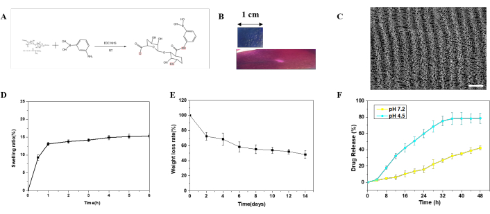
Figure 1: Preparation and characterization of the structural color hydrogel patch (CPG). (A): Reaction between CSMA and PBA; (B): Photograph of CPG, demonstrating the color change upon stretching; (C): SEM characterization of CPG; (D): Swelling behavior of CPG; (E): Degradation profile of CPG; (F): Drug release kinetics from CPG. Scale bar: 100 nm. Error bars represent standard deviation (SD). Six rats were used in the experimental group. Please click here to view a larger version of this figure.
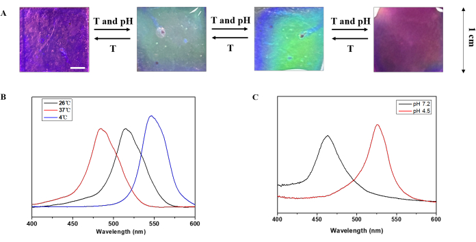
Figure 2: Intelligent sensing of CPG patch. (A): Variation of structural color of CPG patch with changes in environmental temperature and pH; (B): Spectral analysis of CPG patch at different temperatures; (C): Spectral analysis of CPG patch at different pH levels. Scale bar: 1 mm. Data represent mean ± SD, analyzed using one-way ANOVA with Tukey's post-hoc test. Please click here to view a larger version of this figure.
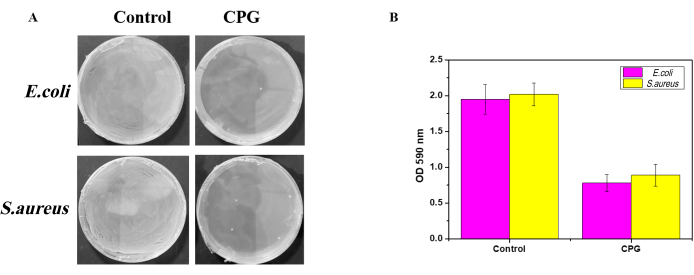
Figure 3: Antibacterial activity of CPG patch. (A): Representative images of co-culture of S. aureus and E. coli with CPG patch; (B): Analysis of optical density (OD) values. Scale bar: 5 mm. Error bars represent SD. Statistical significance was determined by one-way ANOVA with Tukey's post-hoc test. Six replicates were used per group. Please click here to view a larger version of this figure.
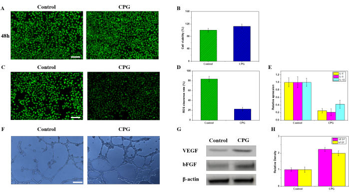
Figure 4: Biological functional evaluation of CPG patch. (A): Viability staining of HUVEC cells co-cultured with CPG patch for 48 h, showing live cells (green) and dead cells (red); (B): Cell proliferation measured by MTT assay; (C): ROS staining of CPG patch; (D): DPPH scavenging rate in the VEGF group; (E): q-PCR analysis of inflammatory factors; (F): Promotion of angiogenesis by CPG patch; (G): Western blot results of VEGF and bFGF expression; (H): Statistical analysis of grayscale values of VEGF and bFGF. Scale bar: 100 µm. Error bars represent SD. Data were analyzed using one-way ANOVA with Tukey's post-hoc test. Six rats were used in each group. Please click here to view a larger version of this figure.
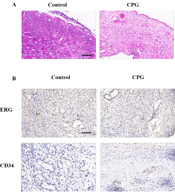
Figure 5: In vivo experiments of the CPG patch. (A): HE staining; (B): Angiogenesis staining. Scale bar: 100 µm. Data represent mean ± SD, analyzed by one-way ANOVA with Tukey's post-hoc test. Each group contained 6 animals. Please click here to view a larger version of this figure.
Discussion
The findings of this study demonstrate the multifunctionality of CPG hydrogel patches, particularly in terms of their structural color changes, temperature sensitivity, pH responsiveness, and antimicrobial properties. These hydrogel patches can undergo visible structural color changes when subjected to mechanical deformation, offering a highly sensitive mechanism for monitoring strain. This feature aligns with previous studies that have explored structural color in materials for sensor applications, where such color shifts are employed as indicators of strain or pressure26,27. Our study extends these findings by utilizing these color changes as a non-invasive means of monitoring wound stress, which is essential for assessing wound healing in real-time. The ability to monitor such mechanical strains provides critical information for clinicians, potentially helping to identify early signs of complications, such as suture dehiscence, thus supporting more targeted and timely interventions. Importantly, the optical readout provides a direct and intuitive sensing mechanism without the need for additional power or imaging devices, offering a decentralized monitoring solution ideal for point-of-care or at-home postoperative care.
The thermoresponsive and pH-sensitive behaviors of the CPG hydrogel patches enhance their potential for clinical use. As wounds heal, fluctuations in temperature and pH are common, particularly in the presence of infection or inflammation28. Elevated temperatures and altered pH values at the wound site are known to be associated with these conditions. In this study, the CPG patches exhibited sensitivity to such changes, enabling real-time monitoring of local wound conditions. This feature is of particular value in clinical settings, where early detection of infection or inflammation is essential for optimizing therapeutic strategies. Monitoring temperature and pH variations is especially important in wound management because they serve as early, non-invasive biomarkers of the wound status29. Elevated temperature may signal the onset of infection before clinical symptoms manifest, while shifts toward alkaline pH often indicate chronicity, microbial colonization, and impaired healing. In contrast, acidic environments are associated with acute wounds and better healing potential24. Thus, integrating temperature and pH-responsive elements into wound dressings allows for timely interventions and continuous assessment of healing progress, which can prevent wound deterioration or progression to systemic infection. The ability of the hydrogel to adapt dynamically to these changes also provides a new tool for clinicians to monitor wound status continuously, offering greater accuracy in tracking the healing process. Moreover, such dynamic responsiveness allows for spatiotemporal control over therapeutic release, ensuring that drug delivery is synchronized with pathological cues, which is a core objective of modern smart biomaterials.
Furthermore, the antimicrobial properties of the CPG hydrogel patches are critical in reducing the bacterial load at the wound site. Chitosan, which was incorporated into the hydrogel's structure, has long been recognized for its antimicrobial properties, due to its cationic nature, which facilitates electrostatic interactions with the negatively charged bacterial cell walls29. However, the antibacterial mechanism of chitosan-based materials is not limited to simple charge attraction. Upon contact with bacterial membranes, chitosan can increase membrane permeability, leading to leakage of intracellular contents such as potassium ions, proteins, and nucleic acids. Additionally, chitosan can chelate divalent metal ions (e.g., Ca2+ and Mg2+), disrupting enzyme activity and metabolic functions. Low molecular weight chitosan may also penetrate the bacterial cell wall and bind to DNA, inhibiting mRNA transcription and protein synthesis. These multifaceted mechanisms have been extensively documented. The porous structure of the hydrogel scaffold further amplifies these effects by enhancing surface area and bacterial contact, resulting in synergistic antibacterial efficacy. This property is particularly relevant in the treatment of chronic infections, such as osteomyelitis, where biofilm formation complicates the healing process25,30. The results of this study demonstrate that the CPG hydrogel patches were effective in inhibiting bacterial growth, reducing the bacterial load at the wound site, and potentially creating a more favorable environment for tissue regeneration. This antimicrobial activity, combined with the other properties of the hydrogel, positions the patches as a promising candidate for managing chronic wounds, which are often plagued by bacterial contamination and delayed healing. Additionally, by inhibiting local bacterial colonization, the hydrogel indirectly prevents the upregulation of pro-inflammatory cytokines that are often sustained in infected chronic wounds, thereby promoting a pro-regenerative microenvironment.
While ROS are typically associated with oxidative stress and tissue damage, it is widely recognized that controlled generation of ROS plays a crucial role in bacterial eradication. ROS, such as hydroxyl radicals and superoxide anions, can oxidize bacterial proteins, lipids, and DNA, ultimately leading to membrane rupture and cell death. Therefore, the presence of ROS at moderate levels serves as a critical component of innate antimicrobial defense mechanisms. In the context of chronic osteomyelitis, where bacterial colonization and biofilm formation persist, eliminating pathogens is a prerequisite for meaningful tissue regeneration. The CPG hydrogel, by suppressing excessive ROS while maintaining effective antibacterial activity through CMCSMA-mediated membrane disruption and biofilm penetration, offers a balanced approach. This dual function ensures that inflammation is not prolonged by persistent bacterial presence, allowing for a transition from immune activation to a regenerative microenvironment. Thus, rather than indiscriminately scavenging ROS, this system modulates the oxidative microenvironment to support both bacterial clearance and tissue healing.
In addition to antimicrobial activity, the CPG hydrogel patches exhibited biological functionality that is crucial for tissue repair and regeneration31. In vitro assays demonstrated that the hydrogel supported cell viability and proliferation, as well as angiogenesis. These findings are consistent with previous studies that highlighted the importance of angiogenesis in wound healing32. Angiogenesis is critical for providing adequate oxygen and nutrient supply to the wounded tissue, facilitating tissue regeneration. Additionally, the pro-angiogenic effects of the hydrogel are particularly important for treating chronic wounds, as these wounds often suffer from poor vascularization, which hinders the healing process33,34. By promoting blood vessel formation, the CPG hydrogel not only contributes to tissue regeneration but also creates a more conducive environment for the long-term survival of newly formed tissue. Moreover, the hydrogel's ability to promote osteoblast migration and differentiation adds significant value in the context of bone healing, especially for patients with chronic bone infections or bone defects35. The enhancement of osteogenesis is a promising avenue for improving outcomes in osteomyelitis and related bone conditions, where healing is often compromised. Such osteoinductive functionality, integrated with localized infection control, addresses a central challenge in musculoskeletal tissue engineering: the need for convergent solutions that promote both antimicrobial defense and tissue regeneration in a single platform.
The in vivo results further corroborate the therapeutic potential of the CPG hydrogel patches, showing their ability to support tissue repair, reduce infection, and promote osteogenesis. In chronic wound models, these hydrogels effectively facilitated wound closure and tissue regeneration. In bone regeneration models, the CPG hydrogel promoted osteoblast migration and differentiation, enhancing bone healing. These findings align with those of other studies that have explored hydrogel-based wound healing and bone regeneration strategies36. The ability of CPG hydrogel patches to simultaneously address infection, promote angiogenesis, and support osteogenesis provides a powerful platform for comprehensive wound care, making it a promising candidate for clinical use.
Despite these promising results, there are several limitations to the current study. First, while the in vitro and in vivo models demonstrated positive outcomes, the preclinical studies were conducted primarily in murine models. While mice are widely used for wound healing studies, they may not fully replicate the complexity of human wound healing. Therefore, further validation in larger animal models, as well as human clinical trials, is necessary to confirm the safety and efficacy of these hydrogel patches in real-world clinical settings. Second, although the CPG hydrogels exhibited good mechanical properties and biological functionality in the short term, their long-term stability and degradation behavior in vivo remain unclear. Hydrogels can degrade over time, and their degradation products may affect the healing process or even induce adverse reactions. Future studies should focus on optimizing the degradation rate of these hydrogels to ensure that they degrade in a controlled manner, without releasing harmful byproducts. Additionally, the controlled release of bioactive agents incorporated within the hydrogel should be further refined to ensure sustained therapeutic effects over time, especially for chronic wound treatment.
While this study focuses on a hydrogel-based supramolecular scaffold, alternative approaches to address the same therapeutic hypothesis include electrochemical biosensors, microfluidic wound-on-chip systems, and smart bandages incorporating electronic components for environmental sensing. However, unlike those technologies that often require power sources and complex interfaces, the hydrogel patch developed here provides a self-contained, visually interpretable platform without external instrumentation, making it especially attractive for resource-limited settings.
The importance of this work lies in its translational potential. Beyond osteomyelitis, the CPG platform can be customized for other inflammation-related conditions, such as diabetic foot ulcers, surgical site infections, and post-tumor resection monitoring. The integration of diagnostic and therapeutic functionalities into a single hydrogel matrix offers a unique opportunity for the development of next-generation smart wound dressings. Additionally, the photonic crystal-based structural color system presents opportunities for real-time, colorimetric biosensing applicable in personalized medicine. Moreover, the modularity of the CPG design permits adaptation for future integration with stem cells, exosomes, or gene delivery vectors to further extend its regenerative capabilities, aligning with current trajectories in precision regenerative medicine.
Looking ahead, future research should focus on several key areas. First, optimizing the composition and structure of the CPG hydrogel patches will be crucial for enhancing their long-term stability and performance. Modifying the crosslinking density and incorporating other bioactive agents, such as growth factors or cytokines, could improve the therapeutic potential of the hydrogel for specific clinical applications. Second, expanding the range of applications for these hydrogels beyond chronic wounds to other types of tissue regeneration, such as nerve or cartilage repair, could further demonstrate their versatility. Research into how CPG hydrogels can be tailored to specific wound types, such as diabetic ulcers or pressure sores, is needed to expand their clinical applicability. Third, integrating advanced therapies, such as stem cell therapy or gene therapy, with hydrogel patches may enhance their regenerative properties, allowing for more comprehensive and effective treatments. Finally, large-scale, multicenter clinical trials and real-world application studies will be necessary to evaluate safety, manufacturability, user compliance, and regulatory readiness of the CPG platform, thereby supporting its eventual clinical translation.
In conclusion, CPG hydrogel patches represent a promising platform for advanced wound care and tissue regeneration. Their ability to monitor wound stress, respond to temperature and pH changes, exhibit antimicrobial properties, and promote tissue regeneration offers a multifaceted approach to treating chronic wounds. While the results from preclinical studies are encouraging, further optimization and clinical validation are necessary to fully realize the potential of these hydrogel patches in clinical settings. Future research directions should include improvements in the material composition, broader applications, integration with advanced therapeutic strategies, and clinical trials to establish their efficacy and safety in humans.
In this experiment, we utilized a combination of methyl CSMA-PBA as the inverse opal scaffold, temperature-responsive GelMA hydrogel as the filler, and loaded it with SA to create a CPG integrated diagnostic and therapeutic smart structural color hydrogel patch. The experimental results demonstrate that this hydrogel patch exhibits excellent mechanical properties and adhesive strength. It can inhibit bacterial growth, significantly upregulate the expression of vascular endothelial growth factor, and downregulate the levels of inflammatory factors, thereby providing significant clinical prospects for the treatment of COM. Moreover, importantly, based on CPG's ability to detect changes in temperature and pH at the site of infection, it serves a monitoring role in the postoperative recovery of chronic osteomyelitis.
Disclosures
The authors declare no conflicts of interest related to this study. There were no financial or personal relationships that could influence the work presented in this manuscript. The authors confirm that no AI tools were used during the preparation of this manuscript. All authors have approved the final version of the manuscript for submission.
Acknowledgements
This work was supported by the National Health Commission Scientific Research Fund—Major Health Science and Technology Plan of Zhejiang Province (NO. WKJ-ZJ-2419) and Zhejiang Clinovation Pride (Clinical Innovation Team for Traumatic Osteomyelitis) (NO. CXTD202501009). We would like to express our sincere gratitude to everyone who supported and contributed to this research. Your guidance, assistance, and feedback have been invaluable in completing this work.
DATA AVAILABILITY:
The raw data supporting the findings of this study are available from the corresponding author upon reasonable request. Additionally, all data have been uploaded to Zenodo and are publicly available at the following link: https://doi.org/10.5281/zenodo.16785606
AUTHOR CONTRIBUTION:
Xiaoyu Han and Qiaofeng Guo: Writing-review and editing; Xiang Wang and Bingyuan Lin: conceptualization; Yiyang Liu and Haiyong Ren: methodology; Kai Huang and Huajun Yu: visualization and supervision. All authors have read and agreed to the published version of the manuscript.
Materials
| Name | Company | Catalog Number | Comments |
|---|---|---|---|
| Acrylamide (AM, 99%) | Sigma-Aldrich | A8887 | Monomer for hydrogel polymerization |
| Acryloyl Chloride (AC, 99%) | TCI Chemicals | A0376 | Used in chemical modification reactions |
| Alkaline Alumina (200-300 mesh) | Aladdin | A112336 | Used to purify methyl methacrylate |
| Anti-bFGF antibody | Santa Cruz | sc-9084 | Immunofluorescence staining for growth factor detection |
| Anti-CD31 antibody | Abcam | ab28364 | Immunohistochemistry for vascular marker detection |
| Anti-EGF antibody | Sigma-Aldrich | E9644 | Immunohistochemistry staining |
| Anti-VEGF antibody | Santa Cruz | sc-7269 | Immunofluorescence staining for angiogenesis |
| Bis-acrylamide (BIS, 97%) | Sigma-Aldrich | M7279 | Crosslinking agent in hydrogel network |
| Cell Counting Kit-8 (CCK-8) | Dojindo | CK04 | Assessment of cell viability |
| Chitosan, Methylacrylated (CSMA-PBA) | Sigma-Aldrich | C3641 | Scaffold formation in hydrogel patches |
| DCFH-DA Probe | Beyotime | S0033 | Reactive oxygen species (ROS) detection |
| Dimethyl Sulfoxide (DMSO) | China National Pharmaceutical Group Corporation | DMSO-AR | Solvent for hydrogel formulation |
| Escherichia coli (E. coli) | ATCC | - | Used in antibacterial testing |
| Fluorescence Microscope | Olympus | BX53 | Immunofluorescence and cell imaging |
| Gelatin Methacrylate (GelMA) | Sigma-Aldrich | G1894 | Temperature-responsive hydrogel component |
| Hydrochloric Acid (HCl) | Chongqing Tianyun | HCl-AR | pH adjustment in synthesis |
| Hydrogen Peroxide (H2O2) | Sigma-Aldrich | H1009 | Induces oxidative stress in cells |
| Hydrofluoric Acid (HF, 4%) | Sinopharm | H814912 | Etching silica colloidal crystals |
| Instron Universal Tensile Testing Machine | Instron | 3345 | Mechanical and adhesive strength testing |
| L929 Cells | ATCC | CCL-1 | Used for cytotoxicity and biocompatibility assays |
| Leica Microtome | Leica | RM2235 | Used for paraffin sectioning of tissues |
| Male Sprague-Dawley Rats (8 weeks) | Reagan Biotechnology | - | In vivo chronic osteomyelitis model |
| Masson’s Trichrome Stain Kit | Solarbio | G1340 | Histological staining for fibrosis |
| Methyl Methacrylate (MMA, 99%) | Sigma-Aldrich | M27805 | Monomer for scaffold formation |
| MTT Assay Kit | Beyotime | M5655 | Cell proliferation and cytotoxicity evaluation |
| Paraformaldehyde (4%) | Servicebio | G1101 | Tissue fixation |
| Paraffin | Sigma-Aldrich | P1003 | Embedding medium for histology |
| PBS Buffer | Gibco | 10010023 | Used in cell and hydrogel processing |
| Phenylalanine (99%) | Sigma-Aldrich | P5482 | Used in chemical modification |
| Phenylboronic Acid (PBA) | TCI Chemicals | P0157 | pH-sensitive component in hydrogel |
| Potassium Persulfate (KPS) | Beijing Baolingwei | KPS-AR | Radical initiator in polymerization |
| PrimeScript RT reagent Kit | Takara | RR047A | Used for cDNA synthesis |
| Reactive Oxygen Species (ROS) Assay Kit | KeyGen Biotech | KGAF019 | Antioxidant activity assessment |
| Real-Time PCR System | Applied Biosystems | StepOnePlus | Gene expression quantification |
| RNAiso Plus | Takara | 9108 | RNA extraction reagent |
| Rheometer (HR-1) | TA Instruments | HR-1 | Rheological characterization of hydrogel |
| Salicylic Acid (SA) | Sigma-Aldrich | S3254 | Drug molecule in hydrogel |
| Scanning Electron Microscope (SEM) | JEOL | JSM-7500F | Morphological analysis of hydrogel structure |
| Silica Nanoparticles (12 nm) | Sigma-Aldrich | 637246 | Formation of silica colloidal crystal templates |
| Sodium Hydroxide (NaOH) | China National Pharmaceutical Group Corporation | NaOH-AR | pH adjustment |
| Staphylococcus aureus (S. aureus) | ATCC | - | Used in antibacterial testing |
| SYBR Green PCR Master Mix | Applied Biosystems | 4367659 | qPCR detection reagent |
| UV Light Source (365 nm, 3 mW/cm²) | Herolab | UVT-28 | Used for UV polymerization |
| UV Spectrophotometer | Shimadzu | UV-2600 | Drug release measurement |
References
- Kim, S. M., et al. Tissue engineering treatment in osteomyelitis of the jaws. Tissue Eng Regen Med. 12 (S1), 11-26 (2015).
- Masters, E. A., et al. Evolving concepts in bone infection: redefining "biofilm", "acute vs chronic osteomyelitis", "the immune proteome" and "local antibiotic therapy". Bone Res. 7, 20(2019).
- Shnol, H., LaPorta, G. A. 3D printed total talar replacement: A promising treatment option for advanced arthritis, avascular osteonecrosis, and osteomyelitis of the ankle. Clin Podiatr Med Surg. 35 (4), 403-422 (2018).
- Loc-Carrillo, C., et al. Local intramedullary delivery of vancomycin can prevent the development of long bone Staphylococcus aureus infection. PLoS One. 11 (7), e0160187(2016).
- Braun, J., et al. Toxic effect of vancomycin on viability and functionality of different cells involved in tissue regeneration. Antibiotics (Basel). 9 (5), 238(2020).
- Spohn, R., et al. Integrated evolutionary analysis reveals antimicrobial peptides with limited resistance. Nat Commun. 10 (1), 4538(2019).
- Kang, J., et al. Antimicrobial peptide LL-37 is bactericidal against Staphylococcus aureus biofilms. PLoS One. 14 (6), e0216676(2019).
- Li, L., et al. Cathelicidin LL37 promotes osteogenic differentiation in vitro and bone regeneration in vivo. Front Bioeng Biotechnol. 9. 9, 638494(2021).
- Pereira, I., et al. In vivo systemic toxicity assessment of an oxidized dextrin-based hydrogel and its effectiveness as a carrier and stabilizer of granular synthetic bone substitutes. J Biomed Mater Res A. 107 (8), 1678-1689 (2019).
- Szili, E. J., et al. On plasma activated acetyl donors: Comparing the antibacterial efficacy of tetraacetylethylenediamine and pentaacetate glucose. Plasma. 5 (4), 423-435 (2022).
- Ning, J., et al. Argon mitigates impaired wound healing process and enhances wound healing in vitro and in vivo. Theranostics. 9 (2), 477-490 (2019).
- Nain, A., et al. Copper sulfide nanoassemblies for catalytic and photoresponsive eradication of bacteria from infected wounds. ACS Appl Mater Interfaces. 13 (7), 7865-7878 (2021).
- Feng, L., et al. Magnetic targeting, tumor microenvironment-responsive intelligent nanocatalysts for enhanced tumor ablation. ACS Nano. 12 (11), 11000-11012 (2018).
- Zhu, Y., et al. A multifunctional pro-healing zwitterionic hydrogel for simultaneous optical monitoring of pH and glucose in diabetic wound treatment. Adv Funct Mater. 30, 1905493(2020).
- Mirpour, S., et al. Cold atmospheric plasma as an effective method to treat diabetic foot ulcers: A randomized clinical trial. Sci Rep. 10 (1), 10440(2020).
- Sabrin, S., et al. Opportunities of electronic and optical sensors in autonomous medical plasma technologies. ACS Sens. 8 (3), 974-993 (2023).
- Liu, Z., et al. A novel antifungal plasma-activated hydrogel. ACS Appl Mater Interfaces. 11 (26), 22941-22949 (2019).
- Chen, J., et al. Plasma-activated hydrogels for microbial disinfection. Adv Sci (Weinh). 10 (14), e2207407(2023).
- Ghimire, B., et al. The influence of a second ground electrode on hydrogen peroxide production from an atmospheric pressure argon plasma jet and correlation to antibacterial efficacy and mammalian cell cytotoxicity. J Phy D Appl Phys. 55 (12), 125207(2022).
- Mohamed, A. A. H., et al. The effect of a second grounded electrode on the atmospheric pressure argon plasma jet. Plasma Res Express. 2, 015011(2020).
- Maho, T., et al. Antibacterial action of plasma multi-jets in the context of chronic wound healing. Appl Sci. 11 (20), 9598(2021).
- Tharmalingam, N., et al. Mefloquine reduces the bacterial membrane fluidity of Acinetobacter baumannii and distorts the bacterial membrane when combined with polymyxin B. mBio. 16 (4), e0401624(2025).
- Fu, Z., et al. Enhanced photothermal therapy for oral cancer using benzothiadiazole-based nanoparticle-loaded hydrogels. Artif Cells Nanomed Biotechnol. 53 (1), 345-360 (2025).
- Chiang, C. Y., et al. Synthesis of photoresponsive hybrid alginate hydrogel with photo-controlled release behavior. Carbohydr Polym. 119, 18(2015).
- He, L., et al. Multifunctional dynamic chitosan-guar gum nanocomposite hydrogels in infection and diabetic wound healing. Carbohydr Polym. 354, 123316(2025).
- Jaishi, L. R., et al. Metal-Organic Frameworks (MOFs)-based piezoelectric-colorimetric hybrid sensor for monitoring green leaf volatiles. ACS Sens. 9 (12), 6553-6562 (2024).
- Wang, J., et al. Cellulose nanocrystals-based optical organohydrogel fiber with customizable iridescent colors for strain and humidity response. Int J Biol Macromol. 275 (Pt 1), 133501(2024).
- Su, W., et al. Intelligent response agarose/chitosan hydrogel wound dressing: Synergistic chemotherapy, PDT and PTT effects against bacterial infection. Int J Biol Macromol. 298, 139927(2025).
- Haidari, H., et al. Bacteria-activated dual pH- and temperature-responsive hydrogel for targeted elimination of infection and improved wound healing. ACS Appl Mater Interfaces. 14 (46), 51744(2022).
- Dong, J., et al. Vacancies-rich Z-scheme VdW heterojunction as H2S-sensitized synergistic therapeutic nanoplatform against refractory biofilm infections. Biomaterials. 320, 123258(2025).
- Tang, J., et al. Revolutionizing pressure ulcer regeneration: Unleashing the potential of extracellular matrix-derived temperature-sensitive injectable antioxidant hydrogel for superior stem cell therapy. Biomaterials. 314, 122880(2025).
- Qiu, L., et al. Ultrasound-activated piezoelectric heterojunction drives nanozyme catalysis to induce bacterial cuproptosis-like death and promote bone vascularization and osseointegration. Biomaterials. 320, 123249(2025).
- Lai, Y., et al. Advanced healing potential of simple natural hydrogel loaded with sildenafil in combating infectious wounds. Int J Pharm X. 9, 100328(2025).
- Chen, S., et al. Quaternized chitosan-based photothermal antibacterial hydrogel with pro-vascularization and on-demand degradation properties for enhanced infected wound healing. Carbohydr Polym. 355, 123350(2025).
- Shan, J., et al. End-tail soaking strategy toward robust and biomimetic sandwich-layered hydrogels for full-thickness bone regeneration. Bioact Mater. 49, 486-501 (2025).
- Zheng, Y., et al. Hydrogel promotes bone regeneration through various mechanisms: a review. Biomed Tech (Berl). 70 (2), 103-114 (2024).