This study retrospectively analyzed eight orbital apex tumor patients who underwent endoscopic endonasal resection. All achieved complete tumor removal, with 62.5% cavernous hemangiomas, 25% schwannomas, and 12.5% metastatic carcinoma. Postoperatively, 62.5% showed improved vision, with no complications or benign tumor recurrences. The approach proved safe and effective for selected cases.
Case Report
Retrospective Case Analysis of Transnasal Endoscopic Resection of Orbital Apex Tumor
* These authors contributed equally
In This Article
Summary
Abstract
Endoscopic endonasal surgery has gained increasing recognition as an effective approach for the management of orbital tumors. In this study, we retrospectively analyzed the clinical data of eight patients with orbital apex tumors (OATs) who underwent endoscopic endonasal resection at the Orbital Disease and Ophthalmic Oncology Department of Shenzhen Eye Hospital between April 2021 and September 2024. The cohort consisted of one male and seven female patients, with a mean age of 41.7 ± 14.2 years. All patients successfully underwent complete tumor resection by the endoscopic endonasal approach, followed by standardized postoperative treatment including anti-inflammatory therapy, corticosteroids, and neurotrophic medications. Histopathological examination revealed five cases of cavernous hemangioma (62.5%), two cases of schwannoma (25%), and one case of metastatic carcinoma (12.5%). During the follow-up period of at least 6 months, visual acuity improved in five patients (62.5%), decreased in two patients (25%), and remained unchanged in one patient (12.5%). No postoperative complications were observed, including ocular motility disorders, visual field defects, cerebrospinal fluid leakage, or hemorrhage. All patients with benign tumors showed no evidence of recurrence, while the patient with a malignant tumor received adjuvant radiotherapy. Endoscopic endonasal resection represents a safe, minimally invasive, and effective surgical option for selected OATs. The procedure demonstrates significant advantages in terms of clinical outcomes and complication avoidance. However, careful patient selection based on tumor characteristics and precise surgical planning remain crucial for optimal results. These findings support the clinical application of this technique for the management of orbital apex tumors.
Introduction
Orbital apex tumors (OATs) are a complex surgical problem, in part because OATs are difficult to access due to the narrow, complex anatomy of their situation1, which is between the orbit and intracranial space that houses structures, such as the optic canal (OC), superior orbital fissure (SOF), and inferior orbital fissure (IOF), forming an opening to the orbit2. OATs are rare but can give rise to a variety of symptoms, such as compromised visual acuity, restricted motion of extraocular muscles, diplopia, and pain3,4. OATs include mainly cavernous hemangioma, meningioma, schwannoma, neurofibroma, and fibrous tumors5. Magnetic resonance imaging (MRI) and computed tomography (CT) are the primary imaging modalities used for OATs6.
In the treatment of OATs, there are several traditional orbital apex surgery techniques, such as extended lateral orbitotomy, medial transconjunctival/transcaruncular orbitotomy, and frontotemporal craniotomy, which are difficult and particularly challenging in OATs located inferior to the optic nerve. In addition to removing the tumor, these techniques can result in damage to the optic nerve, muscles, and blood vessels in the orbital apex7. In the past 30 years, with the continuous expansion of nasal endoscopy technology, transnasal endoscopy surgery is now considered an innovative approach to the biopsy, debulk, and resect of orbital apex lesions, and is convenient to expose the medial intraorbital structures, the orbital apex, and the optic canal, thus providing a safer, less invasive approach. Transnasal endoscopy surgery has therefore been gaining popularity slowly over traditional external approaches8,9,10.
Transnasal endoscopy is usually the suitable approach for OATs found within or external to the muscle cone, when it is located in the nasal side of the optic nerve. However, even an experienced transnasal endoscopic surgeon could not avoid the risks of removing OATs located in the temporal side of the optic nerve, especially when the tumor is adjacent to the optic nerve11. A global multicenter clinic trial in 2015 revealed that 65.2% of patients had visual impairment after endoscopic endonasal resection of an orbital cavernous hemangioma12.
In this study, we include certain types of OATs, including cavernous hemangioma, neurilemmoma, and metastatic carcinoma. We analyzed retrospectively the therapeutic effects of transnasal endoscopic surgery on OATs, to further discuss the challenges of transnasal endoscopic surgery in the treatment of OATs and to discuss tentatively possible standards for patient selection, surgical planning, and surgical techniques to further consider surgical choices and intraoperative precautions. The overall goal of this methodological approach was to critically assess the efficacy of transnasal endoscopic surgery for OATs and to develop a standardized framework for its application.
Case presentation:
This study reports on a 44-year-old female patient who was admitted to Shenzhen Eye Hospital between January and June 2022, presenting with a progressive decline in vision in her right eye over the course of 4 years. She reported no blurred vision, diplopia, ocular dryness, or pain. Her medical history included hypertension for 8 years, with poor blood pressure control due to irregular medication use.
Diagnosis, Assessment, and Plan:
The patient underwent a comprehensive ophthalmic examination, which led to the following diagnostic assessments: A diagnosis of orbital cavernous hemangioma (OD) was supported by imaging (CT/MRI) that revealed an intraorbital, retrobulbar tumor causing displacement of the medial rectus muscle and optic nerve, along with mild right globe protrusion (exophthalmometry: 17 mm OD versus 14 mm OS). Additionally, ametropia (OU) was confirmed through refractive testing, with a baseline refraction of -1.00 DS in the right eye and a corrected visual acuity of 1.0 with -0.75 DS / -0.50 DC x 98° in the left eye.
The examination also assessed the impact of the tumor on visual function:
Visual acuity (VA) in the right eye was severely reduced to hand motion at close range, while the left eye was correctable to 1.0.
Fundus examination revealed temporal pallor of the right optic disc and slightly attenuated retinal arteries, suggesting optic nerve compromise.
Optical coherence tomography (OCT) showed thinning of the temporal retinal nerve fiber layer (RNFL) in the right eye, consistent with optic neuropathy.
Fundus fluorescein angiography (FFA) demonstrated hypofluorescence of the right optic disc with sharp margins.
Visual evoked potentials (VEP) were severely to moderately abnormal in the right eye under both low and high spatial frequency conditions, and mildly abnormal in the left eye, indicating bilateral (asymmetric) visual pathway dysfunction, worse on the right.
In summary, the diagnostic findings confirm the presence of an orbital cavernous hemangioma causing significant visual impairment and optic nerve damage in the right eye, alongside refractive errors in both eyes (Figure 1).
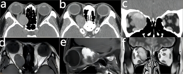
Figure 1: Magnetic resonance imaging (MRI) and computed tomography (CT) of a patient with cavernous hemangioma of the orbital apex. (A-C) CT soft-tissue window shows the tumor in the coronal and sagittal positions. (D-F) MRI T2-weighted images showed an isointensity tumor in the right orbital apex, whereas T2-weighted-enhancement images showed a high-intensity tumor in the coronal and sagittal positions. Please click here to view a larger version of this figure.
Plan: The treatment plan consists of the following surgical steps to be performed under endoscopic guidance: Preoperative imaging, including CT and MRI scans, will be utilized to identify the orbital tumor and delineate its precise relationship to the optic nerve and other critical structures. Using endoscopic visualization, the surgical approach will begin with a comprehensive definition of the middle meatus and ethmoid sinus anatomy. A grinding drill will be employed to meticulously remove bone from the lamina papyracea (orbital lamina), followed by an incision of the orbital fascia along the longitudinal axis of the eye. With the assistance of endoscopes providing various angles of view, the orbital tumor will be thoroughly and precisely excised.
Protocol
In this study, we reviewed the medical and surgical records of eight patients who underwent transnasal endoscopic resection after a diagnosis of OATs. Clinical data of inpatients selected from the Department of Orbital Diseases and Ophthalmic Oncology of Shenzhen Eye Hospital (China) from April 2021 to September 2024 were used. The ethics committees of the two hospitals (the seventh Affiliated Hospital of Sun Yat-Sen University and Shenzhen Eye Hospital) do not require such articles to seek ethical approval, and all aspects of the study were conducted in accordance with the Declaration of Helsinki. All patients had preoperative CT and/or MRI scanning with image guidance protocols, and informed consent was obtained from all participants prior to their inclusion in this study.
1. Patient selection
- Inclusion criteria: Patients with tumors primarily located in the intraorbital retrobulbar region (medial, superior, or inferior to the optic nerve) were selected. A clearly defined anatomical relationship between the tumor and surrounding structures (optic nerve, extraocular muscles, or orbital apex) was established. The presence of radiological features highly suggestive of benign or low-grade malignant tumors (e.g., cavernous hemangioma, schwannoma, meningioma, inflammatory pseudotumor) was established. It was established that MRI/CT demonstrates no extensive invasion of the skull base, intracranial compartment, or infratemporal fossa, and no encasement of the internal carotid artery. An absence of extensive bony destruction (metastatic carcinomas were excluded) was also confirmed.
- Exclusion criteria: Patients with high-grade malignancies (e.g., lymphoma, sarcoma, metastatic carcinoma) were excluded. Tumors with extensive extraorbital extension (intracranial, pterygopalatine fossa, or infratemporal fossa involvement) were also excluded. Tumors encasing >50% of the optic nerve or ophthalmic artery were also excluded. Patients with severe comorbidities (e.g., cardiac disease, diabetes mellitus) that contraindicate surgery were also excluded. Patients with coagulopathy or hemorrhagic diathesis were also excluded. Patients with active systemic infection or poor general health status precluding surgical intervention were also excluded. Patients who refused the proposed surgical procedure were also excluded.
2. Preoperative preparation, operative position, and anesthesia
- The patient was asked to adhere to a low-fat, low-salt, and low-sugar diet the day before the surgical operation and to refrain from consuming food or water for at least 8 h prior to the surgery.
- The patient was placed in a supine position with the head elevated and slightly tilted to the right.
- General anesthesia was administered using 1%-4% sevoflurane, cyclopofol (10 mg/mL), cisatracurium (2 mg/ mL), and sufentanil (5 µg/mL). Endotracheal intubation was performed. The anesthetic effect was evaluated based on the patient's post-anesthesia and intraoperative conditions, including the presence of a complete anesthesia block, the absence of additional drugs during the procedure, and the stability of vital signs.
3. Surgical procedure (Figure 2)
- Local anesthesia of the nasal mucosa was achieved using a mixture of 1% lidocaine and 0.1% epinephrine.
- The uncinate process was resected using a sickle-shaped uncinectomy knife, and its caudal end was thoroughly removed with mucosal forceps. A longitudinal incision was made along the medial edge of the bulla ethmoidalis, which was then completely excised using an electric microdebrider. The basal lamella of the middle turbinate was also removed.
- After removal of the anterior ethmoid sinuses, the posterior ethmoid sinuses were further resected following a medial-to-lateral excision strategy. The sphenoid sinus ostium was identified approximately 1.5 cm above the posterior choanal margin. The bony structures of the medial and inferior walls of the sphenoid sinus were preferentially removed to expose its internal anatomy. Resection was extended medially until the optic nerve canal was fully exposed.
- The bony structure at the superior aspect of the anterior wall of the sphenoid sinus was completely removed to establish continuity between the roof of the sphenoid sinus and the roof of the posterior ethmoid sinuses. The medial orbital wall was exposed, and the prominence of the optic nerve canal could be identified at the junction between the upper and middle thirds of the lateral wall of the most posterior ethmoid cell or the sphenoid sinus.
- The ethmoidal orbital plate was thinned using a sinus drill until the bone became nearly transparent. The thinned bony layer was carefully elevated with a nasal septum dissector. Complete removal of the ethmoidal orbital plate, particularly in the region near the orbital apex, prior to incision of the orbital fascia, allowed adequate exposure of the deep orbital fascia. This facilitated complete tumor resection after incision of the orbital fascia.
- An exit cut slightly smaller than the tumor mass was incised on the orbital fascia, revealing the tumor in the space between the medial and inferior rectus muscles. Complete resection of the tumor was carried out. Following incision of the orbital septum, significant herniation of intraorbital fat typically occurs. Carefully debulk the prolapsed fat in a piecemeal fashion using a manual microdebrider (an instrument shaped similarly to a sphenoid punch but featuring an internal suction channel connected to wall suction, allowing simultaneous removal of resected tissue).
NOTE: The use of a powered microdebrider is discouraged due to the high risk of excessive fat resection. This approach provides adequate exposure of the medial rectus muscle and the intraorbital tumor. Crucially, fat must not be retracted outward, as this can lead to bleeding or inadvertent over-resection of orbital fat. - The orbital fascial tissue was carefully repositioned and trimmed, followed by meticulous hemostasis using low-power monopolar electrocoagulation.
- The nasal cavity was copiously irrigated with sequential solutions of diluted povidone-iodine (0.05%) and 0.9% normal saline.
- Absorbable nasal packing was placed in the ethmoid and sphenoid sinuses to facilitate proper repositioning of the orbital fat. The incised orbital fascia does not need to be sutured; it only needs to be restored to its original position through a filler.
- The final intraoperative assessment confirmed that bilateral pupils were isocoric (3 mm in diameter) and reactive, marking the completion of the procedure.
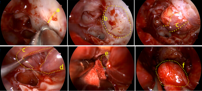
Figure 2: Transnasal endoscopy surgical removal process. (A) The ethmoid sinuses were fully opened, and the orbital board was removed. (B) Orbital fascia exposed after orbital board removal. (C) Bulging of the medial rectus muscle after incision of the orbital fascia. (D) Exposed inferior rectus. (E) The tumor was revealed in the space between the internal rectus and the inferior rectus. (F) Complete removal of the tumor. Please click here to view a larger version of this figure.
Results
All patient medical information is listed in Table 1 along with a summary of basic conditions, pre- and post-operative visual acuity, and postoperative pathology of all patients. A total of eight patients with eight eyes (two right eyes, six left eyes) were collected. There were seven females (87.5%) and one male (12.5%). The average age was 41.7 ± 14.2 years, ranging from 12 to 58 years. The preoperative symptoms of three patients (37.5%) were vision loss, two patients (25%) had exophthalmos, two patients (25%) had exophthalmos combined with vision loss, and one patient (12.5%) was asymptomatic before surgery. The decision to proceed with surgical intervention in the one asymptomatic patient (12.5%) was based on a multifaceted clinical rationale, consistent with standard neurosurgical and orbital oncology principles. The absence of subjective symptoms does not preclude the necessity for treatment, particularly in the anatomically complex and high-stakes region of the orbital apex. All patients had a CT examination that found intraorbital lesions. Pre- and post-operative visual acuity comparison showed that visual acuity decreased in two patients (25%), showed no change in one patient (12.5%), and improved in five patients (62.5%). Postoperative pathology determined that five patients (62.5%) had cavernous hemangioma, two patients (25%) had schwannoma, and one patient (12.5%) had metastatic nasopharyngeal. There were no cerebrospinal fluid leaks or postoperative hemorrhages. The eight patients were followed up for 6 months or more after surgery, and there was no eye movement disorder or visual field loss. No recurrence was observed in the seven patients with a postoperative pathologic benign condition, whereas the one patient with a malignant condition received postoperative radiotherapy.
| Case | Age (years) | Involved eye | Preoperative vision | Postoperative vision | Vision change | Main symptom/s | Postoperative pathology |
| 1/F | 12 | OS | 1 | 0.8 | Decline | Eyeball prominent | Schwannoma |
| 2/F | 58 | OS | 1 | 0.6 | Decline | No obvious symptoms | Cavernous hemangioma |
| 3/F | 45 | OS | 0.1 | 0.1 | Invariant | Vision decreased | Schwannoma |
| 4/F | 44 | OD | 0.1 | 0.8 | Enhance | Eyeball prominent | Cavernous hemangioma |
| 5/M | 51 | OS | 0.6 | 0.9 | Enhance | Eyeball prominent, vision decreased | Metastatic nasopharyngeal carcinoma |
| 6/F | 44 | OD | HM | 0.9 | Enhance | Vision decreased | Cavernous hemangioma |
| 7/F | 31 | OS | 0.6 | 1 | Enhance | Vision decreased | Cavernous hemangioma |
| 8/F | 49 | OS | 0.6 | 0.9 | Enhance | Eyeball prominent, vision decreased, | Cavernous hemangioma |
Table 1: Patient medical information. Abbreviations: F = female; HM = hand motion; M = male; OD = oculus dexter; OS = oculus sinister.
Discussion
There is a wide variety of orbital tumors, including cavernous hemangioma, meningioma, schwannoma, neurofibroma, and fibrous tumors5. Surgical treatment is the primary treatment for most of these. Different orbital tumors have different properties, locations, and adjacencies; therefore, the surgical methods also vary. The orbital apex is a relatively narrow conical bony space containing many important structures, such as nerves, blood vessels, and muscles13. OATs regularly cause exophthalmos, eye movement disorders, vision loss, and other adverse consequences. Therefore, surgery to remove orbital tumors is complicated, urgent, and more likely to cause serious surgical complications. The complications of surgery are related to anatomy, pathology, surgical methods, and surgical techniques. Therefore, it is necessary to select appropriate surgical methods and surgical approaches based on detailed preoperative examination and comprehensive evaluation of a tumor's location, size, and nature. Common surgical procedures can be divided into anterior orbital opening, lateral orbital opening, intranasal orbital opening, combined internal and external orbital opening, transcranial orbital opening, and orbital contents resection14.
The endoscopic endonasal approach (EEA) has emerged as a pivotal technique for selected OATs. The success of this protocol hinges on several critical steps. First, a wide sphenoethmoidectomy is performed to fully expose the medial orbital wall from the lacrimal sac to the orbital apex, providing the necessary working space and landmarks15,16. The subsequent crucial step is the precise bony removal of the lamina papyracea, focusing initially on the thinnest portion over the orbital lesion. Following this, a meticulous incision of the periorbita is required, parallel to the medial rectus muscle, to avoid vascular injury and control herniation of orbital fat. Finally, dissection within the intraconal space necessitates working through the natural corridor between the medial and inferior rectus muscles to access the lesion while minimizing traction on the optic nerve17,18,19.
The anatomy of the nose and eye is closely related. Except for the lateral wall of the orbit, the other walls are surrounded by the sinuses, and many structures are common to the nose and eye. The bone of the medial wall of the eye is composed of the frontal process of the maxillary sinus, lacrimal bone, and ethmoid cardboard. The main structures of the medial wall of the eye from front to back are the anterior lacrimal ridge, dacryocystic fossa, posterior lacrimal ridge, ethmoid and related blood vessels, nerves, orbital orifice of the optic canal, and the optic nerve. The posterior ethmoid sinus and sphenoid sinus are anterior to the optic chiasma and pituitary gland, and the lateral optic nerve is located at the superior lateral wall of the sphenoid sinus. As a result of this anatomical adjacency, many nasal diseases are susceptible to ocular symptoms involving the orbit, and many ocular diseases, especially those close to the nose, are treated with an intranasal approach, which is straightforward and simple16.
Kennedy et al.(15) first reported that transnasal endoscopy could be used for optic nerve decompression of thyroid-associated ophthalmopathy. In 1999, the first case of transnasal endoscopy in the removal of an orbital cavernous hemangioma was reported by Herman et al.(18). With the development of nasal-related disciplines and the deepening of minimally invasive concepts, as well as the continuous development of nasal endoscopic technology, transnasal endoscopic surgery has been developed significantly and applied. It has been used in diseases of the lacrimal duct system, repair of orbital fractures, resection of medial orbital tumors, orbital decompression, and optic nerve decompression17,19.
In our experience, we have incorporated specific modifications to enhance safety and efficacy. A key modification was the routine use of multi-angle endoscopes (45° and 70°) in addition to the 0° scope, which provided a panoramic view around anatomical corners, crucial for visualizing the superior and lateral aspects of the orbital apex. When encountering significant bleeding from the orbital venous plexus, troubleshooting involves controlled bipolar cautery and the use of hemostatic agents, rather than aggressive suction. Furthermore, for firm, encapsulated tumors, we employed a technique of incising the capsule and performing intracapsular debulking before delivering the capsule, thereby minimizing the required working corridor14.
Compared with the traditional transorbital approach, transnasal endoscopic resection is more convenient for the treatment of OATs, especially optic neuropathy. This approach makes use of the potential pathway between the medial rectus and inferior rectus to enter into the muscular cone and orbital apex, exposing the intraorbital segment of the optic nerve and the intraorbital segment of the ophthalmic artery, facilitating the treatment of intraorbital lesions located below the optic nerve20,21,22. It avoids excessive intraoperative squeezing of the eyeball and stretching of the optic nerve and causes little damage to the important tissues in the orbit. Facial scarring is also avoided23,24. The primary significance of EEA with respect to existing methods lies in providing direct access to the medial orbital apex without requiring brain retraction or skin incisions, thus avoiding facial scarring and reducing post-operative recovery time20,21. Compared to lateral micro-orbitotomy, it minimizes manipulation of the optic nerve and globe, potentially reducing the risk of post-operative ptosis, diplopia, and optic neuropathy17,18.
However, transnasal endoscopic resection also has certain limitations in the treatment of OATs. The most significant limitation is its restricted access to lesions located lateral or superior to the optic nerve, making it unsuitable for pathologies in these compartments without risking iatrogenic injury19. Additionally, the technique offers a narrow surgical corridor, which can make instrument maneuverability challenging for large tumors. Furthermore, reliance on intraoperative image-guidance systems has limitations; after opening the periorbita, the shift of orbital contents can lead to a loss of registration accuracy, meaning surgical anatomy, rather than navigation alone, must guide the final dissection24.
Because it cannot be operated across the optic nerve, it is not suitable for the treatment of lateral and external lesions of the optic nerve22. In our medical records, nasal endoscopes with 0°, 45°, and 70° angles were used during the surgery, enabling us to have a broader field of vision. For OATs, we also need to use a different size drill to better expose the surgical field.
Owing to the special orbital anatomy, orbital operation is prone to a variety of complications, such as visual impairment, ptosis, eye movement disorders, orbital bleeding, and pupil changes. Transnasal endoscopic resection also increases the risk of nasogenic orbital infection. The occurrence of surgical complications is not only related to the nature, location, and size of the lesion, but also closely related to the choice of surgical approach25. Preoperative orbital CT and MRI can determine the location of the lesion and determine whether most orbital tumors are benign or malignant26. The image navigation system can conduct three-dimensional reconstruction of preoperative CT and MRI images of patients, and locate the operative area accurately through the electromagnetic induction positioning system, assisting doctors to judge the lesion adjacency and safety boundary accurately27. The image navigation system has an important role in the surgery of the orbitonasal base, orbital skull base tumors, and resection of a nasal skull base tumor. But we do not think we can rely entirely on the image navigation system during surgery. In addition to systematic errors in navigation, the orbital pressure changes after the orbital fascia is cut through the nose and part of the orbital fat is removed, and the position of the orbital lesion relative to the surrounding structure also changes to varying degrees. If the surgery continues to follow the guidance of the image navigation system, it is likely that different degrees of complications will occur. An image navigation system is more helpful for orbital bone tumor resection or foreign body removal, but is limited for non-bone tumor resection.
The most significant limitation is its restricted access to lesions located lateral or superior to the optic nerve, making it unsuitable for pathologies in these compartments without risking iatrogenic injury19. Additionally, the technique offers a narrow surgical corridor, which can make instrument maneuverability challenging for large tumors. Furthermore, reliance on intraoperative image-guidance systems has limitations; after opening the periorbita, the shift of orbital contents can lead to a loss of registration accuracy, meaning surgical anatomy, rather than navigation alone, must guide the final dissection24.
Malignant tumors accounted for a relatively low proportion of orbital tumors but were prone to recurrence and metastasis. For malignant tumors, targeted radiotherapy and chemotherapy are also needed after surgery. In this study, a patient with malignant lesions had a history of radiotherapy for nasopharyngeal carcinoma. Preoperative consideration was given to nasopharyngeal carcinoma recurrence and orbital metastasis, which was also confirmed by postoperative pathology.
Finally, it is paramount to emphasize that these procedures are optimally performed by a cohesive multidisciplinary team. This surgery is not the domain of a single specialty but requires collaboration between experienced Otolaryngologists (ENTs) and Ophthalmologists (Oculoplastic Surgeons). The ENT surgeon brings expertise in endoscopic nasal anatomy and navigation, while the ophthalmic surgeon provides critical knowledge of orbital dynamics and physiology. Pre-operative planning and intraoperative decision-making are vastly enhanced by this partnership, which is now considered the standard of care at high-volume skull base centers.
Disclosures
The authors have nothing to disclose.
Acknowledgements
The authors have no acknowledgments.
Materials
| Name | Company | Catalog Number | Comments |
|---|---|---|---|
| Integrated Power Console | Medtronic America | 1898001 | Sterile,dry heat sterilized, reusable |
| NasoPore | Stryker Instruments America | 5400-010-004 | Sterile, ethylene oxide sterilized, disposable |
| Sinoscopes and Accessories | KARLSTORZ Germany | 7230AA | Sterile,dry heat sterilized, reusable |
References
- Chastain, J. B., Sindwani, R. Anatomy of the orbit, lacrimal apparatus, and lateral nasal wall. Otolaryngol Clin North Am. 39 (5), 855-864 (2006).
- Abuzayed, B., Tanriover, N., Gazioglu, N., Eraslan, B. S., Akar, Z. Endoscopic endonasal approach to the orbital apex and medial orbital wall: anatomic study and clinical applications. J Craniofac Surg. 20 (5), 1594-1600 (2009).
- Bleier, B. S., Healy, D. Y. Jr, Chhabra, N., Freitag, S. Compartmental endoscopic surgical anatomy of the medial intraconal orbital space. Int Forum Allergy Rhinol. 4 (7), 587-591 (2014).
- Kim, B. S., Im, Y. S., Woo, K. I., Kim, Y. D., Lee, J. I. Multisession Gamma Knife Radiosurgery for Orbital Apex Tumors. World Neurosurg. 84 (4), 1005-1013 (2015).
- Mendoza-Santiesteban, E., et al. Diagnosis and surgical treatment of orbital tumors. Semin Ophthalmol. 25 (4), 123-129 (2010).
- Rose, G. E., Verity, D. H. Neuro-ophthalmology of orbital disease. Handb Clin Neurol. 102, 467-491 (2011).
- Calandriello, L., et al. Cavernous venous malformation (cavernous hemangioma) of the orbit: Current concepts and a review of the literature. Surv Ophthalmol. 62 (4), 393-403 (2017).
- Murchison, A. P., Rosen, M. R., Evans, J. J., Bilyk, J. R. Endoscopic approach to the orbital apex and periorbital skull base. Laryngoscope. 121 (3), 463-467 (2011).
- Sia, D. I., Chan, W. O., Wormald, P. J., Davis, G., Selva, D. Decompression of benign orbital apex lesion via medial endoscopic approach. Orbit. 31 (5), 344-346 (2012).
- Kent, J. S., Allen, L. H., Rotenberg, B. W. Image-guided transnasal endoscopic techniques in the management of orbital disease. Orbit. 29 (6), 328-333 (2010).
- Cohen, L. M., Grob, S. R., Krantz, K. B., Feldman, K. A., Rootman, D. B. Combined Endonasal and Orbital Approach for Resection of Orbital Apical Tumors. Ophthalmic Plast Reconstr Surg. 38 (4), 393-400 (2022).
- Bleier, B. S., et al. Endoscopic endonasal orbital cavernous hemangioma resection: global experience in techniques and outcomes. Int Forum Allergy Rhinol. 6 (2), 156-161 (2016).
- Castelnuovo, P., Turri-Zanoni, M., Battaglia, P., Locatelli, D., Dallan, I. Endoscopic Endonasal Management of Orbital Pathologies. Neurosurg Clin N Am. 26 (3), 463-472 (2015).
- Cho, K. J., Paik, J. S., Yang, S. W. Surgical outcomes of transconjunctival anterior orbitotomy for intraconal orbital cavernous hemangioma. Korean J Ophthalmol. 24 (5), 274-278 (2010).
- Kennedy, D. W., Goodstein, M. L., Miller, N. R., Zinreich, S. J. Endoscopic transnasal orbital decompression. Arch Otolaryngol Head Neck Surg. 116 (3), 275-282 (1990).
- Stuck, B. A., et al. Rhinosinusitis guidelines--unabridged version: S2 guidelines from the German Society of Otorhinolaryngology, Head and Neck Surgery. Hno. 60 (2), 141-162 (2012).
- Levy, J., et al. Endoscopic orbital decompression for Graves' ophthalmopathy. Isr Med Assoc J. 6 (11), 673-676 (2004).
- Herman, P., et al. Transnasal endoscopic removal of an orbital cavernoma. Ann Otol Rhinol Laryngol. 108 (2), 147-150 (1999).
- Locatelli, M., Carrabba, G., Guastella, C., Gaini, S. M., Spagnoli, D. Endoscopic endonasal removal of a cavernous hemangioma of the orbital apex. Surg Neurol Int. 2, 58(2011).
- De Rosa, A., et al. Endoscopic endo- and extra-orbital corridors for spheno-orbital region: anatomic study with illustrative case. Acta Neurochir (Wien). 161 (8), 1633-1646 (2019).
- Lee, J. Y., Ramakrishnan, V. R., Chiu, A. G., Palmer, J., Gausas, R. E. Endoscopic endonasal surgical resection of tumors of the medial orbital apex and wall. Clin Neurol Neurosurg. 114 (1), 93-98 (2012).
- Düz, B., Secer, H. I., Gonul, E. Endoscopic approaches to the orbit: a cadaveric study. Minim Invasive Neurosurg. 52 (3), 107-113 (2009).
- Stokken, J., Gumber, D., Antisdel, J., Sindwani, R. Endoscopic surgery of the orbital apex: Outcomes and emerging techniques. Laryngoscope. 126 (1), 20-24 (2016).
- Yao, B. S., Bleier, B. S. Endoscopic management of orbital tumors. Curr Opin Otolaryngol Head Neck Surg. 24 (1), 57-62 (2016).
- Purgason, P. A., Hornblass, A. Complications of surgery for orbital tumors. Ophthalmic Plast Reconstruct Surg. 8 (2), 88-93 (1992).
- Ben Simon, G. J. Rethinking orbital imaging establishing guidelines for interpreting orbital imaging studies and evaluating their predictive value in patients with orbital tumors. Ophthalmology. 112 (12), 2196-2207 (2005).
- Galletti, B., Gazia, F., Galletti, C., Galletti, F. Endoscopic treatment of a periorbital fat herniation caused by spontaneous solution of continuity of the papyracea lamina. BMJ Case Rep. 12 (4), e229376(2019).
Reprints and Permissions
This article has been published
Video Coming Soon