This study details a rat fixation method suitable for photographing acupuncture and infrared thermography. Furthermore, it demonstrates that electroacupuncture at sensitized acupoints inhibits local inflammatory pain.
Method Article
A Visualized Sensitized Acupoint Procedure for Knee Osteoarthritis: Behavioral, Thermal, and Molecular Assessments
* These authors contributed equally
In This Article
Summary
Abstract
Knee osteoarthritis (KOA), a leading cause of musculoskeletal pain and functional disability worldwide, remains challenging to manage due to the limited efficacy of current pharmacological and surgical interventions in achieving sustained pain relief. Although acupuncture has demonstrated clinical promise in alleviating KOA-related pain, its underlying mechanisms require further elucidation. Application of acupuncture to sensitized acupoints can markedly enhance the effectiveness of therapy. This protocol establishes a rat model of KOA through intra-articular injection of sodium monoiodoacetate (MIA) and systematically describes an acupuncture protocol targeting sensitized acupoints, including restraint methods, acupoint localization, and acupuncture parameters. Therapeutic efficacy was comprehensively evaluated through pain behavioral assessments, infrared thermographic analysis of the knee joint, inflammatory cytokine detection in serum, and histopathological examination of cartilage tissue. Results indicate that acupuncture at sensitized acupoints significantly alleviates pain phenotypes in MIA-induced model rats by inhibiting the release of pro-inflammatory mediators, blocking nociceptive signal transmission, improving blood microcirculation, regulating local joint temperature, and delaying joint degeneration.
Introduction
Knee osteoarthritis (KOA), a prevalent degenerative joint disorder, is characterized by chronic inflammation and whole-joint pathology, including cartilage erosion, subchondral bone remodeling, osteophyte development, and synovial inflammation1,2. Clinically, patients typically present with pain, muscle weakness, joint instability, transient morning stiffness, and progressive functional limitations. KOA imposes a substantial global disease burden, affecting approximately 365 million individuals with functional disability, with epidemiological models predicting a 74.9% escalation in case numbers from 2020 baselines by mid-century 20503. Notably, while pain serves as the primary treatment motivator4, it simultaneously exacerbates functional disability, profoundly compromising multiple quality-of-life domains through impaired mobility and reduced daily activity capacity. Consequently, therapeutic strategies aim to alleviate pain, restore function, and delay disease progression5. Current first-line pharmacotherapy includes analgesics, non-steroidal anti-inflammatory drugs, chondroitin and glucosamine sulfate, and intra-articular corticosteroids to reduce pain and inflammation6. However, prolonged use of these agents is constrained by significant iatrogenic risks, particularly gastrointestinal complications and cardiovascular adverse events7, thereby underscoring the critical need for safer, more sustainable therapeutic alternatives.
Acupuncture and electroacupuncture have been extensively utilized in osteoarthritis management, demonstrating efficacy in alleviating knee pain and improving joint functionality8,9, with the World Health Organization (WHO) recognizing arthritis as a priority condition for acupuncture therapy. As both stimulation targets and effectors, acupoints remain central to acupuncture research, where their morphological appearance, spatial dimensions, and functional responsiveness undergo dynamic changes during disease states, a phenomenon termed "acupoint sensitization"10,11. Critically, this sensitization process modulates the receptive field area and therapeutic responsiveness of acupoints, thereby determining treatment efficacy. Previous preclinical studies12,13demonstrated sensitization at Yanglingquan (GB34, located in the depression anterior and inferior to the head of the fibula, near the lateral side of the hindlimb) and Zusanli (ST36, located on the anterior side of the leg, the lateral border of the patellar ligament is depressed to the anterior midpoint, 3/16 of the ankle joint) in KOA animal models. Laser speckle imaging revealed elevated local blood perfusion at these acupoints, while photoacoustic imaging paradoxically showed no significant structural alterations in microcirculation, suggesting that microcirculatory changes during acupoint sensitization in KOA are primarily functional rather than structural. Investigations into underlying mechanisms require the use of rat models; however, poor procedural compliance in animals results in considerable time investment. To address these constraints, a rat immobilization apparatus was developed, specifically designed to facilitate access to lower limb acupoints and enhance the efficiency of electroacupuncture procedures.
Infrared thermography, as a functional imaging modality, utilizes non-contact infrared sensors to detect thermal radiation from predefined regions of interest, with computer-processed data generating pseudocolor thermograms for quantitative analysis14,15. This technique enables non-invasive monitoring of cutaneous hemodynamic changes by measuring temperature variations associated with blood flow fluctuations in cutaneous, vascular, and muscular tissues16, thereby facilitating visual assessment of functional microcirculatory alterations at acupoints. In this study, pain sensitization at GB34 and ST36 acupoints was first validated using an electronic algometer, followed by the combined application of safranin O fast green staining, IL-1β and TNF-α analysis, infrared thermographic imaging, and pain-related behavioral assessments to investigate the analgesic effects of electroacupuncture stimulation at GB34 and ST36 acupoints in monosodium iodoacetate-induced (MIA) knee osteoarthritic rats.
Protocol
All experimental protocols were approved and rigorously supervised by the Animal Ethics Committee of Beijing University of Chinese Medicine (ID: bucm-2024091102-3197), and all procedures strictly adhered to the National Institutes of Health (NIH) Guide for the Care and Use of Laboratory Animals. Thirty male Sprague-Dawley rats (weight range: 210-250 g) were used in this study. The reagents and equipment used are listed in the Table of Materials.
1. Experimental animals and groups
- House the animals in the dedicated animal facility.
- Maintain ambient temperature at 22 °C ± 2 °C, relative humidity at 50%-60%, and a 12-h light/dark cycle. Provide ad libitum access to food and water throughout the study.
- After a 7-day acclimatization period, randomly divide rats into five experimental groups (n = 6 per group): normal control (Con) group, Sham operation (Sham) group, Model (MIA) group, non-fixed (NF) group, and electroacupuncture (EA) group.
2. Establishment of a monoiodoacetate-induced KOA model
- Place rats in an induction chamber. Anesthetize using 4% isoflurane in oxygen for induction, then maintain under 1.5%-2.0% isoflurane delivered via a facemask (following institutionally approved protocols).
- Fix rats in left lateral recumbency. Remove hair using a gentle depilatory cream on the right knee joint and surrounding areas.
- Disinfect the right knee joint three times with povidone iodine from inside to outside, followed by repetition of the same steps with 75% ethanol.
- Maintain the right knee at 90° flexion. Identify the inferior patellar border and tibial tuberosity. Advance a 22-G needle (0.7 mm × 30 mm) 3-5 mm into the joint space via the lateral or medial border of the patellar ligament at a 30°-45° cutaneous approach angle. Confirm intra-articular placement and slowly infuse 50 µL of MIA solution (1 mg/50 µL). For sham controls, inject an equivalent volume of sterile saline.
- Perform five passive range-of-motion manipulations to ensure uniform intra-articular drug dispersion. Return the animal to its home cage for postoperative recovery.
3. Procedure for fixing rats
- Secure the rat in a black restraint sleeve. Create a 1.5-cm-diameter circular aperture in the lower right quadrant through which the right hindlimb is exteriorized, as shown in Figure 1A.
- Secure paired nylon cable ties (5 mm × 300 mm), spaced 6.8 cm apart, through a plastic mesh frame. Position the superior restraint at the axillary region and anchor the inferior restraint at the pelvic girdle, as illustrated in Figure 1B,C,E.
NOTE: The NF group does not undergo the procedures described in step 3.2.
4. Electroacupuncture treatment
- Begin electroacupuncture intervention on the fourth day following a consecutive three-day fixation process.
- Perform aseptic preparation at the GB34 and ST36 acupoint regions. Refer to Figure 1D for standardized surface anatomical mapping of these loci.
- Insert disposable sterile acupuncture needles (diameter: 0.25 mm; length: 13 mm) into the acupoints. Connect GB34 to the anode and ST36 to the cathode according to the electrostimulation protocol (Figure 1G).
- After a 20-min electroacupuncture session, deactivate the device. Detach the electrodes from the needle handles and carefully remove the acupuncture needles to complete the therapeutic procedure. Administer interventions daily over two consecutive weeks.
NOTE: Except for the NF group, the remaining three groups received no treatment but were immobilized for 20 min daily in the same manner as the EA group.
5. Mechanical Withdrawal Threshold (MWT) test
- In a quiet environment at a room temperature of 24 °C ± °C, place rats on a foot test platform with a stainless steel mesh bottom and an organic plastic cage body.
- Stimulate the center area of the right hind paw or designated acupuncture points vertically using the 0. mm diameter probe of the Electric Von Frey electronic pain detector. Gradually and uniformly increase the intensity of stimulation until a paw withdrawal avoidance response is observed. Record the value displayed at this moment as the mechanical withdrawal threshold.
NOTE: Each rat is measured once at 10-min intervals, with the mean of three consecutive trials designated as the final result.
6. Paw Withdrawal Latency (PWL) test
- Acclimate rats on the thermal platform for 5 min before formal assessment.
- Maintain the platform surface temperature at 55.0 °C ± 0. °C during testing. Initiate timing when the right hind paw contacts the heated surface and stop timing upon observation of the first positive reaction (paw licking, jumping, or rapid withdrawal). Define the recorded duration as the PWL.
NOTE: Perform triplicate measurements per rat with 5-min intertrial intervals. Calculate the mean value as the representative PWL.
7. Infrared thermal imaging
NOTE: Depilate the hind limbs the day before thermography to eliminate the influence of hair on temperature measurement.
- Maintain experimental conditions at 2 °C ambient temperature with 60% relative humidity under sound-attenuated and diffuse lighting.
- Restrain the rat in a black restraint sleeve with the right hindlimb extended through a circular hole. Place on the operating table for 10 min.
NOTE: If the rat is not calm after 10 min, extend the waiting time until it becomes quiet before proceeding to the next step. - Acquire lateral and ventral stifle joint thermal images using the thermal imager at a 5 cm distance, configured at 0.97 emissivity, 9 Hz frame rate, and 160 × 120 pixel IR resolution.
- Process the acquired infrared thermographic data using dedicated analytical software to enable precise quantification of thermal parameters through computer-based spectral analysis.
8. Enzyme-Linked Immunosorbent Assay (ELISA)
- Anesthetize rats by intraperitoneal injection of 20% urethane solution (1.2 g/kg). Open the abdominal cavity and insert the blood sampling needle into the abdominal aorta. Insert the end of the needle into a vacuum blood collection vessel without an anticoagulant.
- Allow blood samples to clot for 30 min. Centrifuge at 1006. x g for 15 min at °C. Quantify serum protein levels using the BCA assay.
- Measure IL-1β and TNF-α concentrations using commercial ELISA kits according to the manufacturer's protocols.
9. Safranin fast green staining
NOTE: Following abdominal aortic blood collection, excise the right stifle joint using surgical scissors, preserving 1 mm osteoarticular segments of both the proximal femur and distal tibia. Carefully debride periarticular soft tissues.
- Immerse stifle joints in 4% paraformaldehyde (PFA) for 72 h. Rinse thoroughly with phosphate-buffered saline (PBS) under gentle agitation to ensure complete PFA removal.
- Immerse samples in ethylenediamine tetraacetic acid (EDTA) decalcification solution at °C for 4 weeks. Replace the EDTA solution every 2 days. Prick the tissue lightly with a needle; absence of resistance indicates completion of decalcification. Rinse decalcified specimens with PBS for 4-6 h to remove residual EDTA.
- Dehydrate samples using an automatic dehydrator. Embed in paraffin and allow to solidify into rigid blocks. Section paraffin-embedded blocks using a microtome to obtain 4-µm-thick slices. Transfer slices to a water bath. Once expanded, retrieve sections and place them on glass slides. Dry in a temperature-controlled chamber for 12 h.
- Place slides in xylene (I) for 10 min and xylene (II) for another 10 min. Soak sequentially in anhydrous ethanol (I), anhydrous ethanol (II), 90% ethanol, and 75% ethanol for 5 min each. Rinse with ultrapure water.
- Stain slides with Fast Green solution for 2 min. Rinse under tap water until cartilage appears glass-like and transparent with clearly visible lacunae under microscopic observation. Differentiate in 1% acetic acid solution for 10 s, monitoring until the residual blue tint is completely removed. Rinse immediately with ultrapure water.
- Stain slides with Safranin O solution for 15 min until the cartilage matrix shows uniform, intense ruby-red coloration while bone tissue appears pale pink.
- Soak slides sequentially in anhydrous ethanol (I), (II), (III), and (IV) for 10 s each. Immerse in freshly prepared xylene (I) and xylene (II) for 10 min each.
- Remove slides and apply neutral resin to the slide surface, carefully avoiding tissue contact. Place the edge of the coverslip on the slide and lower slowly to prevent bubble formation. Wipe off excess xylene and resin. Air-dry slides overnight at room temperature. Place dried sections under an optical microscope for imaging.
10. Statistical analysis
- Use SPSS software for statistical analysis. Express data as mean ± standard deviation.
- Analyze MWT and PWL data using repeated-measures ANOVA. Compare other datasets across groups using one-way ANOVA.
- Express results as mean ± standard deviation (SD). Define statistical significance as p < 0.05.
Results
No animals died during the entire experiment. Results of the operation procedure duration and needle detachment count for each rat in both the NF and EA groups indicated that the EA group, following restriction with nylon cable ties, experienced reduced operation time and fewer needle detachments (P < 0.001; Figure 1H,I). Analysis of acupoint pain thresholds revealed no statistically significant differences in mechanical pain sensitivity between GB34, ST36, and non-acupoint regions prior to modeling (P > 0.05). Fourteen days after MIA injection, pain sensitivity increased at all three sites, with GB34 and ST36 exhibiting significantly higher sensitivity compared to non-acupoint regions (P < 0.05 or P < 0.01; Figure 1J). Results of mechanical withdrawal threshold (MWT) and paw withdrawal latency (PWL) assessments showed that baseline thresholds were consistent across groups. Two weeks after injection of normal saline or MIA, no significant differences were observed between the Sham and Con groups (P > 0.05). After two weeks of treatment, the EA group exhibited a significantly improved pain threshold compared with the MIA group (P < 0.001; Figure 2).
Infrared thermal imaging revealed that in the Con group, the average skin temperature of the lateral and ventral surfaces of the knee joint decreased significantly after anesthesia compared to pre-anesthesia levels (P < 0.01 or P < 0.001; Figure 3). Compared with the Con group, no significant difference in knee skin temperature was observed in the Sham group (P > 0.05). However, the MIA group showed significantly higher average skin temperature in both regions compared to the Sham group (P < 0.001 or P < 0.01). The EA group demonstrated significantly lower average skin temperature than the MIA group (P < 0.05; Figure 4).
Histological analysis with safranin O fast green staining showed severe bone destruction in the knee joint cartilage of the MIA group, characterized by paler cartilage staining and a reduced number of cartilage cells. In contrast, the EA group exhibited alleviated cartilage damage, with slightly lighter cartilage color and improved morphology and quantity of cartilage cells (Figure 5).
ELISA results showed no significant differences in serum levels of inflammatory cytokines IL-1β and TNF-α between the Con and Sham groups (P > 0.05). Compared with the Sham group, the MIA group exhibited significantly elevated serum levels of TNF-α and IL-1β (P < 0.001). EA intervention significantly reduced these cytokine levels compared with the MIA group (P < 0.001; Figure 6).
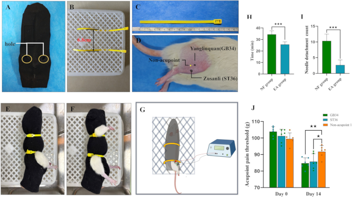
Figure 1: Electroacupuncture intervention method. (A) Physical image of a rat's restraint device. (B) Diagram showing placement of nylon strapping. (C) Actual length of the nylon cable fixed in place. (D) Surface anatomical locations of GB34 and ST36 acupoints in rats. GB34 (Yanglingquan) is located in the depression anterior and inferior to the head of the fibula, near the lateral side of the hindlimb. ST36 (Zusanli) is situated on the anterior side of the leg, along the lateral border of the patellar ligament, approximately 3/16 of the distance from the knee to the ankle joint. The non-acupoint site is located 4 mm lateral to ST36. (E) Diagram of rat restraint. (F) Restraint diagram showing the upper nylon cable positioned away from the cervical region. (G) Diagram illustrating the electroacupuncture stimulation procedure. (H) Statistical comparison of operation time between the two fixation methods (***P < 0.001). (I) Frequency of acupuncture needle detachment in the two groups (n = 6 per group, ***P < 0.001). (J) Comparison of mechanical pain thresholds between acupoints and non-acupoints in the EA group (n = 6 per group, *P < 0.05, **P < 0.01). Please click here to view a larger version of this figure.
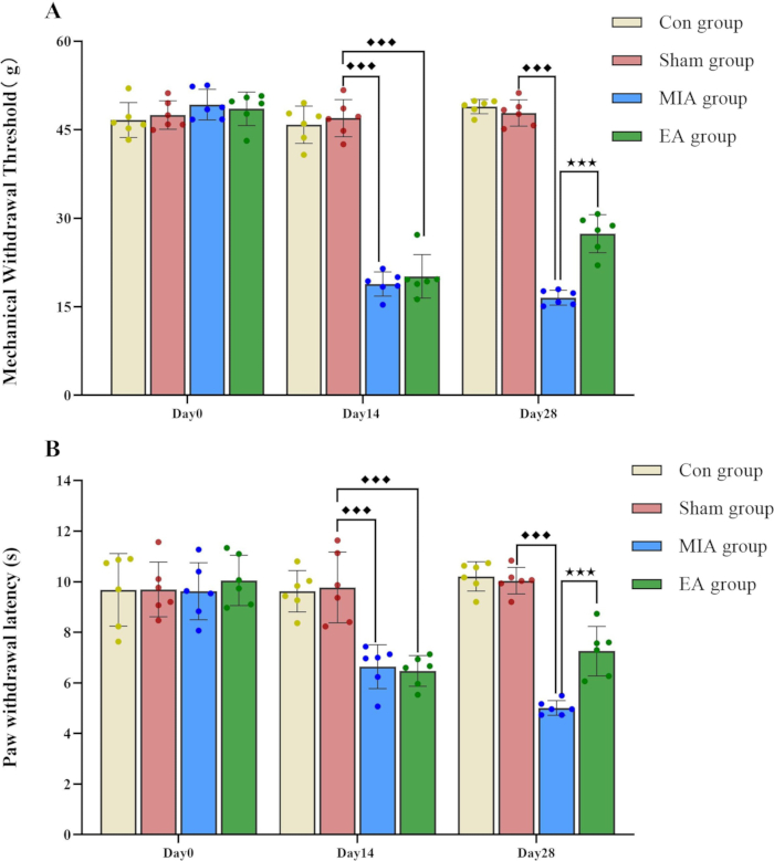
Figure 2: Comparison of mechanical and thermal pain thresholds among four experimental groups of rats (n = 6 per group). (A) Statistical results of mechanical withdrawal threshold (MWT). (B) Statistical results of paw withdrawal latency (PWL). Compared to the sham group, ♦♦♦P < 0.001; compared to the MIA group, ***P < 0.001. Please click here to view a larger version of this figure.
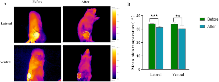
Figure 3: Comparison of stifle joint temperatures in awake and anesthetized states (n = 6 per group). (A) Representative infrared thermal images of the control group under awake and anesthetized conditions. (B) Mean skin temperatures at lateral and ventral stifle joint regions before and after anesthesia (**P < 0.01, ***P < 0.001). Please click here to view a larger version of this figure.
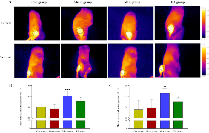
Figure 4: Comparison of stifle joint temperature among four experimental groups (n = 6 per group). (A) Representative infrared thermal images from each group. (B,C) Mean skin temperature at lateral and ventral stifle joint regions across groups. Compared to the sham group, ♦♦P < 0.01, ♦♦♦P < 0.001; compared to the MIA group, *P < 0.05. Please click here to view a larger version of this figure.
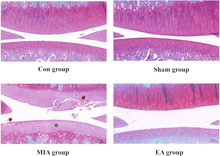
Figure 5: Comparative analysis of histopathological features in rat knee joints among four experimental groups (Safranin O-Fast Green staining, n = 3 per group). In the MIA group, a marked reduction in articular cartilage is observed (green arrow), along with pronounced cartilage damage (red arrow). Scale bar: 100 µm. Please click here to view a larger version of this figure.
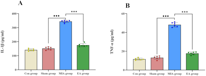
Figure 6: Comparison of serum IL-1β and TNF-α expression among four experimental groups (n = 6 per group). (A) Serum IL-1β expression levels. (B) Serum TNF-α expression levels. Compared to the sham group, ♦♦♦P < 0.001; compared to the MIA group, ***P < 0.001. Please click here to view a larger version of this figure.
Discussion
MIA, a metabolic inhibitor, selectively disrupts cellular aerobic glycolysis, thereby inducing chondrocyte apoptosis17,18,19. Intra-articular MIA administration results in chondrocyte depletion and subsequent articular cartilage degeneration, characterized by morphological irregularities and histopathological alterations that closely resemble human OA pathology20. Thus, it is widely utilized in research on OA pain mechanisms21. Iodoacetic acid (IAA) injection is also commonly used; however, it is not easily soluble in water, and the resulting solution typically becomes acidic. This acidity can exert an additional stimulating effect on joint tissues. Currently, the sodium salt form of IAA, known as MIA, is commonly used22. Consequently, the MIA-induced knee arthritis model was adopted in this study to conduct a visual investigation of the mechanism underlying acupuncture sensitization at specific acupoints. Safranin O-fast green staining has been widely used to demonstrate pathological changes in joint diseases. Therefore, this staining method was employed to verify successful induction of the KOA model23,24.
Both peripheral and central sensitization mechanisms critically mediate pain processing in KOA25,26. Joint injury induces sustained exposure of nociceptors to inflammatory mediators within the articular microenvironment, promoting receptor upregulation and neuronal hyperexcitability27. This persistent peripheral input drives central sensitization via enhanced synaptic transmission in spinal dorsal horn neurons, leading to chronic pain characterized by hypersensitivity28. Acupoint sensitization is defined as a dynamic biological process wherein specific body surface acupoints (or periacupoint regions) transition from a "resting" to an "activated" state under pathological conditions29. This transition is characterized by significant alterations in sensory processing, including lowered detection thresholds, heightened nociceptive hypersensitivity (allodynia/hyperalgesia), and measurable changes in local tissue physicochemical properties30. Notably, periarticular acupoints in KOA exhibit localized accumulation of algogenic substances, correlating with mechanical hyperalgesia and aberrant nociception. Clinical evidence supports targeting these sensitized acupoints31,32, which may act as homeostatic trigger points, to enhance therapeutic efficacy. Previous experimental investigations in KOA models revealed pathological mast cell infiltration and active degranulation at GB34 and ST36 acupoints, accompanied by substantial release of pronociceptive mediators, including serotonin (5-HT) and substance P (SP)11,30,33. The present study demonstrated that acupuncture at sensitized acupoints significantly elevated mechanical and thermal pain thresholds, confirming potent analgesic efficacy. Histopathological analysis further revealed ameliorated cartilage lesions, characterized by preserved chondrocyte architecture and reduced matrix degradation, indicating structural preservation alongside pain relief.
In this study, a rat restraint protocol was introduced for acupuncture intervention and infrared thermographic imaging. The use of dark visual blockage effectively reduced anxiety in rats caused by environmental stimuli. The flexible fiber composition of the restraint device allowed easy adaptation to the animal's skin, and its high breathability significantly lowered the risk of suffocation during treatment. The black cloth sleeve outperformed cable-tie fixation alone in minimizing stress responses in rats, supporting its selection. Integration of platform fixation with cable ties prevented the dislodging of acupuncture needles, thereby ensuring treatment efficacy. The protocol's advantages, derived from its simple materials and ease of operation, facilitate large-scale acupuncture procedures and reduce experimenter workload.
Importantly, thermographic measurements obtained from unanesthetized rats eliminated interference from anesthetic metabolism and temperature fluctuations, providing a refined method for infrared thermographic imaging in conscious animals. Infrared thermal imaging studies conducted in conscious and restrained laboratory mice yielded results consistent with those of Bjorn Redfors et al.34, who reported that vasodilation and core temperature changes induced by isoflurane anesthesia led to reduced peripheral tissue temperatures in rodent models. These findings support the present approach in minimizing anesthesia-related artifacts, allowing more accurate quantification of physiological temperature changes associated with knee joint disorders and offering valuable guidance for future research.
However, the restraint device has certain limitations. Its design obstructs access to acupoints in other body regions, thus limiting its use to the rat's lower extremities. Additionally, the device does not completely eliminate physical or psychological stress in the animals. As stress-related biomarkers were not measured in this study, the safety and stability of the restraint device could not be fully established. Future experiments will include systematic evaluation of stress-related biomarkers such as cortisol, along with behavioral assessments including the elevated plus maze and open field tests. This integrated approach will enable more comprehensive and objective assessment of the device's safety and stability, providing a stronger scientific foundation for its application.
In KOA, IL-1β and TNF-α promote disease progression and are closely associated with joint pain35,36,37. Inflammation in the affected region alters local temperature, which can be visually recorded via infrared thermography to capture temperature fluctuations indicative of the nature, severity, and extent of pain31. Findings from this study indicated increased skin temperature on both the underside and lateral aspects of the right knee joint in MIA rats, accompanied by heightened peripheral pain sensitivity. Levels of inflammatory factors were elevated compared to those in the control surgery group. It is important to note that earlier studies reported increased local blood flow at these acupoints, while the microcirculatory structure remained largely unchanged. Experimental findings in this study further support the view that changes in vascular microcirculation observed during the sensitization phase in KOA model animals are primarily functional12,13.
Moreover, infrared thermography enables dynamic monitoring of KOA progression, reducing the need for invasive assessments and potentially detecting subclinical joint changes earlier than radiographic imaging, thereby providing visual evidence for evaluating acupuncture efficacy38. Clinical studies have shown that joint temperature changes correlate with pain characteristics, which may contribute to advancing research in traditional Chinese medicine (TCM) syndrome differentiation and treatment20,39. However, application of this technique faces limitations, including strict environmental controls (humidity, temperature, airflow), specific measurement distance requirements, and technical challenges in mapping three-dimensional thermal variations on non-planar acupoint surfaces. Despite these constraints, the capability of infrared thermography to visualize inflammation in real time warrants continued exploration in musculoskeletal research.
In summary, this study protocol provides detailed procedures for combining acupuncture and infrared thermography. Findings indicate that acupuncture applied to sensitized acupoints can suppress the generation of inflammatory pain in local joints and the nervous system, slow the transition from acute to chronic pain, and achieve effective pain relief. This study offers a scientific and feasible research framework for investigating the mechanism of acupuncture in the treatment of knee joint diseases.
Disclosures
All authors have declared no potential conflicts of interest.
Acknowledgements
This research was supported by the National Key R&D Program of China (grant 2023YFC3502700) and the Fundamental Research Funds for the Central Universities (2025-JYB-XJSJJ014)
Materials
| Name | Company | Catalog Number | Comments |
|---|---|---|---|
| 0-10ul Adjustable-Volume Pipettor | Servicebio | IC012395160823 | |
| 0-200ul Adjustable-Volume Pipettor | Servicebio | IC021029160323 | |
| 1.5 ml eppendorf tubes | Servicebio | EP-150-M | |
| 10ul micropipette tip | Servicebio | TP-10 | |
| 1ml syringe | Shanghai Zhiyu Medical Equipment Co., LTD | ZSQ-1 | |
| 200ul micropipette tip | Servicebio | TP-200 | |
| 22-gauge needle | Kruuse | 112416c | |
| 2ml eppendorf tubes | Servicebio | EP-150-M | |
| 5ml red blood tube | Shanghai Xinle Biotechnology Co. LTD | CXG-5R | |
| 75% alcohol | Shandong Anjie Gaoke Disinfection Technology Co. LTD | 75-500ml | |
| Adherent slide | CITOTEST | 188105 | |
| Automatic Plate Washer | Rayto | RT-3100C | |
| Blood collection needle | Jiangxi Ruibang Industrial Group Co. LTD | CXZ-07 | |
| Bone tissue safranin and solid green staining solution | Shanghai Yuan Ye Biotechnology Co., Ltd. | R32730 | |
| Cover glass | Citotest Labware Manufacturing Co.,Ltd | 10212432C | |
| Dehydrator | DIAPATH | Donatello | |
| Electronic balance | Mettler-Toledo | ME203E/02 | |
| Electrothermal blast drying oven | Labotery | GEL-70 | |
| Embedding machine | Wuhan Junjie Electronics Co., Ltd | JB-P5 | |
| Environmental Friendly Dewaxing Transparent Liquid | Servicebio | G1128-1L | |
| Enzyme label detector | BioTeK | Epoch | |
| Ethanol | SCRC | 100092683 | |
| Ethylenediamine tetraacetic acid | Macklin | 60-00-4 | |
| Fat-free cotton ball | Caoxian Hualu Sanitary Material Co. LTD | TZMQ-500 | |
| High speed refrigerated centrifuge | Dragon | D3024R | |
| High Speed Tissue Grinder | Servicebio | KZ-III-F | |
| Hot Plate Analgesiometer | Kew basis | KW-CT-1 | |
| Imaging system | Nikon | NIKON DS-U3 | |
| Infrared thermal imager | Testo AG | Testo 865 | |
| Iodophor | Shandong Anjie Gaoke Disinfection Technology Co. LTD | DF-500 | |
| IR Soft | Testo AG | ||
| Isoflurane | RWD life science | R510-22-10 | |
| Latex gloves | Beijing Ruijing Latex Products Co. LTD | RJJC-S | |
| Mechanical Paw Poking Test Device | Kew basis | KW-RB | |
| MIA | Sigma | 206-165-7 | |
| Neutral gum | SCRC | 10004160 | |
| Normal butanol | SCRC | 100052190 | |
| Normal saline solution | SHIMEN | SLYS-500ml | |
| Paraffin liquid | Macklin | 8012-95-1 | |
| Paraformaldehyde Fixative (Neutral) | Servicebio | G1101 | |
| Pathology slicer | Shanghai Leica Instrument Co., Ltd | RM2016 | |
| Rat IL-1 beta ELISA Kit GER0002-96t | Servicebio | AC30253754 | |
| Rat TNF-alpha ELISA Kit GER0004-96t | Servicebio | AC40253711 | |
| SD rats | Beijing Sibefei Biotechnology Co., Ltd | SCXK (jing) 2024-0001 | |
| Small Animal Anesthesia Machine | YuYAN | ABM | |
| Surgical mask | winner | WJKZ-1 | |
| Tissue spreader | Zhejiang Kehua Instrument Co., Ltd | KD-P | |
| Ultrapure Water Polishing System | Aiken water El | AK-RO-C2 | |
| Upright optical microscope | Nikon | NIKON ECLIPSE E100 | |
| Urethane | Macklin | 2621-79-6 | |
| Vortex Mixer | Servicebio | MV-100 | |
| Xylene | SCRC | 10023418 |
References
- Giorgino, R., et al. Knee osteoarthritis: Epidemiology, pathogenesis, and mesenchymal stem cells: What else is new? An update. Int J Mol Sci. 24 (7), 6405(2023).
- Li, J., et al. MRI-based texture analysis of infrapatellar fat pad to predict knee osteoarthritis incidence. Radiology. 304 (3), 611-621 (2022).
- Yang, G., et al. Burden of knee osteoarthritis in 204 countries and territories, 1990-2019: Results from the global burden of disease study 2019. Arthritis Care Res (Hoboken). 75 (12), 2489-2500 (2023).
- Vincent, T. L. Peripheral pain mechanisms in osteoarthritis. Pain. 161 Suppl 1 (1), S138-S146 (2020).
- Kolasinski, S. L., et al. American College of Rheumatology/arthritis foundation guideline for the management of osteoarthritis of the hand, hip, and knee. Arthritis Care Res (Hoboken). 72 (2), 149-162 (2020).
- Kongtharvonskul, J., et al. Efficacy and safety of glucosamine, diacerein, and NSAIDs in osteoarthritis knee: A systematic review and network meta-analysis. Eur J Med Res. 20 (1), 24(2015).
- Brophy, R. H., Fillingham, Y. A. Aaos clinical practice guideline summary: Management of osteoarthritis of the knee (nonarthroplasty), third edition. J Am Acad Orthop Surg. 30 (9), e721-e729 (2022).
- Corbett, M. S., et al. Acupuncture and other physical treatments for the relief of pain due to osteoarthritis of the knee: Network meta-analysis. Osteoarthritis Cartilage. 21 (9), 1290-1298 (2013).
- Ahsin, S., Saleem, S., Bhatti, A. M., Iles, R. K., Aslam, M. Clinical and endocrinological changes after electroacupuncture treatment in patients with osteoarthritis of the knee. Pain. 147 (1-3), 60-66 (2009).
- Wang, X., et al. Knowledge mapping of acupoint sensitization and acupoint specificity: A bibliometric analysis. Front Neurosci. 17, 1292478(2023).
- Ding, N., et al. Mast cells are important regulator of acupoint sensitization via the secretion of tryptase, 5-hydroxytryptamine, and histamine. PLoS One. 13 (3), e0194022(2018).
- Ding, N., et al. Laser speckle imaging of sensitized acupoints. Evid Based Complement Alternat Med. , 7308767(2018).
- Ding, N., et al. Lack of association between acupoint sensitization and microcirculatory structural changes in a mouse model of knee osteoarthritis: A pilot study. J Biophotonics. 12 (6), e201800458(2019).
- Schiavon, G., et al. Infrared thermography for the evaluation of inflammatory and degenerative joint diseases: A systematic review. Cartilage. 13 (2_suppl), 1790s-1801s (2021).
- Tattersall, G. J. Infrared thermography: A non-invasive window into thermal physiology. Comp Biochem Physiol A Mol Integr Physiol. 202, 78-98 (2016).
- Zheng, S. Y., et al. Lumbar temperature change after acupuncture or moxibustion at weizhong (bl40) or chize (lu5) in healthy adults: A randomized controlled trial. J Integr Med. 23 (2), 145-151 (2025).
- Pitcher, T., Sousa-Valente, J., Malcangio, M. The monoiodoacetate model of osteoarthritis pain in the mouse. J Vis Exp. (111), e53746(2016).
- Bove, S. E., et al. Weight bearing as a measure of disease progression and efficacy of anti-inflammatory compounds in a model of monosodium iodoacetate-induced osteoarthritis. Osteoarthritis Cartilage. 11 (11), 821-830 (2003).
- Kwon, M., Nam, D., Kim, J. Pathological characteristics of monosodium iodoacetate-induced osteoarthritis in rats. Tissue Eng Regen Med. 20 (3), 435-446 (2023).
- De Sousa Valente, J. The pharmacology of pain associated with the monoiodoacetate model of osteoarthritis. Front Pharmacol. 10, 974(2019).
- Neugebauer, V., Han, J. S., Adwanikar, H., Fu, Y., Ji, G. Techniques for assessing knee joint pain in arthritis. Mol Pain. 3, 8(2007).
- Wang, J., Jia, R., Zheng, X., Liu, R., Zong, W. Superoxide dismutase response and the underlying molecular mechanism induced by iodoacetic acid. Chemosphere. 234, 513-519 (2019).
- Hyllested, J. L., Veje, K., Ostergaard, K. Histochemical studies of the extracellular matrix of human articular cartilage--a review. Osteoarthritis Cartilage. 10 (5), 333-343 (2002).
- Pauli, C., et al. Comparison of cartilage histopathology assessment systems on human knee joints at all stages of osteoarthritis development. Osteoarthritis Cartilage. 20 (6), 476-485 (2012).
- Ohashi, Y., Uchida, K., Fukushima, K., Inoue, G., Takaso, M. Mechanisms of peripheral and central sensitization in osteoarthritis pain. Cureus. 15 (2), e35331(2023).
- Arendt-Nielsen, L., Skou, S. T., Nielsen, T. A., Petersen, K. K. Altered central sensitization and pain modulation in the CNS in chronic joint pain. Curr Osteoporos Rep. 13 (4), 225-234 (2015).
- Kim, Y., et al. The effects of intra-articular resiniferatoxin on monosodium iodoacetate-induced osteoarthritic pain in rats. Korean J Physiol Pharmacol. 20 (1), 129-136 (2016).
- Neugebauer, V., Schaible, H. G. Evidence for a central component in the sensitization of spinal neurons with joint input during development of acute arthritis in cat's knee. J Neurophysiol. 64 (1), 299-311 (1990).
- Zhang, M., et al. Acupoint sensitization is associated with increased excitability and hyperpolarization-activated current (i(h)) in c- but not aδ-type neurons. Neuroscience. 404, 499-509 (2019).
- Li, R., Liu, M., Lv, Y., Jing, F. Mast cells in acupoint sensitization: Mechanisms and research advances. Am J Chin Med. 53 (5), 1265-1284 (2025).
- Tu, J. F., et al. Thermal sensitization of acupoints in patients with knee osteoarthritis: A cross-sectional case-control study. J Integr Med. 23 (3), 289-296 (2025).
- Li, S., et al. Metabolic profiling of dialysate at sensitized acupoints in knee osteoarthritis patients: A study protocol. Medicine (Baltimore). 98 (45), e17843(2019).
- Yang, J., et al. Effects of acupuncture on serotonin, histamine, substance P, and tryptase levels at sensitized points in model rats with knee osteoarthritis. J Tradit Chin Med Sci. 11 (04), 500-512 (2024).
- Redfors, B., Shao, Y., Omerovic, E. Influence of anesthetic agent, depth of anesthesia and body temperature on cardiovascular functional parameters in the rat. Lab Anim. 48 (1), 6-14 (2014).
- Wu, J., et al. Engineering exosomes derived from tnf-α preconditioned IPFP-MSCs enhance both yield and therapeutic efficacy for osteoarthritis. J Nanobiotechnology. 22 (1), 555(2024).
- Liao, C. R., et al. Advanced oxidation protein products increase tnf-α and il-1β expression in chondrocytes via NADPH oxidase 4 and accelerate cartilage degeneration in osteoarthritis progression. Redox Biol. 28, 101306(2020).
- Kapoor, M., Martel-Pelletier, J., Lajeunesse, D., Pelletier, J. P., Fahmi, H. Role of pro-inflammatory cytokines in the pathophysiology of osteoarthritis. Nat Rev Rheumatol. 7 (1), 33-42 (2011).
- De Marziani, L., et al. Symptoms and joint degeneration correlate with the temperature of osteoarthritic knees: An infrared thermography analysis. Int Orthop. 49 (1), 101-108 (2025).
- De Marziani, L., et al. Infrared thermography in symptomatic knee osteoarthritis: Joint temperature differs based on patient and pain characteristics. J Clin Med. 12 (6), 2319(2023).
Reprints and Permissions
Request permission to reuse the text or figures of this JoVE article
Request Permission