This protocol presents a method for assessing parameters of postocclusive reactive hyperemia in the skin of an anesthetized rat using laser speckle contrast imaging and invasive blood pressure measurement.
A subscription to JoVE is required to view this content. Sign in or start your free trial.
Method Article
Evaluation of Postocclusive Reactive Hyperemia in Rat Paw Skin Using Laser Speckle Contrast Imaging
* These authors contributed equally
In This Article
Summary
Abstract
Laser Speckle Contrast Imaging (LSCI) is a modern, non-invasive technique for assessing microvascular perfusion in humans and laboratory animals. The combination of LSCI with invasive blood pressure (BP) measurement and a vascular occlusion test allows the study of postocclusive reactive hyperemia (PORH), providing an assessment of microvascular blood flow reserve and the vascular reactivity. This article demonstrates the technique of using the LSCI to record and visualize PORH in the hind paw of a rat under general anesthesia. A special pneumatic cuff is inflated to create a temporary (180 s) vascular occlusion at the level of the tibia. Evaluation of the subsequent PORH (peak hyperemia and kinetic indices) can provide additional information on the mechanisms and severity of the pathological process under study (circulatory shock, heart failure, hypertension, etc.) or identify vascular effects of a new drug that are not apparent when assessing perfusion at rest. Invasive BP monitoring allows calculation of cutaneous vascular conductance and consideration of changes in systemic hemodynamics, which is particularly important in experimental models of critical illness. The use of this approach to identify microvascular dysfunction in a rat model of hemorrhagic shock will also be demonstrated.
Introduction
Microvascular dysfunction is involved in the pathogenesis of many cardiovascular, endocrine and other diseases, compromising tissue perfusion and oxygenation, and disrupting the vasomotor regulatory mechanisms of systemic hemodynamics (for example, in hypertension)1. Microcirculation disorders also have great pathophysiological significance in the mechanisms of organ dysfunction in critically ill patients with trauma, sepsis and various types of shock (hemorrhagic, septic, cardiogenic, etc.)2.
Currently, several laser-based methods are used in clinical and preclinical studies for non-invasive assessment of tissue perfusion and microvascular function: Laser Doppler Flowmetry (LDF), Laser Doppler Imaging (LDI/LDPI), and Laser Speckle Contrast Imaging (LSCI). Among them, LSCI is the most promising for non-invasive assessment of skin perfusion in the clinic. It has clear advantages over LDF and LDI/LDPI as the method is straightforward to put into practice and offers visualization of the whole area of investigation with both high spatial and temporal resolution3. LSCI is a technique that uses laser light to create speckle patterns, which are then analyzed to visualize blood flow dynamics. Real-time, high-resolution assessments of microcirculation are provided, which are essential for various research areas, such as neuroscience4, abdominal surgery5, dermatology6, and other vascular research studies7,8, both in clinical and experimental settings.
A simple assessment of skin perfusion values at rest is often uninformative for identifying microvascular disorders. This is due to both the intrinsic functional characteristics of skin circulation (high variability, dependence on temperature, stress, and other factors) and "falsely normal" tissue perfusion during the body's compensatory response (for example, an increase in cardiac output in the early stages of septic shock). The additional use of standard functional load allows for a better assessment of microcirculation and the identification of microvascular dysfunction hidden under resting conditions1. It is also known that LSCI is useful in the assessment of skin microcirculation under pharmacological stimuli9.
The combination of LSCI with a vascular occlusion test allows a non-invasive assessment of the phenomenon of postocclusive reactive hyperemia (PORH), which reflects important functional parameters of the microcirculation, in particular the blood flow reserve and the reactivity of the microvasculature10. This approach is widely used in clinical studies to identify functional microcirculatory abnormalities not apparent from baseline perfusion measurements11, but assessment of PORH is rare in preclinical animal studies12,13. Compared with clinical studies, the use of a vascular occlusion test in an in vivo experiment provides the researcher with additional opportunities to assess the mechanisms of PORH, the pathogenesis of severe diseases, and the pharmacodynamics of vasoactive drugs. Of course, these opportunities must be within the limits of modern bioethical standards for humane treatment of animals.
This protocol presents a method for assessing parameters of PORH in the skin of an anesthetized rat using LSCI and invasive blood pressure measurement. The latter involves catheterization of the rat's carotid artery and allows measurement of mean arterial pressure (MAP), and pulse rate. Additionally, arterial blood samples may be collected for laboratory testing (e.g. arterial blood gas analysis). It is worth noting that this protocol is primarily oriented toward an acute experiment that does not involve awakening the animal from anesthesia after completion of the experimental procedures (for example, modeling a critical illness). The method we propose for assessing microcirculation in rats is relatively simple and will be particularly useful in studies of comparative vascular physiology and pathophysiology, including assessment of endothelial dysfunction, as well as in preclinical testing of new vasoactive drugs.
Access restricted. Please log in or start a trial to view this content.
Protocol
The procedures described below were performed as part of a protocol approved by the Ethical Review Board of the Federal Research and Clinical Center of Intensive Care Medicine and Rehabilitology.
1. Preparation of the pneumatic cuff for vascular occlusion
- Use a pneumatic cuff from a noninvasive blood pressure (NIBP) measurement system for rats to create transient vascular occlusion on the hind paw of a rat.
- Connect the cuff to the aneroid manometer and the rubber pump of a conventional mechanical tonometer for measuring blood pressure in humans. Use three segments of transparent 3 mm plastic tubing (1 m of which is included in the NIBP system) and a T-connector of the appropriate size (Figure 1).
- Check the functionality and tightness of the assembled pneumatic system before using it on an animal. Confirm that the cuff pressure is maintained at 200-220 mmHg for several minutes when inflated.
2. Preparation of arterial catheter, workstation, and surgical instruments
- Treat the work area with 70% alcohol. Cover the operating table with a sterile drape. Gas sterilize catheters and instruments (in survival procedures and chronic experiments).
- For carotid artery catheterization, use a PE-50 catheter (OD 0.9 mm, ID 0.6 mm) 12 cm long with a locking ring 25 mm from the end. To prevent arterial perforation, use a catheter with smooth edges.
- Attach a blunt needle 22 G and a 2 mL syringe filled with heparinized saline (5 IU heparin/1 mL) to the outer end of the catheter. Flush the catheter with heparinized saline (0.1-0.2 mL) to ensure patency and prevent clot formation.
3. Anesthesia and presurgery handling
NOTE: All anesthetic agents can depress respiration and circulation. Proper dosing based on animal species, strain, age, and health status is critical to avoid fatal overdose. Personnel must be trained in animal anesthesia and monitoring vital signs. Procedures must be approved by the relevant Institutional Animal Care and Use Committee (IACUC) or equivalent ethics body. Caution must be exercised when handling needles, scalpels, and other sharp instruments. Always use a sharps container for immediate disposal. Never recap needles. Be aware of the risk of needlestick injuries, which can transmit zoonotic agents or cause physical trauma.
- Anesthetize the rat by an intraperitoneal (IP) injection of a mixture of tiletamine (10 mg/kg) + zolazepam (10 mg/kg) 2% solution in saline, and xylazine (5 mg/kg) 0.5% solution in saline. Ensure that anesthesia is adequate by checking the absence of reflexes (toe pinch).
- Shave fur from the anterior and posterior areas of the neck (including the interscapular region). Treat the skin in these areas with a skin antiseptic (70% alcohol). Place the animal in a supine position on a heated surgical table.
- Monitor the condition of the anesthetized animal by regularly assessing the depth of anesthesia (spontaneous movements, response to painful stimuli), respiratory pattern (frequency, depth, rhythm, wheezing), the color of the skin and mucous membranes. Use additional injections of tiletamine + zolazepam 10 mg/kg IP as needed.
- Avoid significant hypothermia by maintaining the animal's rectal temperature at 36.5-37.0 oC using a heating table and insulating blankets.
4. Left carotid artery catheterization
- Make a 2 cm midline skin incision on the ventral surface of the neck. Using a hemostat and forceps, bluntly dissect longitudinally the muscles and fascia of the neck to expose the left carotid artery and isolate a 5-7 mm section of the vessel. Carefully separate the vagus nerve from the carotid artery, avoiding injury to it.
- Using a 4-0 silk suture, place a loose tie on the caudal part of the artery, and ligate the cranial end of the vessel. Use the jaws of microsurgical forceps placed under the artery to gently stretch it to prevent bleeding following the incision.
- Using a microsurgical scissors angled at 45° cranially, make a transverse incision on the artery between the two ligatures (to a depth no greater than half the diameter of the vessel).
- After making a small incision in the artery, insert the catheter towards the heart up to the fixing ring with the assistance of forceps. While holding the catheter in the lumen of the vessel, tighten the caudal ligature and tie additional knots above the fixing ring of the catheter.
- Check the patency of the catheter by aspirating a small amount of blood into a syringe and flush the catheter again. Clamp the catheter with a smooth needle holder 2-3 cm from the fixing ring. Remove the blunt needle and syringe from the catheter.
- Carefully turn the rat into a prone position. Make a 0.5-1.0 cm midline skin incision between the scapulae using surgical scissors. Tunnel the hemostatic clamp subcutaneously through the dorsal incision towards the ventral one. Grasp the outer end of the catheter and, carefully passing it through the subcutaneous tunnel, bring it out into the interscapular region.
- Insert a blunt needle with a 2 mL syringe attached to the outer end of the catheter.
- Close the ventral and dorsal surgical wounds using an atraumatic needle and 4-0 monofilament suture.
5. Invasive blood pressure measurement
- Fill the calibrated transducer from the invasive BP measurement system with heparinized saline (5 IU heparin/1 mL).
- Place the animal in the ventral position. Disconnect the 2 mL syringe from the blunt needle and connect the catheter to the transducer (Figure 2). Remove the clamp from the catheter and flush the blood pressure line with 0.1-0.2 mL of heparinized saline.
- Use a data acquisition system to measure and record BP and heart rate. Well-defined pulsation on the blood pressure waveform (amplitude of 10-15 mm Hg and higher) indicates the patency and correct positioning of the catheter.
NOTE: To accurately measure blood pressure, the transducer must be positioned at the level of the animal's heart. If air bubbles appear in the BP measurement line, remove them to avoid air embolism of internal organs. - Stabilize the anesthetized animal for 15-20 min before measuring mean arterial pressure (MAP) and other baseline physiological variables.
- Open the data acquisition software application. From the File menu, select New.
- Ensure the correct physical input (e.g., Input 1) is selected. Click the dropdown menu arrow on the corresponding channel (e.g., Channel 1) header and select Channel Settings.
- In the dialog box, adjust the voltage range (±100 mV) to accommodate the expected signal. Set the display units to mmHg.
- Adjust Sampling Rate: Click on the Sampling Rate button in the toolbar. For invasive BP, set the sampling rate to 200 Hz (200 samples/second). If the study objectives include pulse wave morphology analysis, increase the sampling rate to 1 kHz.
- Click the Start button to begin recording the experimental data.
- Once the desired data segment has been captured, click the Stop button. Save the data file via File | Save As... Give it a unique filename.
6. Skin perfusion measurement using LSCI
- Wipe the plantar surface of the rat's hind paw with gauze soaked in water to clean the skin.
NOTE: Do not use alcohol or other solutions that can cause hyperemia of the skin. - Place the pneumatic cuff on the rat's hind paw at the level of the tibia and gastrocnemius muscle.
NOTE: Be careful when inserting the fingers and ankle through the lumen of the cuff to avoid damaging the inflatable membrane and compressing the soft tissues of the knee. - Place the rat's foot on a piece of gauze and additionally secure the toes with a strip of adhesive tape to minimize movement artifacts (Figure 2).
- Prepare data acquisition hardware and software to record skin perfusion using LSCI method.
NOTE: In the presented work, hardware consists of high-speed near-infrared (NIR) monochrome camera, camera objective, NIR linear polarizer, and laser source with the set of diffusers. Software consists of a video recording tool that captures frames at a specified frame rate ( 90 frames per second) and exposure time ( 11 ms). The actual dimensions of the area that is the camera's field of view are 16 x 16 mm. - Make sure that the area of interest (tarsus and metatarsus) is in the focal point of the camera objective.
- Adjust the laser source so that the illumination is uniform and not excessive over the entire area of interest.
- Adjust the linear polarizer so that the specular reflection from the measured object is minimized.
- Make sure the parameters in a video recording tool are set correctly, then click the Capture button to record raw monochrome images for 30 s (baseline recording, could be more or less). Use the images to calculate Speckle Perfusion values (see formula (2) below).
NOTE: In this study, a specially developed experimental setup was used, but any commercially available device that implements the LSCI method can be utilized to perform measurements according to this protocol. It is recommended to perform LSCI measurements in a darkened room to minimize the influence of external lighting on the results obtained.
7. Vascular occlusion test and PORH assessment
- Close the relief valve of the rubber pump.
- After capturing baseline skin perfusion (at rest), rapidly inflate the pneumatic cuff on the animal's limb to a pressure greater than the animal's systolic blood pressure (usually 200-220 mmHg to reliably occlude arterial and venous blood flow without causing limb injury).
- Continue recording the skin LSCI signal for 180 s (30-300 s depending on the objectives of the study) to confirm vascular occlusion and subsequently determine the "biological zero" of speckle perfusion.
- Rapidly deflate the cuff after reaching the target occlusion time and continue recording the skin LSCI signal for another 180 s (could be more or less) to record the PORH dynamics.
- Record the invasive arterial pressure waveform simultaneously with the cutaneous perfusion assessment before, during, and after vascular occlusion. Use these data to calculate MAP and cutaneous vascular conductance (see below).
- Obtain Speckle Perfusion values using an algorithm written in a software environment. The first step is to transform raw monochrome images into speckle contrast images. Calculate the average speckle contrast using formula ( 1):
 (1)
(1)
Where Intensity is the intensity of each pixel at a given time.
NOTE: The number of images over which the contrast is calculated could greatly influence the outcome of the processing. In this study, a temporal LSCI algorithm that averages over 90 images was used. - Convert Speckle Contrast values to Speckle Perfusion values using formula (2):
 (2)
(2) - Select a rectangular region that includes the area of interest (tarsus and metatarsus) for averaging Speckle Perfusion values (inside this rectangular region). Use the time evolution (graph) of the Speckle Perfusion obtained by such a procedure for further calculation of the studied parameters.
- Based on the measured skin perfusion values, calculate the main PORH parameters ( Figure 3).
- Calculate cutaneous vascular conductance (CVC) using formula (3):
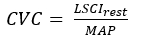 (3)
(3)
Where LSCIrest is the average value of skin speckle perfusion recorded at baseline (arbitrary units = AU); MAP is the mean arterial pressure (mmHg) recorded over the same time period. - Calculate peak cutaneous vascular conductance (CVCmax) using the formula ( 3) and substituting the appropriate values for peak hyperemia (LSCImax) and MAP.
8. Experimental procedures and repeated measurements
- After recording skin perfusion, PORH, and other physiological variables of interest at baseline, perform the experimental intervention according to the approved study protocol (for example, modeling of the pathological process and/or administration of a vasoactive drug).
- Re-evaluate PORH and other physiological variables at the next study time point after the development of the simulated pathological process or achievement of the effect of the administered drug.
9. End of the experiment
- Upon completion of the acute experiment, euthanize the rat by intra-arterial injection of 2% lidocaine (2 mL) under deep general anesthesia (tiletamine + zolazepam and xylazine). Depending on local laws and protocols, use an alternative method of humane euthanasia for laboratory rodents that has been approved by the Institutional Animal Care and Use Committee (IACUC).
NOTE: For survival procedures and planned recovery of the animal from anesthesia, provide appropriate care and regular assessment of the animal's condition, including prevention of hypothermia and adequate postoperative analgesia (e.g., ketoprofen 5-10 mg/kg IM).
Access restricted. Please log in or start a trial to view this content.
Results
The above-described method for assessing microvascular function was applied by us in a series of experiments with modeling of hemorrhagic shock (blood loss of 35% of the calculated blood volume) in Wistar male rats weighing 250-300 g. In the group of animals with hemorrhage (HS, n = 13), the assessment of PORH parameters with simultaneous monitoring of invasive BP was performed 30 min after the completion of hemorrhage. In the control group of sham-operated animals (Sham, n = 10), hemorrh...
Access restricted. Please log in or start a trial to view this content.
Discussion
Here we describe a modernized technology for assessing cutaneous microcirculation and vascular reactivity in an in vivo experiment in rodents. For successful and standardized use of this method in physiological and pharmacological studies, several additional considerations should be taken into account. In the above protocol, rats are anesthetized with a combination of tiletamine + zolazepam + xylazine, which is dictated mainly by local standards for working with laboratory animals and limited access to buprenorphine, ket...
Access restricted. Please log in or start a trial to view this content.
Disclosures
The authors have no conflicts of interest to declare.
Acknowledgements
This work was supported by the Russian Science Foundation under Project 24-25-00310.
Access restricted. Please log in or start a trial to view this content.
Materials
| Name | Company | Catalog Number | Comments |
|---|---|---|---|
| Atraumatic needle with surgical suture nylon, 4C-0.5x16-4/0-N | Medtekhnika, Russia | N/A | Blue. Length 75 cm. USP 4/0 (MP1.5). For suturing a surgical wound |
| Catheter for rat's carotid artery with blunt-end syringe needle 22 G | SciCat, Russia | N/A | PE-50, length 12 cm, OD=0.9 mm, ID= 0.6 mm.With one fixing ring |
| Deltran Disposable Pressure Transducer | Utah Medical Products, US | 6069 | 12" cable |
| DiscofixC 3-way stopcock for infusion therapy and monitoring | B. Braun, Germany | 16494C | |
| Heparin | Belmedpreparaty, Belarus | N/A | Heparin sodium 5000 IU/ml, ampoules 5 mL |
| Lidocaine | Velpharm, Russia | N/A | Solution for injection 20 mg/mL, ampoules 2 mL |
| Pneumatic cuff "Systole" | Neurobotics, Russia | N/A | Part of non-invasive blood pressure measurement for rodents. Type 5-10 mm tail (rats) |
| Sodium chloride | Pharmasyntez, Russia | N/A | Solution for infusion 0,9%, 250 mL |
| Software LabChart8 | ADInstruments, Australia | N/A | Version 8.1.13 |
| Software MATLAB | The MathWorks Inc., US | N/A | Version R2019b |
| Sterile syringe for single use, 1 mL, 3-part | Huaian City Hengchun Medical Product Co. Ltd., China | N/A | 27 G needle |
| Sterile syringe for single use, 2 (2.5) mL, 3-part | Vogt Medical Vertrieb GmbH, Germany | 1310524 | 23 G needle |
| Suture SILK-S | Polytechmed, Russia | N/A | Non-absorbable, braided,made of natural silk with a silicone coating, black in cassette. USP 4/0 (MP1.5). Length 100 m. For ligating vessels |
| Xyla | Interchemie, Netherlands | N/A | Xylazine 20 mg/mL, 50 mL |
| Zoletil 100 | Virbac, France | N/A | Tiletamine HCl 50 mg/mL, zolazepam HCl 50 mg/mL, 5 mL |
References
- Roustit, M., Cracowski, J. Non-invasive assessment of skin microvascular function in humans: an insight into methods. Microcirculation. 19 (1), 47-64 (2012).
- Duranteau, J., et al. The future of intensive care: the study of the microcirculation will help to guide our therapies. Crit Care. 27 (1), 190(2023).
- Basak, K., Manjunatha, M., Dutta, P. K. Review of laser speckle-based analysis in medical imaging. Med Biol Eng Comput. 50 (6), 547-558 (2012).
- Goldberg, J., et al. Intraoperative laser speckle contrast imaging to assess vessel flow in neurosurgery: a pilot study. Neurosurgery. 94, 983-992 (2024).
- Paramasivam, R., et al. Laser speckle contrast imaging for intraoperative assessment of intestinal microcirculation in normo- and hypovolemic circulation in a porcine model. Eur Surg Res. 65 (1), 1-8 (2023).
- Linkous, C., et al. Applications of laser speckle contrast imaging technology in dermatology. JID Innov. 3 (5), 100187(2023).
- Golubova, N., et al. Effect of thinned-skull cranial window on monitoring cerebral blood flow using laser speckle contrast imaging. IEEE J Sel Top Quantum Electron. 31 (4), 1-8 (2025).
- Golubova, N., Potapova, E., Seryogina, E., Dremin, V. Time-frequency analysis of laser speckle contrast for transcranial assessment of cerebral blood flow. Biomed Signal Process Control. 85, 104969(2023).
- Escobar, S., et al. Evaluation of systemic endothelial-dependent and endothelial-independent microvascular reactivity in metabolically healthy obesity: an observational study. Microvasc Res. 148, 104553(2023).
- Shirazi, B. R., Valentine, R. J., Lang, J. A. Reproducibility and normalization of reactive hyperemia using laser speckle contrast imaging. PLoS One. 16 (1), e0244795(2021).
- Tew, G. A., Klonizakis, M., Crank, H., Briers, J. D., Hodges, G. J. Comparison of laser speckle contrast imaging with laser Doppler for assessing microvascular function. Microvasc Res. 82 (3), 326-332 (2011).
- Yuan, X., et al. A comparison of the cutaneous microvascular properties of the spontaneously hypertensive and the Wistar-Kyoto rats by spectral analysis of laser Doppler. Clin Exp Hypertens. 41 (4), 342-352 (2019).
- Dubensky, A., et al. Post-occlusive reactive hyperemia variables can be used to diagnose vascular dysfunction in hemorrhagic shock. Microvasc Res. 152, 104647(2024).
- Meglinski, I. V., Kal'chenko, V. V., Kuznetsov, Y. L., Kuznik, B. I., Tuchin, V. V. Towards the nature of biological zero in the dynamic light scattering diagnostic modalities. Dokl Phys. 58, 323-326 (2013).
- Feng, J., et al. Catheterization of the carotid artery and jugular vein to perform hemodynamic measures, infusions and blood sampling in a conscious rat model. J Vis Exp. (95), e51881(2015).
- Henninger, N., Heimann, A., Kempski, O. Electrophysiology and neuronal integrity following systemic arterial hypotension in a rat model of unilateral carotid artery occlusion. Brain Res. 1163, 119-129 (2007).
- Souza-Silva, E., Ascenso, R., Tonussi, C. R., Da Silva-Santos, J. E. Detection of blood flow perfusion and post-occlusive reactive hyperemia in the skeletal muscle of rats. Life Sci. 278, 119571(2021).
- Roustit, M., Millet, C., Blaise, S., Dufournet, B., Cracowski, J. L. Excellent reproducibility of laser speckle contrast imaging to assess skin microvascular reactivity. Microvasc Res. 80 (3), 505-511 (2010).
- Nilsson, M., et al. Day-to-day variability in cutaneous microcirculation measured with multi-exposure laser speckle contrast imaging and multispectral imaging. Microvasc Res. 160, 104819(2025).
Access restricted. Please log in or start a trial to view this content.