Primary amoebic meningoencephalitis (PAM) can be complicated by acute myocarditis, which may rapidly progress to circulatory failure and is associated with a poor prognosis. Hence, in PAM patients, cardiac evaluation should be initiated promptly to identify potential myocardial injury. Early diagnosis and immediate treatment are crucial upon confirmation of cardiac involvement.
Case Report
A Case of Primary Amebic Meningitis with Acute Myocarditis
1Department of Pediatrics, West China Second Hospital, Sichuan University, 2Key Laboratory of Birth Defects and Related Diseases of Women and Children (Sichuan University), Ministry of Education, 3Department of Cardiology, Chengdu Second People's Hospital, 4University of Electronic Science and Technology Affiliated Hospital, Sichuan Provincial People's Hospital, 5Organ Transplant Center, Sichuan Provincial People's Hospital, School of Medicine, University of Electronic Science and Technology of China, 6School of Medicine, University of Electronic Science and Technology of China, 7Women and Children's Hospital of Tibet Autonomous Region, 8Xizang Region Child Development Clinical Medical Research Center
Opens in a new tab
In This Article
Summary
Loading...
$$\rightleftharpoonup{xx}$$
$$\longleftharp{xx}$$,
$$\longrightharp{xx}$$,
Abstract
Loading...
$$\rightleftharpoonup{xx}$$
$$\longleftharp{xx}$$,
$$\longrightharp{xx}$$,
Primary amebic meningitis is a rare, acute, fatal central nervous system disease caused by Naegleria fowleri infection. In the past, the resulting damage to the central nervous system has received more attention, whereas the impact on the heart has been less extensively described. This case involved a patient with a diagnosis of amoebic meningitis, in whom severe myocarditis was subsequently discovered. After ruling out alternative pathogens, the myocarditis was established as being secondary to the amoebic infection.
The patient was admitted to the hospital due to fever, headache, and vomiting, and gradually became unconscious. Cerebrospinal fluid examination revealed haemorrhagic suppurative meningitis, and Hongkiin second-generation sequencing of cerebrospinal fluid and blood pathogens resulted in the detection of Naegleria fowleri; thus, a diagnosis of primary amebic meningitis was made. Electrocardiogram, myocardial injury marker analysis, and bedside transthoracic echocardiography were performed to diagnose acute myocarditis. Despite active treatment, respiratory and circulatory failure, shock, and eventually brain herniation and brain death rapidly occurred.
Introduction
Loading...
$$\rightleftharpoonup{xx}$$
$$\longleftharp{xx}$$,
$$\longrightharp{xx}$$,
Primary amebic meningitis (PAM) is an explosive, haemorrhagic, and necrotic meningitis caused by infection with Naegleria fowleri1. Because this infection can cause severe encephalitis, the mortality rate is more than 95%2. PAM has an incubation period of 2-15 days and typically causes death 3-7 days after the onset of symptoms3. Because the disease progresses rapidly and the patient's condition deteriorates in a short time, more attention is paid to the damage and treatment of the central nervous system; damage to organs other than the central nervous system is easy to miss. If acute myocarditis is combined, it can lead to circulatory failure in a short period of time, making the disease more dangerous. Therefore, it is particularly important to make a clear diagnosis and carry out comprehensive treatment as soon as possible.
Case Presentation:
The patient was admitted to the hospital due to fever, headache, and vomiting, and gradually became unconscious. Cerebrospinal fluid examination revealed haemorrhagic suppurative meningitis, and Hongkiin second-generation sequencing of cerebrospinal fluid and blood pathogens resulted in the detection of Naegleria fowleri; thus, a diagnosis of primary amebic meningitis was made. The molecular diagnostic technology for rapid detection of pathogens involves conducting unbiased sequencing of nucleic acids in samples, and then identifying pathogenic microorganisms through bioinformatics analysis. Electrocardiogram, myocardial injury marker analysis, and bedside transthoracic echocardiography were performed to diagnose acute myocarditis. Despite active treatment, respiratory and circulatory failure, shock, and eventually brain herniation and brain death rapidly occurred.
Diagnosis, Assessment, and Plan:
The patient was 6 years old, previously healthy, and had no other history of central nervous system or myocardial damage. Naegleria fowleri was detected in the patient's cerebrospinal fluid and peripheral blood, and other pathogens causing meningitis and myocarditis were excluded.
The initial diagnosis of primary meningitis was based on symptoms and signs of high fever and increased intracranial pressure, along with cerebrospinal fluid showing hemorrhagic and purulent meningitis. Genetic sequencing of the cerebrospinal fluid confirmed an infection with Entamoeba histolytica. The diagnosis of acute myocarditis was based on significantly elevated markers of myocardial injury, an ECG showing significant ST-segment abnormalities and pathological Q waves, echocardiography indicating left ventricular enlargement and decreased cardiac function, and no other cardiac diseases were found, leading to the diagnosis of acute myocarditis. Further viral antibody tests showed no abnormalities, and second-generation metagenomic sequencing of venous blood pathogens identified Entamoeba histolytica, leading to the diagnosis of Entamoeba histolytica myocarditis.
In the treatment, oxygen intake, electrocardiogram, blood pressure, and oxygen saturation monitoring were used to maintain the stability of the internal environment. Amphotericin B and metronidazole were added to combat infections, along with mannitol and glycerol fructose to reduce intracranial pressure. Sodium creatine phosphate and L-carnitine were administered to nourish the myocardium. Respiratory and circulatory failure gradually developed, necessitating tracheal intubation and mechanical ventilation, veno-arterial extracorporeal membrane oxygenation (VA-ECMO) support, and the use of vasoactive drugs (epinephrine and norepinephrine).
Protocol
Loading...
$$\rightleftharpoonup{xx}$$
$$\longleftharp{xx}$$,
$$\longrightharp{xx}$$,
This protocol is in accordance with the ethical principles outlined in the Declaration of Helsinki and has been approved by the Ethics Committee of (ethics approval number: 2024-159) of West China Second Hospital of Sichuan University. Written informed consent from the parents of the patient was obtained prior to the diagnostic and therapeutic procedures. All procedures were part of routine clinical care, and no experimental interventions were performed.
1. Patient selection
- A 6-year-old female patient presented to a local hospital with fever, headache, and vomiting for 2 days.
- During the course of the disease, her body temperature reached 40.5 °C, and ibuprofen was used to reduce her fever; however, she still experienced fever, persistent severe headache, and vomiting. Three hours before admission, she developed a consciousness disorder, delirium, and irritability. The patient gazed upwards, and her limbs shook involuntarily.
- The patient was in good health and had no history of exposure to swimming water before the onset of illness.
- The patient's axillary temperature was 39.2 °C, heart rate was 150 beats/min, the respiratory rate was 36 breaths/min, the blood pressure was 112/74 mmHg, and the blood oxygen saturation was 98%.
- The patient had a score of 4 (E1M2V1) on the Glasgow coma scale, the double pupil diameter was 5 mm, and the patient had a slow response to light. There was neck resistance; no cardiac murmurs were heard. Both the Babinski sign and Brudzinski's sign were positive.
2. Standardized laboratory testing
- Venous blood (10 mL) was collected into ethylenediaminetetraacetic acid (EDTA) and serum separator tubes.
- A white blood cell count was subsequently performed, with neutrophil fractionation analyzed by flow cytometry. Levels of C-reactive protein, procalcitonin, and interleukin-6 were measured using chemiluminescence immunoassay.
- Cerebrospinal fluid (CSF) analysis was performed using microscopy and biochemical analysis. Protein was quantified by the pyrogallol red-molybdate complex method, and glucose was measured by the hexokinase method.
- Biomarkers of myocardial injury were measured by electrochemiluminescence immunoassay.
- Rapid molecular assay was used for the detection of virus in serum samples.
3. Imaging protocol
- Electrical activity of the heart was evaluated using a 12-Lead ECG (paper speed 25 mm/s, amplitude 10 mm/mV, record limb leads [II, III, aVF] and precordial leads [V4-V6]).
- The blood flow to the heart was evaluated using a cardiac color Doppler ultrasound at the bedside. The parameters set were as follows: Rest in supine position, CDFI M 2-DE imaging mode, frequency of the child's probe 5.0 - 7.0 MHz, Frame rate: ≥ 30 frames per second, Speed range: 15-40 cm/s.
- On the third day after admission, a head CT (non-contrast, horizontal position, 120 kV, slice thickness 5 mm) was performed.
4. Pathogen gene sequencing and diagnosis
- Metagenomic next-generation sequencing
- Nucleic acid extraction
- A 3 mL cerebrospinal fluid (CSF) sample was collected via lumbar puncture under aseptic conditions, sealed, and immediately transported on dry ice to the molecular biology laboratory for analysis.
- For nucleic acid extraction, a 2-mL aliquot of cerebrospinal fluid (CSF) was centrifuged at 21130 × g for 5 min. The DNA was extracted and purified by taking 200 µL of the cell-free supernatant sample according to the instructions of the DNA isolation kit.
- DNA concentration and quality were checked through a Fluoremeter and agarose gel electrophoresis.
- Library generation and sequencing
- DNA library construction was performed according to the library construction kit operating instructions. Library quality control was performed by a fluoremeter and a bioanalyzer.
- Qualified DNA libraries with different barcode tags were pooled and then sequenced using a sequencing platform and an SE75bp sequencing strategy.
- Bioinformation pipeline
- After obtaining the sequencing data, high-quality data were generated by filtering out connectors, low-quality, low-complexity, and shorter sequences.
- Next, human-derived sequences matching the human reference database (hg38) were eliminated by using the SNAP software. The remaining data were then aligned to the microbial genome database using Burrow-Wheeler Alignment. This database includes a large collection of microbial genomes from NCBI, covering more than 30,000 microorganisms, including 17,748 bacterial species, 11,058 viral species, 1,134 fungal species, and 308 parasitic species. The microbial composition of the samples was finally determined.
- Nucleic acid extraction
- Second-generation sequencing of venous blood pathogens
- A 5 mL whole blood sample was collected and aseptically sealed. It was then transported at low temperature to the laboratory for immediate processing.
- A 2 mL aliquot of the blood sample was first centrifuged at 845 g for 10 min to separate the plasma from the blood cells.
- The plasma supernatant was subsequently centrifuged at 21130 g for 10 min to pellet any remaining cellular debris.
- Subsequent steps for DNA extraction, purification, library preparation, sequencing, and bioinformatic analysis were identical to those described for the cerebrospinal fluid sample.
- Run the gene sequencing platform using the following settings: lon torrent S5, sequencing length 76 cycles; detection content includes bacteria, archaea, mycoplasma, chlamydia, rickettsia, spirochetes, viruses, fungi, and tens of thousands of other known microorganisms.
5. Therapeutic Interventions
- The patient was treated with amphotericin (11.2 mg IV once a day, a total of 4 days; concentration of 0.1 mg/mL) and rifampicin (0.225 g nasal feeding once a day, a total of 3 days) to counter the infection and with mannitol (55 mL IV every 6 h, a total of 4 days, concentration of 200 mg/mL) and glycerol fructose (150 mL IV once a day, a total of 4 days) to reduce the patient's cranial pressure.
- The treatment plan was adjusted on Day 2 to include creatine phosphate sodium (1 g IV once a day, concentration of 20 mg/mL), vitamin C (4 g IV once a day, concentration of 100 mg/mL), and L-carnitine (1 g Nasogastric feeding twice a day, concentration of 100 mg/mL) to support myocardial function.
- On Day 3, mechanical ventilation via tracheal intubation, veno-arterial extracorporeal membrane oxygenation (VA-ECMO), and vasoactive medications (epinephrine and norepinephrine) were initiated to manage the worsening cardiopulmonary function. Innovative ventilator-assisted ventilation (pressure control): FiO2: 45%, PEEP: 10 cmH2O, respiratory rate: 10 times/min.
- The right internal carotid artery and vein were entered on the right side, and heparin anticoagulation was performed in the peripheral veins with ACT monitoring. Parameters involved were: Centrifugation force: 909 × g (3227 r/min), flow rate 1.5 L /min, FIO2 100%, airflow 2 L/min.
- Standard nursing care after admission included oxygen therapy, intensive care, electrocardiogram monitoring, blood pressure monitoring, blood oxygen saturation monitoring, close monitoring of vital signs (including temperature, consciousness, pupil dilation, and others), maintenance of water and electrolyte balance, and acid-base balance.
- Specialized nursing care following admission included stomach tube retention, artificial feeding, sputum aspiration, tracheal intubation care, arteriovenous catheter care, hypothermia treatment, etc.
Results
Loading...
$$\rightleftharpoonup{xx}$$
$$\longleftharp{xx}$$,
$$\longrightharp{xx}$$,
The examinations revealed significant increases in the following parameters: WBC count (17.23 × 10⁹/L); neutrophil classification (92.3%) via a flow cytometer; C-reactive protein (20.86 mg/L); procalcitonin (7.08 ng/mL); and interleukin-6 (6.372 pg/mL) using chemiluminescent immunoassay. The results from the CSF were as follows: pressure 200 mmH2O; light red, positive protein qualitative test ++; red blood cell count, 3200 × 106/L; nucleated cell count, 960 × 106/L, and a large number of neutrophils. Cerebrospinal fluid biochemistry results revealed a protein concentration of greater than 10,000.0 mg/L and a glucose level of greater than 4.76 mmol/L.
The patient's myocardial injury markers were already elevated: troponin I (TNI) at 15.33 ng/mL, myoglobin (Myo) at 163.2 ng/mL, and CK-MB at 42.82 ng/mL. The serum sample showed negative results for Epstein-Barr virus (EBV) IgG and IgM antibodies, six respiratory viruses (respiratory syncytial virus, adenovirus, influenza A virus, influenza B virus, parainfluenza virus, and human metapneumovirus), and influenza A/B virus nucleic acid (A/B flow nucleic acid). These data are summarized in Table 1.
However, despite these interventions, the patient's electrocardiogram (ECG) showed an evolution from ST-T segment changes to the development of necrotic Q waves (Figure 1, Figure 2, and Figure 3). Concurrently, the heart became dilated, and the ejection fraction was markedly reduced (Table 2). The patient exhibited no improvement in cardiac function, and her neurological status continued to decline. A head CT performed on the third day after admission revealed extensive cerebral edema and the formation of a cerebral herniation (Figure 4). Neurological examinations indicated the absence of brainstem reflexes, and the patient was diagnosed with brain death shortly thereafter. The family was informed of the prognosis, and after discussions, life-sustaining treatments were withdrawn. An autopsy was not performed.
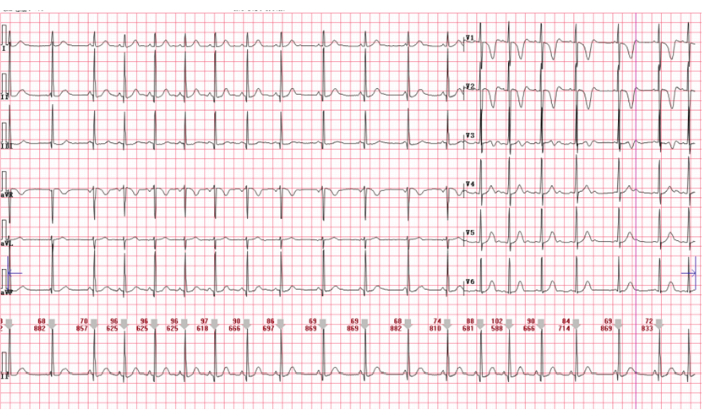
Figure 1: Electrocardiogram (Day 1) revealed sinus arrhythmia with an abnormal T wave. Please click here to view a larger version of this figure.
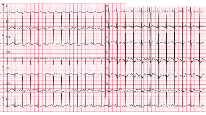
Figure 2: Day 1 ECG. ECG (Day 1, 19:30 after admission) showed sinus tachycardia 150 times/min, right axis deviation +92°, and ST segment changes (I and AVL leads, ST segment elevation greater than 0.1 mV, and II, III, AVF, and v4-v6 leads ST segment depression 0.05-0.2 mV). Indicates myocardial injury. Please click here to view a larger version of this figure.

Figure 3: Day 3 ECG. ECG (Day 3) showed sinus tachycardia (118 beats/min), unskewed electrical axis, atrial premature beat, ST segment changes (ST segment elevation greater than 0.1 mV in leads I and AVLV2-V5; ST segment depression greater than 0.05 mV in leads II, III, AVR, and AVF). The I and AVL leads presented the QR type. Indicates myocardial necrosis. Please click here to view a larger version of this figure.
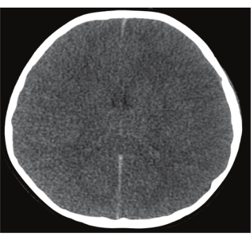
Figure 4: Day 4 head CT. Head CT (Day 4) revealed extensive swelling of the bilateral cerebral hemispheres, an unclear outline of the brainstem boundary, disappearance of the cerebral sulci, narrowing of the bilateral ventricles, and narrowing and disappearance of the third and fourth ventricles, suggesting the formation of a cerebral hernia. Please click here to view a larger version of this figure.
| Items | Result | Reference Range |
| WBC count | 17.23 × 10⁹ /L | 3.5-9.5 × 10⁹ /L |
| Neutrophil classification | 92.30% | 40.0–75.0% |
| C-reactive protein | 20.86 mg/L | 0-5 mg /L |
| Procalcitonin | 7.08 ng/mL | <0.046 CUTOFF: >2.0 |
| Interleukin-6 | 6.372 pg/mL | <7 pg/mL |
| Troponin I (TNI) | 15.33 ng/mL | <0.04 ng/mL |
| Myoglobin (Myo) | 163.2 ng/mL | <70 ng/mL |
| CK-MB | 42.82 ng/mL | 0–4.87 ng/mL |
| Epstein-Barr virus (EBV) IgG | Negative | Negative |
| IgM antibodies | Negative | Negative |
| Six respiratory viruses | Negative | Negative |
| Influenza A/B virus nucleic acid | Negative | Negative |
| Cerebrospinal fluid(CSF) | ||
| Pressure | 200 mmH2O | 40–100 mmH2O |
| Positive protein qualitative | (++)Positive | (-)negative |
| Red blood cell count | 3200 × 106/L | 0–5 × 10⁶/L |
| Nucleated cell count | 960 × 106/L | 0–15 × 106/L |
| Protein | >10000.0 mg/L | 200–400 mg/L |
| Glucose | > 4.76 mmol/L | 2.8–4.5 mmol/L |
Table 1: Standardized laboratory testing data.
| LV (mm) | EF (%) | FS (%) | |
| Day 2 00:34 | 37 | 42 | 20 |
| Day 2 03:00 | 39 | 32 | 15 |
| Day 2 14:14 | 40 | 29 | 14 |
| Day 2 09:09 | 40 | 21 | 9 |
Table 2: Bedside transthoracic echocardiography examination of the heart. LV = Left Ventricle; EF = Ejection Fraction; FS = Fractional Shortening
Discussion
Loading...
$$\rightleftharpoonup{xx}$$
$$\longleftharp{xx}$$,
$$\longrightharp{xx}$$,
PAM is an acute, rapid, fatal disease of the central nervous system caused by infection with Naegleria fowleri, a free-living thermophilic amoeba in freshwater systems; such infections are predominantly prevalent in children and adolescents who are exposed to contaminated ponds or swimming pools4. The incidence of PAM is unknown. To date, cases of PAM have been reported in many countries, including China5,6. PAM is a neglected disease and is often misdiagnosed as bacterial meningitis or viral encephalitis, which shortens the window for administering potentially life-saving treatment7. The diagnosis depends on the patient's history of exposure to freshwater and a strong suspicion by the doctor, which is supported by cerebrospinal fluid examination8. This case occurred in Southwest China, and the symptoms developed after eating roasted meat 3 days prior, which is consistent with the incubation period reported in the literature4. Unlike in previous studies, the patient had no history of exposure to freshwater, which does not exclude the possibility of transmission via food through the digestive tract. The patient first developed headaches, vomiting, and high fever, then fell into a coma, and finally suffered brain death. Head CT revealed diffuse cerebral oedema and cerebellar tonsillar hernia, which is consistent with the experiences of previously reported patients with PAM9. The cerebrospinal fluid revealed haemorrhagic suppurative meningitis. Gene sequencing of cerebrospinal fluid confirmed Naegleria fowleri infection. When a patient is diagnosed with PAM, the following drug combinations are mainly used: amphotericin B, fluconazole, azithromycin, rifampicin, and miltefosine10. The patient was treated with a combination of amphotericin B and rifampicin. Cases in which the patient survived have been reported in recent years7,11, and survival was possibly due to early identification and treatment, use of a combination of antibiotics, and management of the elevated intracranial pressure according to traumatic brain injury principles11.
Although cardiac MRI and endomyocardial biopsy are the gold standards for diagnosing myocarditis, these procedures were not feasible due to the patient's critical condition. Nonetheless, the diagnosis of acute myocarditis was supported by significantly elevated myocardial injury markers (TNI: 15.33 ng /mL, Myo: 163.2 ng /mL, CKMB: 42.82 ng/mL), abnormal ECG findings (pathological Q waves, ST-segment elevation), and echocardiographic evidence of left ventricular enlargement with reduced systolic function (EF as low as 21%). Differentiation from sepsis-induced cardiomyopathy was carefully considered. Sepsis-related myocardial dysfunction typically presents as reversible biventricular systolic dysfunction without marked elevation in myocardial biomarkers and usually improves with sepsis management. In contrast, the patient showed persistent cardiac dysfunction, markedly elevated biomarkers, and no identifiable sources of sepsis or other infections, as confirmed by negative tests for common pathogens like EBV and respiratory viruses. The detection of Naegleria fowleri in both cerebrospinal fluid and blood via next-generation sequencing suggests systemic dissemination and potential direct myocardial involvement. These findings collectively support the diagnosis of myocarditis secondary to primary amebic meningitis rather than sepsis-induced cardiomyopathy.
Myocarditis is a type of heart inflammation caused by infection, autoimmunity, drugs, and other factors12. The most common cause of myocarditis is viral infection, but it can also be caused by infections with bacteria, fungi, and spirochaetes13. Previous studies14 confirmed that central nervous system diseases can affect the heart. Markowitz et al4found through autopsy in 1974 that PAM could cause focal or diffuse inflammatory damage to the myocardium and that infectious diseases of the central nervous system caused significant damage to the myocardium. However, few reports of PAM-related myocarditis exist. Because myocarditis presents with nonspecific symptoms, including chest pain, dyspnoea, and palpitations, it often resembles more common diseases. PAM patients progress rapidly, and cardiovascular manifestations are easily misdiagnosed as simply caused by high cranial pressure and brain oedema, resulting in a missed diagnosis of myocarditis. In some patients, cardiac MRI and endocardial myocardial biopsy can help to identify myocarditis, predict the risk of cardiovascular events, and guide treatment15. In this case, although myocardial MRI and endocardial myocardial biopsy could not be performed because of the critical condition of the child, myocardial damage occurred. Markers of myocardial injury were significantly elevated, and an electrocardiogram indicated obvious abnormalities in the ST segment and pathological Q wave. Heart color ultrasound indicated left ventricular enlargement and decreased cardiac function, and no other heart diseases were found. Therefore, acute myocarditis was considered. Improved antibody tests for related viruses revealed no abnormalities. The second-generation sequencing of venous blood pathogens by Macrogene revealed Naegleria fowleri with high confidence, and the condition was considered Naegleria fowleri myocarditis. The treatment options for myocarditis include anti-infection drugs, myocardial nutrition, improved myocardial metabolism, adrenal glucocorticoids, and other treatments if necessary. In the case of heart failure and cardiogenic shock with severe haemodynamic abnormalities in a short period of time, a mechanical circulatory support device can replace part of the heart and/or lung function, and the survival rate of severe myocarditis patients who are treated with device-assisted therapy(DAT) is 57-80%16.
The management of primary amebic meningitis (PAM) complicated by acute myocarditis presents significant diagnostic and therapeutic challenges. In this case, the combination of amphotericin B and rifampicin was selected based on their proven efficacy against Naegleria fowleri and immediate availability in our clinical setting. While agents such as fluconazole, azithromycin, and miltefosine have been used in other cases, miltefosine was not accessible at the time, and the rapid progression of the disease required urgent initiation of treatment. This highlights the importance of early diagnosis and the need for broad access to recommended therapeutic agents in managing PAM.
The route of infection in this case remains uncertain. Although Naegleria fowleri typically infects through the nasal cavity via contaminated freshwater exposure, the patient had no known exposure to such environments. While gastrointestinal transmission has been hypothesized in isolated cases without freshwater contact, no definitive evidence exists to support this pathway. Given the lack of clear exposure history and supporting data, the route of infection should be considered unknown, underscoring the complexity of diagnosing PAM in atypical presentations.
Furthermore, the early detection of cardiomyopathy in PAM patients is critical but challenging. In retrospect, more proactive cardiac monitoring, including frequent electrocardiogram (ECG), serial myocardial injury marker assessments, and routine bedside transthoracic echocardiography, could have facilitated earlier identification of myocardial involvement. Integrating cardiac evaluations into the standard assessment for PAM patients may enhance early detection and improve outcomes, particularly in cases with rapid clinical deterioration.
The pathogenesis of myocarditis involves the interaction between stimuli and the subsequent host immune response. Infectious agents, especially cardiotropic viruses, are the most common cause. However, autoimmune processes independent of microbial triggers, as well as toxic myocardial damage caused by chemicals, drugs, or metabolic disorders, also contribute to the development of myocarditis through multiple mechanisms17. The pathogenesis of myocarditis caused by PAM is still unclear and may involve a mechanism similar to that of myocardial injury caused by other central nervous system. Studies have shown that infection, immune damage, and drug toxicity are the main causes of myocarditis18. Naegleria fowleri was found in the blood of this patient, confirming that Naegleria fowleri can spread outside the central nervous system. Naegleria fowleri can enter the bloodstream through the damaged blood-brain barrier and travel through the bloodstream to other tissues and organs.
This case confirmed the pathogen of meningitis as Naegleria fowleri through metagenomic sequencing technology. The same technique also detected Naegleria fowleri in peripheral blood, which not only provided a pathogen-based rationale for anti-infective treatment but also confirmed the presence of Naegleria fowleri outside the central nervous system, leading to damage in other organs, including the heart. The case demonstrates that early comprehensive examinations -- including cardiac injury markers, electrocardiograms, and color Doppler echocardiography -- combined with dynamic monitoring, can enable early detection of cardiac damage and prompt adjustment of treatment plans. Limitations of this case include unclear infection route and absence of cardiac MRI and myocardial biopsy examinations (due to critical condition).
Disclosures
Loading...
$$\rightleftharpoonup{xx}$$
$$\longleftharp{xx}$$,
$$\longrightharp{xx}$$,
The authors have no conflicts of interest or financial ties to disclose.
Acknowledgements
Loading...
$$\rightleftharpoonup{xx}$$
$$\longleftharp{xx}$$,
$$\longrightharp{xx}$$,
This research received no external funding.
Materials
| Name | Company | Catalog Number | Comments |
|---|---|---|---|
| Agarose gel electrophoresis | Major Science | UVC1-1100 | |
| Agilent 2100 | Agilent Technologies, Palo Alto, USA | N/A | Bioanalyzer |
| Architect | Aboatt | i2000SR | Automatic CLIA Analyzer |
| Cardiac Color Doppler ultrasound | Mindray | Resona 5S | |
| Cobas | Roche | 8000 e801 | Automatic CLIA Analyzer |
| Electrocardiogram ECG | General Electric Company | MAC5500 | |
| Full-Automatic Hematology and Body Fluid Analyzer | Mindray | BriCyte-E6 | |
| Labospect 008 As | HI TACHI, JPN | N/A | Auto Biochemistry Analyzer |
| Nextseq 550 SE75bp | Illumina, San Diego, USA | N/A | Sequencing platform |
| QIAamp DNA Micro Kit | Qiagen | QIAamp DNA Blood MAXI (50) | |
| Qiagen library construction kit | Qiagen | QIAseq Ultralow Input Library Kit | |
| Quantitative PCR Instrument | Hongshi, CHN | SLAN96 | |
| Qubit 3.0 Fluoremeter | Invitrogen | Q33216 | Fluoremeter |
| Somatom Force | Siemens | VB30 | Computed tomography (CT) |
| Sysmex XN-10 | Sysmex, JPN | N/A | Full-Automatic Hematology and Body Fluid Analyzer |
| Xenios Console Xlung kit230 | Fresenius | N/A | Extracorporeal Membrane Oxygenation ECMO |
References
Loading...
$$\rightleftharpoonup{xx}$$
$$\longleftharp{xx}$$,
$$\longrightharp{xx}$$,
- Heggie, T. W. Swimming with death: Naegleria fowleri infections in recreational waters. Travel Med Infect Dis. 8 (4), 201-206 (2010).
- Jahangeer, M., et al. Naegleria fowleri: Sources of infection, pathophysiology, diagnosis, and management; a review. Clin Exp Pharmacol Physiol. 47 (2), 199-212 (2020).
- Shariq, A., Afridi, F. I., Farooqi, B. J., Ahmed, S., Hussain, A. Fatal primary meningoencephalitis caused by Naegleria fowleri. J Coll Physicians Surg Pak. 24 (7), 523-525 (2014).
- Markowitz, S. M., Martinez, A. J., Duma, R. J., Shiel, F. O. Myocarditis associated with primary amebic (Naegleria) meningoencephalitis. Am J Clin Pathol. 62 (5), 619-628 (1974).
- Zhang, L. L., et al. Identification and molecular typing of Naegleria fowleri from a patient with primary amebic meningoencephalitis in China. Int J Infect Dis. 72, 28-33 (2018).
- Huang, S., et al. A pediatric case of primary amoebic meningoencephalitis due to Naegleria fowleri diagnosed by next-generation sequencing of cerebrospinal fluid and blood samples. BMC Infect Dis. 21 (1), 1251(2021).
- Hall, A. D., Kumar, J. E., Golba, C. E., Luckett, K. M., Bryant, W. K. Primary amebic meningoencephalitis: A review of Naegleria fowleri and analysis of successfully treated cases. Parasitol Res. 123 (1), 84(2024).
- Hara, T., Fukuma, T. Diagnosis of the primary amoebic meningoencephalitis due to Naegleria fowleri. Parasitol Int. 54 (4), 219-221 (2005).
- Capewell, L. G., et al. Diagnosis, clinical course, and treatment of primary amoebic meningoencephalitis in the United States, 1937-2013. J Pediatric Infect Dis Soc. 4 (4), e68-e75 (2015).
- Colon, B. L., Rice, C. A., Guy, R. K., Kyle, D. E. Phenotypic screens reveal posaconazole as a rapidly acting amebicidal combination partner for treatment of primary amoebic meningoencephalitis. J Infect Dis. 219 (7), 1095-1103 (2019).
- Linam, W. M., et al. Successful treatment of an adolescent with Naegleria fowleri primary amebic meningoencephalitis. Pediatrics. 135 (3), e744-e748 (2015).
- Ammirati, E., Moslehi, J. J. Diagnosis and treatment of acute myocarditis: A review. JAMA. 329 (13), 1098-1113 (2023).
- Shauer, A., et al. Acute viral myocarditis: Current concepts in diagnosis and treatment. Isr Med Assoc J. 15 (3), 180-185 (2013).
- Dimant, J., Grob, D. Electrocardiographic changes and myocardial damage in patients with acute cerebrovascular accidents. Stroke. 8 (4), 448-455 (1977).
- Sagar, S., Liu, P. P., Cooper, L. T. Jr Myocarditis. Lancet. 379 (9817), 738-747 (2012).
- Long, B., Robertson, J., Koyfman, A., Brady, W. Left ventricular assist devices and their complications: A review for emergency clinicians. Am J Emerg Med. 37 (8), 1562-1570 (2019).
- Zafeiri, M., Knott, K., Lampejo, T. Acute myocarditis: An overview of pathogenesis, diagnosis and management. Panminerva Med. 66 (2), 174-187 (2024).
- Ammirati, E., Cipriani, M., Lilliu, M., et al. Survival and left ventricular function changes in fulminant versus nonfulminant acute myocarditis. J Am Coll Cardiol. 136 (6), 529-545 (2017).
Reprints and Permissions
Request permission to reuse the text or figures of this JoVE article
Request Permission