Here, we present an optimized workflow for producing Chimeric Antigen Receptor (CAR) T cells with large transgenes exceeding 10 kb using lentiviral vectors.
Method Article
Efficient Production of Chimeric Antigen Receptor (CAR) T Cells with Transgenes Exceeding 10 kb Using Lentiviral Vectors
Opens in a new tab
In This Article
Summary
Loading...
$$\rightleftharpoonup{xx}$$
$$\longleftharp{xx}$$,
$$\longrightharp{xx}$$,
Abstract
Loading...
$$\rightleftharpoonup{xx}$$
$$\longleftharp{xx}$$,
$$\longrightharp{xx}$$,
Engineering multiple receptors into chimeric antigen receptor (CAR) T cells has emerged as a powerful strategy to prevent antigen-negative relapse and reduce on-target/off-tumor toxicities. However, manufacturing multi-receptor CAR T cells remains challenging, as increasing lentiviral transgene sizes significantly reduces viral titers and T cell transduction rates. Current production workflows often rely on cell sorting to enrich transduced T cells from low-yield productions. Yet, cell sorting techniques do not increase the absolute number of CAR T cells and add further complexity to the already elaborate manufacturing process. Consequently, these limitations impede the clinical translation of multi-receptor CAR T cells and restrict the development of next-generation immunotherapies.
Here, we present a detailed, step-by-step production workflow optimized for generating CAR T cells with large lentiviral transgenes. Using this workflow, we demonstrate size-dependent increases in transduction efficiency across a range of transgene sizes, with the most pronounced enhancement of up to 14.8-fold observed for a 10.1 kb lentiviral vector. Importantly, the workflow supports robust T cell expansion and eliminates the need for cell sorting. By overcoming current size limitations in lentiviral gene transfer, this workflow enables the efficient generation of multi-receptor CAR T cells, thereby facilitating the development of advanced immunotherapies.
Introduction
Loading...
$$\rightleftharpoonup{xx}$$
$$\longleftharp{xx}$$,
$$\longrightharp{xx}$$,
Chimeric antigen receptor (CAR) T cells have revolutionized the field of immunotherapy. However, despite their remarkable success, critical challenges remain, including immunosuppression mediated by the tumor microenvironment (TME), antigen-negative tumor relapses, and on-target/off-tumor toxicities1,2. Engineering CAR T cells with multiple synthetic receptors, such as dual CARs, combined CAR and TCR constructs, or cytokine switch receptors, has the potential to overcome these obstacles3,4. In blood cancers, equipping effector T cells with dual CARs allows for the simultaneous targeting of two distinct antigens, such as CD19 and CD205, thereby reducing the risk of tumor antigen escape. In solid tumors, on-target/off-tumor toxicities can be mitigated by incorporating, e.g., synthetic Notch (synNotch) receptor circuits that enable logic-gated CAR expression and enhance targeting precision6,7,8,9,10,11,12,13. However, the production of CAR T cells carrying multiple receptors remains challenging due to the underlying increase in transgene size. Self-inactivating third-generation lentiviral vectors derived from the human immunodeficiency virus (HIV) type 1 are the current gold standard for CAR T cell production at research and clinical scale14. To generate CAR T cells, lentiviral vectors are engineered with customized CAR expression cassettes flanked by long terminal repeat (LTR) sequences, which mediate integration into the T cell genome. The maximal effective packaging capacity (EPC) of current lentiviral vectors is approximately 9.2 kilobases (kb) LTR-to-LTR, corresponding to the native genome size of HIV-1. As lentiviral titers decrease semi-logarithmically with increasing vector length15, the engineering of large transgenes with multiple receptors drastically reduces lentiviral production yields and ultimately impedes CAR T cell manufacture16. While cell sorting techniques, such as magnetic- or fluorescence-activated cell sorting (MACS or FACS, respectively), can be used to enrich CAR T cells from low-yield productions, these methods do not increase the absolute number of CAR T cells and add further complexity to an already elaborate manufacturing process. As a result, manufacturing challenges of CAR T cells with large lentiviral transgenes remain a major barrier to both preclinical research and clinical application.
We recently established a CAR T cell production workflow optimized for effector T cells carrying large lentiviral transgenes, such as the single-vector synNotch (svsNotch) system17. Here, we detail the methodology in a step-by-step protocol to facilitate its broad adoption. As proof of concept, we demonstrate size-dependent improvements in transduction efficiency across lentiviral transgene sizes ranging from 5.7 kb to 9.2 kb and 10.1 kb. Overall, this optimized workflow enhances transduction rates by up to 14.8-fold, thereby eliminating the need for cell sorting and enabling the efficient generation of effector T cells with transgene sizes exceeding 10 kb.
Protocol
Loading...
$$\rightleftharpoonup{xx}$$
$$\longleftharp{xx}$$,
$$\longrightharp{xx}$$,
Human T cells were procured through the University of Pennsylvania Human Immunology Core, which operates under principles of Good Laboratory Practice with established standard operating procedures and/or protocols for sample receipt, processing, freezing, and analysis that conform to MIATA and University of Pennsylvania ethics guidelines.
NOTE: A schematic overview of the optimized workflow for lentivirus and CAR T cell production is shown in Figure 1. A comparison of the key optimization steps with the current standard workflow is summarized in Table 1. All cell cultures were maintained at 37 °C in a humidified incubator with 5% CO2 using T cell Medium (TCM) composed of RPMI-1640 supplemented with 10% heat-inactivated fetal bovine serum (FBS), 10 mM HEPES buffer, 2 mM L-alanyl-L-glutamine (see Table of Materials), 100 U/mL penicillin, and 100 U/mL streptomycin.
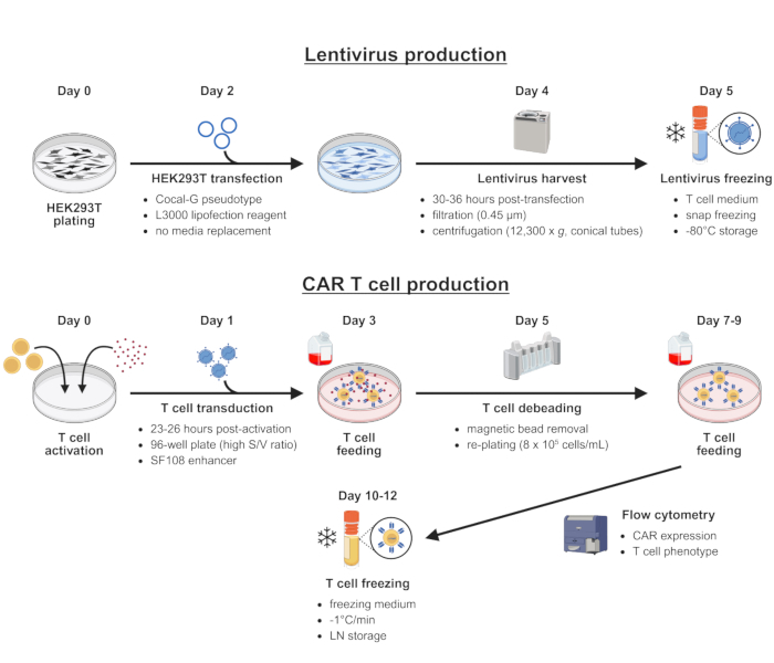
Figure 1: Optimized workflow for lentivirus and CAR T cell production with large transgenes. Schematic representation of lentivirus production (top) and CAR T cell generation (bottom), highlighting key steps and optimizations that enhance viral titers and transduction efficiency. Please click here to view a larger version of this figure.
| Standard workflow | Optimized workflow | ||
| Virus | Viral envelope | VSV-G | Cocal-G |
| Transfection | L2000 | L30001 | |
| LV harvest | 24 h2 + 48 h2 | 30-36 h3 | |
| T-cell | Vessel | T25 flask | 96-well flat bottom plate |
| Surface/Volume ratio | Variable | 5.12 cm2/mL4 | |
| Adjuvant | None | SF1085 | |
| 13 µL L3000 per 1 µg DNA, no media change | |||
| 2centrifugation at 106,800 x g for 2.5 h, round bottom tube | |||
| 3centrifugation at 12,300 x g overnight, conical bottom tube | |||
| 4additional media supplementation after transduction | |||
| 555 µg/mL | |||
Table 1: Comparison of standard and optimized workflows. Key optimizations of the enhanced workflow compared to the current standard workflow. This table has been adapted with permission from Rommel et al.17.
1. Lentivirus production
- Maintenance of HEK293T cells
- Ensure HEK293T cells are evenly distributed, free of clumps, and at 50-70% confluency (typical yield: 1.0-1.5 × 10⁷ cells per T150 flask).
- Aspirate the supernatant, add 20 mL of room-temperature (RT) PBS to the upper side of the flask to avoid dislodging the cells, gently swirl PBS over the cells, and aspirate the wash.
- Add 3 mL of 0.05% Trypsin-EDTA and incubate for 15-30 s at RT while tilting the flask side to side. Monitor cell detachment.
- Add 7 mL of cold TCM, resuspend cells against the bottom of the flask to generate a single-cell suspension, and transfer to a 50 mL tube. Wash the flask with 10-20 mL of cold TCM and add the wash to the same tube.
- Centrifuge the cells (300 × g, 5 min, 4 °C), aspirate the supernatant, resuspend in 10 mL of TCM, and count the cells.
- Plate HEK293T cells in T150 flasks at 3 × 10⁶ cells per flask for passaging every 2 days (e.g., Monday and Wednesday) or at 1 × 10⁶ cells per flask for passaging every 3 days (e.g., plating on Friday for passaging over the weekend) using 30 mL of TCM pre-warmed to 37 °C.
- Transfection of HEK293T cells for lentivirus production
NOTE: Perform a small pilot experiment to determine the seeding density that consistently yields 70-80% confluency after 2 days. Ensure that HEK293T cell confluency does not exceed 80% at the time of transfection (see Discussion). Transfection was optimized using reduced-serum minimal essential medium and commercial lipofection reagents (L3000 and P3000, see Table of Materials).- Harvest HEK293T cells via trypsinization as described above and plate 4 × 10⁶ cells per T150 flask with 30 mL of TCM pre-warmed to 37 °C.
- After 2 days, confirm that the cultures are evenly distributed and 70-80% confluent.
- Warm up 50 mL of reduced-serum minimal essential medium to RT. Thaw lentiviral plasmids, gently mix, and quick spin. Pulse vortex lipofection reagents, quick spin, and leave them at RT.
- Prepare DNA master mixes. For each flask, combine in this order: 2 mL of reduced-serum minimal essential medium, 39 µg of lentiviral packaging plasmids (18 µg of pTRP Gag-Pol, 18 µg of pTRP RSV-Rev, 3 µg of pTRP Cocal-G), 15 µg of transfer plasmid, and 108 µL of P3000 reagent (2 µL of P3000/µg DNA). Scale up as required and increase the final amounts by at least 10% to account for pipetting loss. Pulse vortex, quick spin, and store at RT.
- Prepare L3000 master mixes. For each flask, combine in this order: 1.84 mL of reduced-serum minimal essential medium and 162 µL of L3000 (3 µL of L3000/µg DNA). Scale up as required and increase the final amounts by at least 10% to account for pipetting loss. Pulse vortex, quick spin, and incubate at RT for 5 min.
- After 5 min, add 2 mL of L3000 master mix per flask to the corresponding DNA master mix to achieve an approximate 1:2 dilution. Slowly add the L3000 mix starting from the bottom of the tube without touching its sides. Mix thoroughly by pipetting and incubate 15 min at RT.
- Meanwhile, carefully remove 6 mL of supernatant from each HEK293T culture and label each flask.
- After 15 min, transfect each culture by tilting the flask towards the top and slowly releasing 4 mL of transfection mix into the supernatant on the top side of the flask to avoid dislodging the cells. Slowly pipette up/down twice, return the flask to its original position, gently swirl, place it back into the incubator, and record the time.
NOTE: Leave cultures undisturbed until lentivirus harvest. In contrast to the manufacturer's recommendation to replace the HEK293T supernatant containing the L3000 transfection mix with fresh culture medium after transfection18, omitting this step preserves higher lentiviral titers17.
- Harvest, concentration, and freezing of lentiviral supernatants
NOTE: Keep lentiviral supernatants on ice or at 4 °C during all processing steps to prevent degradation.
CAUTION: Lentiviral vectors are classified as infectious materials. Follow appropriate biosafety precautions, including the use of personal protective equipment (e.g., double gloves, disposable lab coats). Decontaminate all work surfaces with 10% bleach followed by 70% ethanol. Disinfect contaminated consumables and liquid waste with 10% bleach before disposal as biohazardous waste in accordance with institutional biosafety guidelines.- Clean ultracentrifuge equipment (buckets, conical tubes, and conical adapters) with 10% bleach, then rinse with water. Briefly air-dry, followed by spraying with 70% ethanol and air-drying under a tissue culture hood.
- Pre-cool the ultracentrifuge with the inserted rotor to 4 °C.
- Harvest lentiviral supernatants 30-36 hours post-transfection by transferring them into 50 mL tubes. Clarify supernatants by centrifugation (500 × g, 5 min, 4 °C), filter through a 0.45 µm pore filter, and store on ice.
- Insert conical adapters into the buckets, transfer the filtered supernatants into conical ultracentrifuge tubes, and load them into the buckets. Balance opposing buckets with cold TCM, ensuring weight differences are below 0.1 g.
- Concentrate supernatants by ultracentrifugation at 12,300 × g overnight at 4 °C.
- The following morning, carefully remove the buckets and store them on ice.
- Prepare a box with dry ice for freezing and labeled cryovials for aliquoting.
- Under a tissue culture hood, open each bucket and remove the ultracentrifuge tube inside with forceps. For each tube, proceed as follows:
- Carefully aspirate the supernatant until 0.5 inches from the bottom of the tube and decant the remaining liquid into a 6-well plate. Gently tap the tube to remove some of the residual liquid.
- Add 300 µL of cold TCM, gently resuspend for 30 s, and transfer the suspension to a cryovial.
- Wash the tube with 200 µL of cold TCM and add the wash to the same cryovial.
- Gently mix and aliquot 60 µL for lentivirus titering (see step 1.4), several 100 µL aliquots for CAR T cell production, and one additional tube with the remaining volume as leftover. Immediately snap-freeze the aliquots on dry ice. Transfer aliquots to -80 °C for long-term storage.
- Titering of concentrated lentivirus using primary T cells
NOTE: Approximately 1 × 10⁵ T cells are required per dilution per lentivirus. For example, six lentiviruses with eight dilutions each require: 6 × 8 × 1 × 105 = 4.8 × 10⁶ T cells. Additional cells should be prepared to account for pipetting loss. T cells are stimulated using magnetic activation beads coated with anti-CD3 and anti-CD28 antibodies (see Table of Materials).- T cell activation (day 0)
- Prerequisite: Keep purified human CD4+ and CD8+ T cells in TCM ready.
- Count CD4+ and CD8+ T cells and combine them at a 1:1 ratio.
- Wash the activation beads as follows: Add 1 mL of TCM to a tube, resuspend the beads by vortexing, and transfer the required amount to achieve a 3-to-1 ratio of beads-to-T cells. Place the tube in a magnetic stand and allow the beads to form a pellet along the tube wall. After 30 s, carefully aspirate the supernatant, remove the tube from the magnetic stand, and resuspend the beads in 1 mL of TCM. Repeat the wash two more times.
- Add activation beads to the T cells and adjust the final concentration to 1 × 10⁶ cells/mL with TCM pre-warmed to 37 °C.
- Mix well and transfer the suspension to a reagent reservoir. Plate cells in 96-well flat-bottom plates with 100 µL of TCM per well using a multichannel pipette. Thoroughly mix every 1-2 min to prevent bead settling.
- T cell transduction (day 1)
NOTE: For lentiviruses with large transgenes (e.g., 9 kb LTR-to-LTR), prepare 6 conditions using 1:2 serial dilutions (undiluted, 1:2, 1:4, 1:8, 1:16, 1:32). For lentiviruses with smaller transgenes (e.g., 7 kb LTR-to-LTR), 8 conditions with 1:4 serial dilutions are required. Use remaining wells as untransduced (UTD) controls.- Thaw 60 µL of each concentrated lentivirus at RT and store on ice.
- Prepare a dilution series in a 96-well round-bottom plate: Add 60 µL of lentivirus to the top row, add 30 µL of cold TCM to each row below, transfer 30 µL from the top to the second row and mix (1:2 dilution), transfer 30 µL from the second to the third row and mix (1:4 dilution). Continue until six conditions are prepared in total (undiluted, 1:2, 1:4, 1:8, 1:16, 1:32). Use immediately for transduction.
- Between 23-26 h after bead stimulation, transduce T cells with 25 µL per well of lentivirus from the dilution plate using a multichannel pipette. Add 25 µL of TCM to UTD control wells.
- T cell feeding (day 3)
- Gently add 125 µL of TCM pre-warmed to 37 °C to each well using a multichannel pipette.
- T cell debeading and flow cytometry analysis (day 5)
- Prepare three sets of 1.5 mL screw cap tubes for each condition and remove the lids. Place two of the three sets into a magnetic stand.
- Resuspend each well and transfer the cells to the first set of tubes to allow the beads to form a pellet along the tube wall. After 30 s, carefully transfer the supernatants to the second set of tubes. Wait 30 s (the beads should no longer be visible), then transfer the supernatants to the third set of tubes and store on ice.
- Stain T cells for the appropriate transduction marker (e.g., anti-CD19 CAR or anti-HER2 synNotch) with antibodies (see Table of Materials) and analyze marker expression by flow cytometry.
- Identify conditions with transduction rates below 30%, corresponding approximately to the linear range between virus volume and transduction rate.
- Calculate lentiviral titers using the following formula:
Titer (TU/mL) = (initial number of T cells x percentage of transduced cells x dilution factor)/virus volume (mL)
e.g., 20% CAR expression with a 1:32 dilution of the lentiviral stock:
(100,000 cells x 0.2 x 32) / (0.025 mL) = 2.56 × 10⁷ TU/mL
- T cell activation (day 0)
2. CAR T cell production
NOTE: Typical starting amounts for CAR T cell production are 1-5 × 10⁶ T cells per group. Increase the number of cells in the UTD control group by 50%, as this group will also be used to normalize transduction levels in downstream experiments. Expect 5-6 population doublings throughout the expansion. T cell transduction was optimized using a block copolymer as an enhancer (see Table of Materials) and with specific surface-to-volume culture ratios in 96-well flat-bottom plates17.
- Preparation of a 100 mg/mL enhancer stock solution
- Add 5 g of copolymer enhancer to a plastic bottle, then add 45 mL of ultrapure water. Stir overnight at RT.
- Transfer the solution to a graduated cylinder and adjust the volume to 50 mL with ultrapure water.
- Filter the solution through a 0.22 µm pore filter and store at 4 °C.
- T cell activation (day 0)
- Prerequisite: Keep purified human CD4+ and CD8+ T cells in TCM ready.
- Count CD4+ and CD8+ T cells and combine them at a 1:1 ratio.
- Wash activation beads as follows.
- Add 1 mL TCM to a tube, resuspend the beads by vortexing, and transfer the amount needed to achieve a 3-to-1 ratio of beads-to-T cells. Place the tube in a magnetic stand and allow the beads to form a pellet along the tube wall.
- After 30 s, carefully aspirate the supernatant, remove the tube from the magnetic stand, and resuspend the beads in 1 mL of TCM. Repeat the wash two more times.
- Add activation beads to the T cells and adjust the final concentration to 1 × 10⁶ cells/mL with TCM pre-warmed to 37 °C.
- Mix well and transfer the suspension to a reagent reservoir. Plate cells in 96-well flat-bottom plates with 50 µL of TCM per well using a multichannel pipette. Thoroughly mix every 1-2 min to prevent bead settling.
- T cell transduction (day 1)
NOTE: Select lentivirus amounts based on the results of the prior titration, ensuring that final transduction levels are comparable between groups (typically between 20% to 30%). To avoid multiple lentiviral integrations, keep maximal transduction levels ≤ 30%. Lentiviruses with large transgenes generally require either undiluted virus or a 1:4-1:8 dilution. Adjust group sizes to avoid lentivirus leftovers, as each freeze-thaw cycle reduces lentiviral titers. Use lentiviral master mixes immediately after preparation.- Thaw the required amounts of concentrated lentivirus at RT and store on ice.
- Prepare a 2 mg/mL enhancer working stock in cold TCM by mixing, e.g., 100 µL of enhancer (100 mg/mL stock) with 4.9 mL of TCM.
- Prepare lentivirus master mixes based on the titration results and add enhancer to a final concentration of 275 µg/mL. Increase the final volumes by at least 10% to account for pipetting loss. E.g., to achieve a 1:16 dilution with 100 wells:
Total volume: 100 wells × 12.5 µL of lentivirus × 110% = 1,375 µL
1:16 dilution: 1,375 µL/ 16 = 85.9 µL of lentivirus
Enhancer: 189 µL (275 µg/mL final)
Final master mix: 1,100 µL of TCM + 85.9 µL of lentivirus + 189 µL of enhancer = 1,375 µL - Between 23-26 h after bead stimulation, transduce T cells with 12.5 µL per well of lentivirus master mix using a multichannel pipette, resulting in a final enhancer concentration of 55 µg/mL. Add 12.5 µL of TCM to UTD control wells.
- T cell feeding (day 3)
- Gently add 125 µL of TCM pre-warmed to 37 °C to each well using a multichannel pipette.
- T cell debeading (day 5)
NOTE: Prepare a sterile reservoir and three sets of 50 mL tubes for each culture. Limit the total wash volume to ≤ 20% of the T cell culture volume to avoid diluting cells below 8 × 10⁵ cells/mL.- Pool each culture into a sterile reservoir using a multichannel pipette, wash the original plates with TCM, and add the wash to the reservoir. Transfer each cell suspension to the first set of 50 mL tubes.
- Place the tubes into a magnetic stand to allow the beads to form a pellet along the tube wall. After 30 s, carefully transfer the supernatants to the second set of tubes and place them into the magnetic stand. Wait 30 s (beads should no longer be visible), then transfer the supernatants to the third set of tubes and store them in the incubator.
- Count each culture and normalize to 8 × 10⁵ cells/mL with TCM pre-warmed to 37 °C. Plate in the appropriate culture vessel depending on the final volume (Table 2).
- Allow the T cells to rest for 1 day, then begin daily feedings starting on day 7 (see next step).
- T cell feeding (day 7 and daily thereafter)
NOTE: T cells double approximately every 24-36 h and undergo 5-7 population doublings before resting. Monitor T cell volumes using an electrical impedance-based cell counter (see Table of Materials) to identify the optimal time point for cryopreservation.- Pool each culture, mix well, and count the cells.
- Normalize each culture to 8 × 10⁵ cells/mL with TCM pre-warmed to 37 °C and replate in the appropriate culture vessel (Table 2).
- Continue expansion until the mean cell volume drops below 320 µm3, then move to the cryopreservation step.
- Validation of T cell transduction using flow cytometry
NOTE: T cell transduction can be assessed at any point between day 5 and the day of cryopreservation. Expression levels will drop over the course of the expansion so that an early measurement will ensure clear separation of negative and positive fractions during flow cytometry analysis.- Harvest 2 × 10⁵ T cells from each culture.
- Stain T cells for the appropriate transduction marker (e.g., anti-CD19 CAR or anti-HER2 synNotch) with antibodies (see Table of Materials) and analyze marker expression by flow cytometry
- T cell cryopreservation (typically between days 10-12)
NOTE: Cryopreserve expanded T cells right before or after their resting state to improve viability during thawing. A mean volume of 320 µm3 can serve as a cut-off for cryopreservation. Once the cell volume drops below 300 µm3, T cells stop dividing, and their total numbers begin to decline. Ensure that all groups have comparable volumes at the time point of cryopreservation, which may require an additional day of culture for some groups. Freeze cells at a constant rate of -1 °C per minute using appropriate freezing containers (e.g., alcohol-free cell freezing containers, see Table of Materials).- Pool each culture into an appropriate tube (e.g., 500 mL), count the cells, and store them in the incubator.
- Calculate the total number of cells and the desired aliquot amounts.
- Centrifuge cells (300 × g, 10 min, 4 °C), aspirate the supernatants, and resuspend each culture in freezing medium containing 50% serum-free media, 40% heat-inactivated FBS, and 10% dimethyl sulfoxide (DMSO) at 2 × 10⁷ cells/mL (see Table of Materials).
- Aliquot cells into cryovials (250 µL to 1 mL, corresponding to 5-20 × 106 cells per vial). Place the cryovials in a cell freezing container and store them at -80 °C overnight.
- Transfer the cryovials to liquid nitrogen for long-term storage.
- T cell thawing
NOTE: For each T cell group, prepare one tube with 50 mL of TCM pre-warmed to 37 °C to dilute the DMSO in the freezing medium after thawing. Warm up additional TCM for plating.- Quickly thaw cryovials at 37 °C in a water bath or under running warm water.
- As soon as liquid is visible, proceed to a tissue culture hood, add 500 µL of pre-warmed TCM, gently mix, and transfer the cells to the prepared tubes containing 50 mL TCM.
- Wash the cryovial with 500 µL of TCM and add the wash to the same tube.
- Incubate the cells for 10 min at RT.
- Centrifuge cells (300 × g, 10 min, room temperature), aspirate the supernatants, and resuspend the cells at 4 × 10⁶ cells/mL.
- Plate the cells in the appropriate culture vessel (Table 2).
- Rest the cells overnight in the incubator before proceeding with downstream experiments.
| Vessel | Minimal volume | Maximal volume |
| 96-well | 0.1 mL/well* | 0.25 mL/well |
| 48-well | 0.2 mL/well | 1 mL/well |
| 24-well | 0.5 mL/well | 2 mL/well |
| 12-well | 1 mL/well | 4 mL/well |
| 6-well | 2 mL/well | 6 mL/well |
| T25 (horizontal) | 3 mL | 8 mL |
| T75 (horizontal) | 8 mL | 20 mL |
| T150 (horizontal) | 20 mL | 60 mL |
| * culture volume is temporarily reduced to 50 µL/well to enhance T cell transduction | ||
Table 2: Recommended culture media volumes for CAR T cell expansions. Minimum and maximum culture media volumes across commonly used vessel formats during T cell expansion.
Results
Loading...
$$\rightleftharpoonup{xx}$$
$$\longleftharp{xx}$$,
$$\longrightharp{xx}$$,
To demonstrate the enhancements achieved with the optimized workflow (Figure 1) across a wide range of transgene sizes, we selected three lentiviral vector sizes: 5.7 kb (CD19 CAR), 9.2 kb (HER2-MSLN svsNotch), and 10.1 kb (HER2-MSLN-CBG svsNotch) (Figure 2A). Lentiviruses were produced under either standard or optimized conditions (Figure 2B: std vs. opt and Table 1).
For the smallest vector with 5.7 kb, only a moderate improvement in T cell transduction was observed in one production batch (Figure 2B: top, 73% vs. 88% using undiluted virus). Consistently, lentiviral titers increased only modestly by approximately 1.5-fold (Figure 2C: top, 2.86 × 10⁸ TU/mL vs. 4.35 × 10⁸ TU/mL). In contrast, the 9.2 kb vector showed substantially improved performance, with maximal T cell transduction rates increasing by approximately 5.4- or 3.8-fold depending on the batch (Figure 2B: middle, 8.3%/16.6% vs. 45.1%/63.1% using undiluted virus) and functional titers improving by approximately 3.3-fold (Figure 2C: middle; 3.1 × 10⁶ TU/mL vs. 10.1 × 10⁶ TU/mL). The strongest effect was seen with the 10.1 kb vector, where transduction rates increased by approximately 12.4- or 9.8-fold (Figure 2B: bottom, 1.4%/1.5% vs. 17.3%/14.7% using undiluted virus) and functional titers improved by approximately 10-fold (Figure 2C: bottom, 0.24 × 10⁶ TU/mL vs. 2.41 × 10⁶ TU/mL).
Next, T cells from healthy donors were transduced with lentiviral vectors produced under standard or optimized conditions, using the corresponding workflows (Figure 3 and Table 1). To compare the transduction efficiencies, equal amounts of lentivirus were used in each workflow, and the viral dose was adjusted to maintain transduction rates at or below 30% to prevent multiple integration events. Compared with T cells transduced using standard conditions and using standard lentivirus production, transduction increased on average by 5.1-fold for the 5.7 kb vector, 13.3-fold for the 9.2 kb vector, and 14.8-fold for the 10.1 kb vector (Figure 3A and Supplementary Table 1). Selected T cell cultures generated with the optimized workflow were further expanded until they reached a resting state (Figure 3B,C). Population doublings ranged from 5.3 to 6.7, depending on the T cell donor (Figure 3B). Daily T cell volume measurements showed minimal cell debris, indicating no signs of toxicity during the expansions. Prior to cryopreservation, transduction rates were assessed by staining for CAR/synNotch receptor expression or by measuring mTag-BFP219 fluorescence (Figure 3C and Supplementary Figure 1). Transduction rates were 31.2% for the 5.7 kb vector (black; 1:512 lentivirus dilution), 29.8% for the 9.2 kb vector (red; 1:16 lentivirus dilution), and 20.2% for the 10.1 kb vector (blue; 1:4 lentivirus dilution). Additional functional data validating the performance of the generated svsNotch T cells and CAR T cells are presented in our companion study17. Importantly, effector T cells produced with the optimized protocol exhibited no differences in population doublings or T cell phenotype compared to those generated with the standard workflow17.
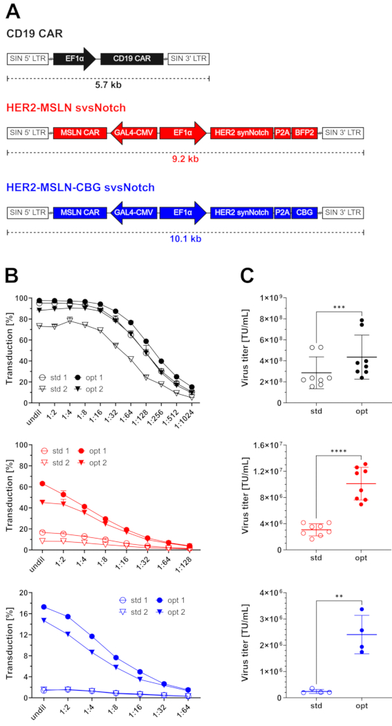
Figure 2: Lentivirus production following standard and optimized workflows. (A) Schematic overview of lentiviral vectors with corresponding transgene sizes17. Top: A CAR against CD19 (CD19 CAR) is expressed by the human elongation factor-1 alpha (EF1α) promoter, with a total transgene size of approximately 5.7 kb. Middle and bottom: A HER2 synNotch receptor is co-expressed with either a blue-fluorescent reporter protein (BFP2) (middle) or a click beetle green luciferase (CBG) (bottom) via a 2A self-cleaving peptide (P2A) by the EF1α promoter. Expression of a mesothelin (MSLN)-targeting CAR (MSLN CAR) is controlled by a GAL4-specific minimal cytomegalovirus (CMV) promoter (GAL4-CMV). Total transgene sizes are approximately 9.2 kb for the HER2-MSLN svsNotch (middle) and 10.1 kb for the HER2-MSLN-CBG svsNotch (bottom). SIN = self-inactivating vector; LTR = long terminal repeat sequence. (B) Transduction of T cells with serial dilutions of lentivirus generated under standard (std) and optimized (opt) conditions using the vectors described in (A). Two independent productions (circles and triangles) per vector with two batches each, and two distinct T cell donors per batch. Transduction rates were averaged across T cell donors. (C) Functional titers of the standard (std, open circles) and optimized (opt, closed circles) virus productions shown in (B). Data were generated from two independent lentivirus productions, each with two batches, and using two distinct T cell donors per batch. All error bars indicate mean ± SD. Statistical significance was calculated using a two-tailed paired t test. *p < 0.03, **p < 0.01, ***p < 0.001, ****p < 0.0001. This figure has been adapted with permission from Rommel et al.17. Please click here to view a larger version of this figure.
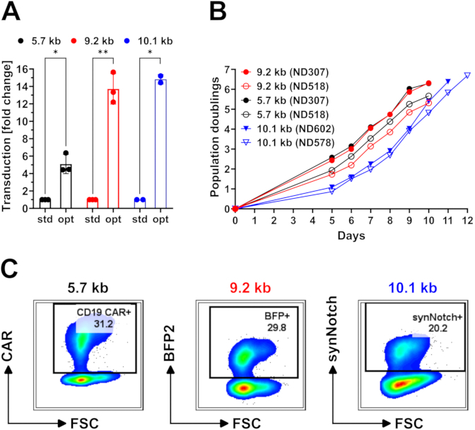
Figure 3: CAR T cell production with standard and optimized workflows. (A) Fold increase in T cell transduction rates with the optimized (opt) workflow compared to the standard (std) workflow (see also Table S1). Data were obtained from two or three independent T cell expansions using distinct donors and two independent lentivirus productions. (B) Population doublings of T cell expansions generated using the optimized workflow. For each vector, two independent T cell expansions from distinct normal donors (ND) are shown, each using a separate lentivirus production. (C) Flow cytometry plots showing CAR, BFP2, and synNotch receptor expression in expanded effector T cells on day 7 (10.1 kb) or day 10 (5.7 kb and 9.2 kb) (see also Supplementary Figure 1). All error bars indicate mean ± SD. Statistical significance was calculated using a two-tailed paired t test. *p < 0.03, **p < 0.01, ***p < 0.001, ****p < 0.0001. This figure has been adapted with permission from Rommel et al.17. Please click here to view a larger version of this figure.
Supplementary Figure 1: Gating strategy for assessing expression of CAR, synNotch receptor, and BFP2, related to Figure 3C. Representative flow cytometry plots showing the gating strategy used to determine CAR, synNotch, and BFP2 expression in expanded T cell cultures. Please click here to download this File.
Supplementary Table 1: Transduction rates across individual T cell expansions, related to Figure 3A. Transduction rates of T cell expansions generated with the standard and optimized workflows. This table has been adapted with permission from Rommel et al.17. Please click here to download this File.
Discussion
Loading...
$$\rightleftharpoonup{xx}$$
$$\longleftharp{xx}$$,
$$\longrightharp{xx}$$,
Here, we present a step-by-step protocol detailing an optimized production workflow for CAR T cells with large lentiviral transgenes, such as svsNotch T cells. This workflow eliminates the need for cell sorting and enables the efficient generation of effector T cells carrying large lentiviral transgenes. Using this workflow, we routinely produce effector T cells with lentiviral transgenes of 9-10 kb and maximal transduction rates of 60-70% (9 kb) or 15-20% (10 kb).
When troubleshooting low viral titers or transduction rates, several key factors should be considered. First, the quality of HEK293T cells. It is important that HEK293T cells are passaged with a consistent schedule and never allowed to grow beyond 50-70% confluency. In our experience, lentiviral vectors produced from HEK293T cells that were previously overgrown exhibit reduced titers, even when the cells appear morphologically recovered. We routinely generate high-titer viral vectors using HEK293T cells beyond passage 30, indicating that higher passaging numbers are tolerated if cells are properly maintained. In addition to cell quality, the confluency of HEK293T cells at the time of transfection is a critical factor. We have observed decreased lentiviral titers when transfecting HEK293T cells at high confluency (90-100%).
Second, strict adherence to the timings, concentrations, and volumes specified in this protocol is essential, as many steps have been thoroughly optimized in a previous study17. For example, the copolymer enhancer (see Table of Materials) has been titrated to maximize transduction rates without detectable toxicity. While higher concentrations can further increase transduction, they also cause toxicities and reduce T cell expansion. Similarly, harvesting lentiviral supernatants outside of the recommended 30-36 h window (e.g., at 24 h or 48 h) yielded lower titers in a corresponding time-course experiment17. In contrast, strict adherence to a 1:1 CD4-to-CD8 T cell ratio is not necessary, as we have not observed significant differences in transduction efficiency when using bulk T cells with variable CD4/CD8 distributions. However, deviations from this ratio may influence the duration of expansion and the number of population doublings. Therefore, we typically maintain the 1:1 CD4-to-CD8 ratio for consistency across experiments.
Future optimization efforts could focus on additional strategies to improve T cell transduction with even larger lentiviral transgenes, such as specialized media formulations or tailored cytokine regimens. Extending this workflow to other immune cell types, including natural killer cells and macrophages, could broaden its therapeutic applications. Importantly, clinical translation of this workflow may also help reduce the manufacturing costs of lentivirus-modified cell therapy products.
The comprehensive step-by-step protocol presented herein provides a practical reference for producing CAR T cells with large lentiviral transgenes and lays the foundation for developing novel CAR T cells against solid and blood cancers.
Disclosures
Loading...
$$\rightleftharpoonup{xx}$$
$$\longleftharp{xx}$$,
$$\longrightharp{xx}$$,
P.C.R. is an inventor on patents and patent applications licensed to Kite Pharma and receives license revenue from such licenses. B.L.L. is an inventor on patents and/or patent applications licensed to Novartis Institutes of Biomedical Research and Kite Pharma and receives license revenue from such licenses. B.L.L. is a scientific founder of Tmunity Therapeutics and Capstan Therapeutics. B.L.L. is a member of the scientific advisory boards of Avectas, Capstan Therapeutics, Cellula Therapeutics, Immuneel Therapeutics, Immusoft, In8bio, Ori Biotech, Oxford Biomedica, Quell Therapeutics, ThermoFisher Pharma Services, and UTC Therapeutics. B.L.L. is a consultant within the past 12 months for AstraZeneca, BioMerieux, Kite Gilead, and Ludwig Institute for Cancer Research. C.H.J. is an inventor on patents and/or patent applications licensed to Novartis Institutes of Biomedical Research, Kite Pharma, Capstan Therapeutics, Dispatch Biotherapeutics, and BlueWhale Bio. C.H.J. is a member of the scientific advisory boards of AC Immune, BluesphereBio, BlueWhale Bio, Cabaletta, Cartography, Cellares, Celldex, Decheng, Qihan Biotech, Shinobi Therapeutics, Verismo, ViTToria Bio, and WIRB-Copernicus.
Acknowledgements
Loading...
$$\rightleftharpoonup{xx}$$
$$\longleftharp{xx}$$,
$$\longrightharp{xx}$$,
We want to thank all members of the June Laboratory, as well as Johannes C. M. van der Loo, Divanshu Shukla, and James L. Riley, for their discussions. In addition, we would like to acknowledge Max Eldabbas, Emileigh Maddox, Tanishk Sinha, and Jiayi Shu of the Human Immunology Core at the Perelman School of Medicine at the University of Pennsylvania for providing purified human T cells. Finally, we would like to acknowledge the Penn Cytomics and Cell Sorting Shared Resource Laboratory for maintaining our flow cytometer instruments. P.C.R. was supported by the National Cancer Institute (grant number 5T32CA009140).
Materials
| Name | Company | Catalog Number | Comments |
|---|---|---|---|
| Antibody - anti-CD19 CAR | Cytoart | 200101 | Anti-CD19 CAR (idiotype), AF647-conjugated, 1:100 |
| Antibody - anti-HER2 synNotch | R&D Systems | FAB95471G | Anti-Trastuzumab (idiotype), AF488-conjugated, 1:80 |
| Bleach | Clorox | Germicidal Bleach | |
| Cell freezing container | Biocision | FTS30 | Alcohol-free |
| Centrifuge (large) | Thermo Scientific | Legend XTR | |
| Centrifuge (mini) | Fisher Scientific | Sprout Plus | |
| Centrifuge (small) | Eppendorf | 5425 | |
| Coulter Counter | Beckman Coulter | Multisizer 4 | Electrical impedance-based cell counter that determines cell numbers and cellular volumes |
| Cryo tubes | Thermo Scientific | 375418 | |
| Dimethyl sulfoxide (DMSO) | Cell Signaling Technology | 12611P | |
| DPBS (no calcium, no magnesium) | Gibco | 14190-136 | |
| Dynabeads (CD3/CD28) | Gibco | 40203D | Activation beads used for T cell stimulation |
| Ethanol | Decon Laboratories | 200 Proof | |
| FBS (heat-inactivated) | Avantor (VWR) | 97068-091 | |
| Flow cytometer | BD | LSRFortessa (647177) | |
| Freezer (-80 °C) | Panasonic | MDFU76VA-PA | |
| Freezer (liquid nitrogen) | MVE HEco | 1500-190 | |
| Fridge (4 °C) | Fisher Scientific | Isotemp | |
| Gloves (Nitrile) | Halyard | 55082 | |
| GlutaMAX Supplement | Gibco | 35050-061 | L-alanyl-L-glutamine |
| HEPES (1 M) | Gibco | 15630-080 | |
| Ice (dry) | n.a. | n.a. | |
| Ice (wet) | n.a. | n.a. | |
| Ice bucket | Fisher Scientific | 07-210-108 | |
| Lab coat (disposable) | Kappler | PVS112WH-MD | |
| Lentiviral packaging plasmids | Proprietary | n.a. | pTRP Gag-Pol, pTRP RSV-Rev, pTRP Cocal-G |
| Lentiviral transfer plasmids | Proprietary | n.a. | e.g. pTRPE svsNotch |
| Lipofectamine 3000 Transfection Reagent | Invitrogen | L3000015 | Commercial lipofection reagent; contains Lipofectamine 3000 (L3000) and its enhancer reagent (P3000) |
| Magnetic stand (large) | Stemcell | EasySep 18103 | For 5 mL/15 mL |
| Magnetic stand (small) | Invitrogen | DynaMag-2 12321D | For 1.5 mL tubes |
| Magnetic stir bar | Fisher Scientific | 14-513-82 | Stir bar kit |
| Magnetic stirring platform | Thermo Scientific | S88857100 | |
| Measuring cylinder (100 mL) | VWR | 76019-316 | |
| MQ water | Merck | Q-POD | Ultrapue water; uses Millipak 40 Express Final Filter, 0.22 Micron (MPGP04001) |
| Multichannel pipette | Fisher Scientific | see comments | 30-300 µL: FBE1200300; 5-50 µL: FBE1200050 |
| Opti-MEM (with L-glutamine, Phenol Red) | Gibco | 31985-070 | Reduced-serum minimal essential medium used for DNA transfection |
| Penicillin-Streptomycin (10,000 U/mL, 10,000 µg/mL) | Gibco | 15140-122 | |
| Pipets | Fisherbrand | see comments | 50 mL: 13-678-11F; 25 mL: 13-678-11; 10 mL: 13-678-11E; 5 mL: 13-678-11D |
| Pipets (aspirating) | Falcon | 357558 | 2 mL |
| Pipette | Gilson | PIPETMAN P10/P20/P200/P1000 | |
| Pipette controller | Hirschmann | Pipetus Z314951 | |
| Pipette tips | Thomas Scientific | see comments | 1000 µL: 1159M42; 200 µL: 1159M40; 20 µL: 1159M43; 10 µL: 1159M41 |
| Plastic storage bottle (500 mL) | Corning | 430282 | |
| Reagent reservoir | Celltreat | 3054-2007 | |
| RPMI-1640 (with L-glutamine, Phenol Red) | Gibco | 11875-085 | |
| Scale | Ohaus | Pioneer | Minimal accuracy of 0.1 g required |
| Steriflip filter (0.45 µm) | Millipore | SE1M003M00 | |
| Synperonic F 108 (SF108) | Sigma-Aldrich | 07579-250G-F | Block copolymer used to enhance T cell transduction |
| Tissue culture flask - T150 | Corning | 430825 | |
| Tissue culture flask - T25 | Corning | 430639 | |
| Tissue culture flask - T75 | Corning | 430641U | |
| Tissue culture hood | Thermo Scientific | 1300 Series A2 | |
| Tissue culture incubator | Thermo Scientific | Heracell 150i | |
| Tissue culture plate - 6-well | Corning | 3516 | |
| Tissue culture plate - 96-well flat-bottom | Falcon | 353072 | |
| Tissue culture plate - 96-well round-bottom | Falcon | 353077 | |
| Trypsin-EDTA (0.05%) | Gibco | 25300-054 | |
| Tubes - 1.5ml screw caps | Sarstedt | 72692005 | |
| Tubes - 15 mL | Falcon | 352099 | |
| Tubes - 5 mL | MSP | 62-1028-2 | |
| Tubes - 500 mL | Corning | 431123 | |
| Tubes - 50ml | Falcon | 352098 | |
| Ultracentrifuge | Beckman Coulter | Optima XPN-100 | |
| Ultracentrifuge rotor with buckets | Beckman Coulter | SW 32 Ti | |
| Ultracentrifuge tube adapters (for conical tubes) | Seton Scientific | 4230 | |
| Ultracentrifuge tubes (conical) | Seton Scientific | 5067 | |
| Vortex | Fisher Scientific | 02215414 | |
| Water bath (37 °C) | Fisher Scientific | Isotemp 210 | Used with Lab Amor Beads (Gibco) instead of water |
| X-VIVO-15 | Lonza | 04-418Q | Serum-free media used for freezing T cells |
References
Loading...
$$\rightleftharpoonup{xx}$$
$$\longleftharp{xx}$$,
$$\longrightharp{xx}$$,
- Sterner, R. C., Sterner, R. M. CAR-T cell therapy: current limitations and potential strategies. Blood Cancer J. 11 (4), 69(2021).
- Wagner, J., Wickman, E., Derenzo, C., Gottschalk, S. CAR T cell therapy for solid tumors: Bright future or dark reality. Mol Ther. 28 (11), 2320-2339 (2020).
- Hong, M., Clubb, J. D., Chen, Y. Y. Engineering CAR-T cells for next-generation cancer therapy. Cancer Cell. 38 (4), 473-488 (2020).
- Teppert, K., et al. Joining forces for cancer treatment: From "TCR versus CAR" to "TCR and CAR". Int J Mol Sci. 23 (23), 14563(2022).
- Aparicio-Perez, C., Carmona, M., Benabdellah, K., Herrera, C. Failure of ALL recognition by CAR T cells: a review of CD 19-negative relapses after anti-CD 19 CAR-T treatment in B-ALL. Front Immunol. 14, 1165870(2023).
- Roybal, K. T., et al. Precision tumor recognition by T cells with combinatorial antigen-sensing circuits. Cell. 164 (4), 770-779 (2016).
- Moghimi, B., et al. Preclinical assessment of the efficacy and specificity of GD2-B7H3 SynNotch CAR-T in metastatic neuroblastoma. Nat Commun. 12 (1), 511(2021).
- Roybal, K. T., et al. Engineering T cells with customized therapeutic response programs using synthetic notch receptors. Cell. 167 (2), 419-432.e16 (2016).
- Srivastava, S., et al. Logic-gated ROR1 chimeric antigen receptor expression rescues T cell-mediated toxicity to normal tissues and enables selective tumor targeting. Cancer Cell. 35 (3), 489-503.e8 (2019).
- Williams, J. Z., et al. Precise T cell recognition programs designed by transcriptionally linking multiple receptors. Science. 370 (6520), 1099-1104 (2020).
- Hyrenius-Wittsten, A., et al. SynNotch CAR circuits enhance solid tumor recognition and promote persistent antitumor activity in mouse models. Sci Transl Med. 13 (591), eabd8836(2021).
- Hernandez-Lopez, R. A., et al. T cell circuits that sense antigen density with an ultrasensitive threshold. Science. 371 (6534), 1166-1171 (2021).
- Choe, J. H., et al. SynNotch-CAR T cells overcome challenges of specificity, heterogeneity, and persistence in treating glioblastoma. Sci Transl Med. 13 (591), eabe7378(2021).
- Dull, T., et al. A third-generation lentivirus vector with a conditional packaging system. J Virol. 72 (11), 8463-8471 (1998).
- Kumar, M., Keller, B., Makalou, N., Sutton, R. E. Systematic determination of the packaging limit of lentiviral vectors. Hum Gene Ther. 12 (15), 1893-1905 (2001).
- Sweeney, N. P., Vink, C. A. The impact of lentiviral vector genome size and producer cell genomic to gag-pol mRNA ratios on packaging efficiency and titre. Mol Ther Methods Clin Dev. 21, 574-584 (2021).
- Rommel, P. C., et al. Engineering single-vector logic-gated CAR T cells with transgene sizes beyond current limitations. J Immunother Cancer. 14, e012318(2026).
- Improve Lentiviral Production Using Lipofectamine 3000 Reagent. , Thermo Fisher Scientific Inc. At https://www.thermofisher.com/us/en/home/life-science/cell-culture/cell-culture-learning-center/cell-culture-resource-library/cell-culture-transfection-application-notes/improve-lentiviral-production-using-lipofectamine-3000-reagent.html (2025).
- Subach, O. M., Cranfill, P. J., Davidson, M. W., Verkhusha, V. V. An enhanced monomeric blue fluorescent protein with the high chemical stability of the chromophore. PLoS One. 6 (12), e28674(2011).
Reprints and Permissions
Request permission to reuse the text or figures of this JoVE article
Request Permission