This study aims to undertake a detailed review of the research advancements in ncRNA for panvascular disease in recent years, from the viewpoints of basic, clinical, and translational medical research.
Review Article
Functional Roles of Non-Coding RNAs in Panvascular Diseases
In This Article
Summary
Abstract
Panvascular disease is a type of multi-systemic vascular disorder, primarily characterized by atherosclerotic vascular lesions as a common pathological feature, encompassing cardiovascular diseases, cerebrovascular diseases, and peripheral vascular diseases, among others. The crucial involvement of non-coding RNA (ncRNA), particularly microRNA, long non-coding RNA, and circular RNA, in different types of panvascular disease has been thoroughly studied. Recent studies have indicated that these regulatory RNA molecules are involved in the mechanisms underlying panvascular diseases by modulating pathophysiological processes such as vascular endothelial function, inflammatory response, apoptosis, and proliferation. However, there is currently no systematic review on the research progress of ncRNA in panvascular disease. Therefore, in order to provide a comprehensive and systematic discussion of the importance of ncRNA in panvascular disease. This study provides a comprehensive synthesis of recent ncRNA research in panvascular disease from basic, clinical, and translational perspectives, aiming to elucidate its specific biological roles and pathogenic mechanisms. This review is conducive to the better development of targeted treatments targeting ncRNA in panvascular disease.
Introduction
Panvascular disease (PVD) is a systemic vascular disorder characterized by endothelial dysfunction, affecting vital organs, including the heart, brain, kidneys, limbs, and aorta1. This comprehensive definition encompasses small vessel, microvascular, and venous diseases, as well as vascular conditions associated with tumors, diabetes, and immune disorders1. Depending on the affected area, PVD can manifest as coronary artery disease (CAD), cerebrovascular disease, peripheral arterial disease, or as polyvascular disease, which affects two or more vascular beds (Figure 1). Given the prevalent lifestyle risk factors, including unhealthy diets, sedentary behavior, and smoking, the incidence of hypertension, dyslipidemia, diabetes, and obesity is on the rise, contributing to an annual increase in the prevalence of PVD.
Currently, the prevention and management systems for PVD are suboptimal. Diagnosis and treatment approaches are primarily derived from the results of studies on single vascular diseases. Due to the insufficient research on multi-vascular conditions, treatments often rely on the secondary prevention strategies and risk management objectives used for localized atherosclerotic diseases. In clinical settings, the management of PVD is fragmented across specialties, including cardiology, neurology, vascular surgery, and interventional medicine, which employ a specialized approach lacking an integrated, multidisciplinary collaboration throughout the disease cycle. The pathobiological mechanisms of PVD involve endothelial dysfunction, chronic inflammation, and the migration and proliferation of vascular smooth muscle cells (VSMCs) (Figure 2). Researchers are actively pursuing pathways or factors that can intervene in these mechanisms to decelerate disease progression. Recently, the advent of bioinformatics has highlighted non-coding RNA (ncRNA) as key detectors and regulators in cardiovascular diseases2. In PVD, ncRNA plays a critical role through various mechanisms.
Compared to traditional protein biomarkers or genomic DNA analyses, ncRNAs offer distinct advantages for studying panvascular diseases. Their expression is often highly tissue-specific and can be dynamically altered in response to disease states, providing a more precise window into pathological processes. Furthermore, their remarkable stability in extracellular fluids like blood and plasma makes them exceptionally suitable as minimally invasive diagnostic and prognostic biomarkers, offering a potential tool for integrated risk assessment across the vascular tree. The translational potential of ncRNAs is rapidly expanding, particularly in emerging areas like RNA editing. Engineered ncRNAs can function as both guide RNAs and components of the editor delivery system, synergistically enhancing editing efficiency, stability, and precision. However, most programs remain in preclinical or early clinical stages, and challenges such as in vivo delivery, dosing optimization, and comprehensive evaluation of editing performance and safety require further resolution.
Review and Perspective
1. Structure and function of ncRNA
The Human Genome Project revealed that only 1.5% of the human genome codes for proteins, with the majority once considered "non-functional"1. Subsequent projects like ENCODE established that a large portion of this non-coding DNA is transcribed into non-protein coding RNAs (ncRNAs), such as miRNAs, lncRNAs, and circRNAs. Advances in high-throughput sequencing have since catalyzed the discovery of these functional ncRNAs, launching a new era in their research. Research on ncRNA and cardiovascular diseases primarily focuses on miRNA, lncRNA, and circRNA1. Besides, ncRNA is categorized by length into long ncRNA (lncRNA, over 200 nucleotides) and short ncRNA. The synthesis of lncRNA, a process mediated by RNA polymerase II through transcription and splicing akin to messenger RNA (mRNA), is fundamentally regulated at the transcriptional level. The lncRNAs regulate chromatin structure and function, gene transcription, and RNA splicing and translation through interactions with DNA, RNA, or proteins. Additionally, lncRNAs participate in the formation and functional regulation of organelles and nuclear condensates3. Mature miRNAs, ranging from 21 to 25 nucleotides, are produced by Dicer processing their precursors, which are approximately 70 to 90 nucleotides long and feature a hairpin structure. The miRNAs, which do not encode proteins, regulate gene expression at the post-transcriptional level by affecting mRNA stability and translation, generally resulting in mRNA degradation or translation inhibition4. However, most circRNAs originate from protein-coding mRNAs, with a minority derived from ncRNAs. CircRNAs production is typically seen as an additional modification in mRNA processing. In this process, pre-mRNA splicing by the typical spliceosome mechanism results in linear mRNAs, while circRNAs form through back-splicing by cis-elements or trans-factors (e.g., ZC3H14). Exonic circRNAs are predominantly found in the cytoplasm, whereas circRNAs that include introns or exons reside mainly in the nucleus5. The circRNAs function by acting as miRNA sponges, protein sponges, or forming circRNA-protein complexes (circRNPs), thereby influencing signaling pathways. Additionally, nuclear circRNAs can enhance RNA polymerase II binding to regulate the transcription of host genes, impacting subsequent biological responses and functions6.
Despite extensive research on ncRNA in various categories of PVD, including CAD, cerebrovascular disease, and peripheral artery disease, systematic reviews examining the relationship between ncRNA and PVD are lacking. Conducting a comprehensive review of ncRNA applications in PVD research is crucial, as it helps summarize existing research and provides new insights and directions for future studies (Figure 3).
2. NcRNA and atherosclerosis
Atherosclerosis is the most common and significant pathophysiological mechanism in PVD and remains a principal focus for researchers. As the relevance of ncRNA emerges, its role in atherosclerosis formation has become a prominent research focus in recent years.
- MiRNA and atherosclerosis
For instance, Wang et al.7found that miR-155 levels were significantly elevated in serum extracellular vesicles derived from monocytes of smokers. Subsequent cellular and animal studies confirmed that miR-155 promotes endothelial cell (EC) dysfunction and accelerates atherosclerosis by targeting BCL2, MCL1, TIMP3, and BCL6, while also activating the NF-κB pathway. Similarly, Guo et al.8 reported that adipose tissue-derived exosomes in obese mice were enriched with miR-132/212, which exacerbates palmitic acid-induced EC apoptosis by targeting G protein subunit alpha12 and enhances PDGF-BB-induced vascular smooth muscle cell (VSMC) proliferation and migration by suppressing PTEN. Notably, melatonin treatment reduced miR-132/212 levels and ameliorated atherosclerosis. In a mouse aortic intima injury model, Rawal et al. discovered that miR-369-3p inhibits Ox-LDL-induced inflammasome activation by targeting the succinate receptor GPR91, thereby attenuating diabetes-aggravated atherosclerosis9. Moreover, Blaser et al. 10 integrated vesicle proteomics and miRNA sequencing of human carotid and aortic valve specimens, revealing that protein and miRNA alterations during disease progression participate in intracellular signal transduction and cell cycle regulation.
In related work, Chen et al.11 showed that small extracellular vesicles (sEVs) released by steatotic hepatocytes impair ABCA1-mediated cholesterol efflux via miR-30a-3p, promoting foam cell formation and atherosclerosis. Thus, inhibiting sEV secretion or targeting miR-30a-3p may represent a therapeutic strategy for nonalcoholic fatty liver disease-associated atherosclerosis. Xie et al. established that Porphyromonas gingivalis triggers VSMC apoptosis and activates the TLR2 pathway, thereby increasing cardiovascular risk. Infected VSMCs secrete high levels of miR-143/145, which are taken up by macrophages, promote Siglec-G transcription, and suppress phagocytosis. Studies in three genetic mouse models confirmed the essential roles of TLR2 and miR-143/145 in this process12. Separately, Cimen et al. observed that miR-206-3p suppresses CXCR4 expression in ECs and VSMCs, affecting cell viability, proliferation, and migration, and proposed that cell-specific blockade of miR-206-3p could offer a novel treatment approach13. Choi et al. revealed that endothelial cell-derived extracellular vesicles enriched in miR-126-5p and miR-212-3p activate monocytes and promote atherosclerosis following radiation injury by mediating inflammatory signals14. Egea et al.15 further demonstrated through mouse models that miRNA let-7f upregulates FPR2, activating the LL-37/FPR2 axis and promoting the recruitment of human mesenchymal stem cells to atherosclerotic plaques, where they secrete cytokines and proteases that influence disease pathology. Collectively, these findings underscore the crucial role of miRNAs in the initiation and progression of atherosclerosis. - LncRNA and atherosclerosis
In recent years, long non-coding RNAs (lncRNAs) have become a major focus in atherosclerosis research owing to their diverse roles in gene expression regulation. For instance, Li et al. demonstrated in mouse models that lncRNA PSMB8-AS1 promotes atherosclerosis by recruiting the transcription factor NONO to activate PSMB9 expression and upregulating VCAM1 and ICAM1 via ZEB1, underscoring the role of lncRNAs in vascular inflammation and adhesion molecule regulation16. Similarly, Jiang et al. showed in an atherosclerosis model that lncRNA NIPA1-SO binds the transcription factor FUBP1 to suppress NIPA1 expression, thereby reducing vascular inflammation, cholesterol accumulation, and plaque formation17. In addition, Winter et al. reported that lncRNA UDT6 downregulates FGF2 expression, thereby inhibiting VSMC migration and proliferation while promoting apoptosis in models of atherosclerosis and abdominal aortic aneurysm18.
Other lncRNAs modulate atherosclerosis through autophagy, pyroptosis, and metabolic signaling. Ding et al. found that homocysteine-induced lncARF binds RRAGD to prevent its ubiquitination, activating PI3K/Akt and MAPK pathways, promoting foam cell autophagy, and attenuating atherosclerotic lesions19. Conversely, Liang et al. revealed that linc00657 acts as a competitive endogenous RNA for miR-106b-5p, elevating TXNIP expression and activating the NLRP3 inflammasome, which induces foam cell pyroptosis and exacerbates disease progression20. Furthermore, Qu et al. observed that heart-specific knockout of lncRNA Gpr137b-ps in ApoE⁻/⁻ mice suppressed amino acid-induced mTORC1 signaling, enhanced macrophage autophagy, and reduced atherosclerosis21. Together, these studies illustrate the multi-level regulatory functions of lncRNAs in atherosclerosis, spanning inflammation, cell proliferation, apoptosis, foam cell dynamics, and autophagy. - CircRNA and atherosclerosis
Circular RNAs (circRNAs), characterized by their covalently closed-loop structure, exhibit high stability against RNase degradation and play crucial roles in the development and progression of atherosclerosis. For example, Tong et al. identified a protective role of circ_0001785 in endothelial cells, showing that it mitigates endothelial damage through a ceRNA mechanism by sponging miR-513a-5p and upregulating TGFBR3 expression, thereby preserving endothelial function22. In a multi-mechanism study, Chen et al. demonstrated that circSQSSTM1 competitively binds miR-23b-3p in the cytoplasm to upregulate Sirt1, while in the nucleus it interacts directly with eIF4A3 to enhance FOXO1 mRNA expression. This dual action activates the Sirt1 promoter, attenuating endothelial inflammation, oxidative stress, and promoting autophagy, collectively slowing atherosclerosis23. Furthermore, Yang et al. reported that IgE stimulation significantly elevates circRNA CDR1as expression, which upregulates VCAM-1 and ICAM-1 via the CDR1as-FUS-phospho-p65 axis, inducing endothelial dysfunction and mast cell activation, thereby exacerbating high-fat diet-induced atherosclerosis24.
Other circRNAs contribute to atherosclerosis through feedback regulation and stress response pathways. Zhang et al. revealed that circ_0086296 promotes atherosclerotic lesion formation by sponging miR-576-3p, which enhances IFIT1 and STAT1 expression and establishes a positive feedback loop via the circ_0086296/miR-576-3p/IFIT1/STAT1 axis25. From a different angle, Holdt et al. discovered that circANRIL binds PES1 to inhibit rRNA processing and ribosome biogenesis, leading to nucleolar stress and p53 pathway activation. This process suppresses proliferation and induces apoptosis in vascular smooth muscle cells and macrophages, ultimately attenuating atherosclerosis26. Collectively, these studies illustrate the diverse functional mechanisms of circRNAs in atherosclerosis, encompassing ceRNA networks, inflammatory activation, feedback pathways, and stress response regulation.
3. NcRNA and CHD
CHD is a leading cause of death worldwide and the most prevalent type of cardiovascular disease. The primary characteristic is plaque accumulation in the coronary arteries, which obstructs blood flow, causing symptoms such as chest pain and shortness of breath. Studies indicate that ncRNA is crucial in the onset and progression of CHD by regulating gene expression and influencing inflammatory responses and cellular functions (Table 1).
- LncRNA and coronary heart disease
With advancing research on ncRNA, an increasing number of lncRNAs have been identified as closely associated with the onset and progression of CHD27,28,29. These lncRNAs play significant roles in the pathology of CHD by regulating gene expression, impacting inflammatory responses, and affecting cellular functions. For instance, Zhang et al.'s study demonstrated a significant upregulation of lncRNA nuclear enriched abundant transcript 1 (NEAT1) in blood samples from CAD patients. Experiments have demonstrated that NEAT1 targets miR-140-3p negatively, and may enhance cell viability and reduce EC apoptosis by activating the miR-140-3p/mitogen-activated protein kinase 1 (MAPK1) pathway, thereby exacerbating CHD30. In myocardial ischemia and reperfusion injury research, Xiao et al. used a mouse model to demonstrate that lncRNA CIRKIL, interacting with Ku70, can worsen myocardial cell apoptosis and cardiac dysfunction, increase myocardial infarction size, and play a detrimental role31. Different lncRNAs may either promote or inhibit disease progression, offering new insights into the pathogenesis of CHD. - MiRNA and coronary heart disease
MiRNAs are crucial in the development and progression of CHD, influencing inflammatory responses, lipid metabolism, and cellular functions. For instance, Qi et al. employed bioinformatics and experimental screening to suggest the miR-338-3p/RPS23 axis as central to CHD progression, offering a new avenue for diagnosis and treatment32. Concurrently, Flinn et al. determined that the hsa-miR-320 family was significantly expressed in the epicardial fat of CHD patients, revealing gender-dependent variations in CHD-related miRNAs and lncRNAs33. Subsequent research analyzing miRNA expression in monocytes from CAD patients indicated that overexpression of miR-21-5p and miR-221-5p was linked with an elevated CHD risk, with studies suggesting metformin as a potential reducer of this risk by normalizing miRNA expression34.
Regarding functional mechanisms, Kumar et al. discovered miR-133b and miR-21 regulated sphingosine-1-phosphate phosphatase 1 (SGPP1), autophagy-related 5(ATG5), and LDL receptor-related protein 6 (LRP6), thus implicating miRNAs in signaling pathway complexities35. Torres-Paz et al. noted a reduction in miR-33a-5p correlated with an increase in ABCA1 levels, suggesting potential cholesterol efflux modulation as a risk reducer for CHD36. Environmental interactions have also been examined, with Shen et al. identifying a synergistic effect between aluminum exposure and changes in miR-4286 levels, heightening acute coronary syndrome risks37. In atherosclerosis research, Liu et al. highlighted that miR-127-3p is significantly upregulated in advanced stages, exacerbating lesion progression through the miR-127-3p/stearoyl-CoA desaturase-1 (SCD1)/unsaturated fatty acids (UFAs) pathway38. Lastly, Mo et al., through case-control studies, found that miR-140-3p levels were notably reduced in CHD patients, positing it as a potential biomarker39. Furthermore, exogenous MecciND2 supplementation preserves mitochondrial membrane potential and curbs ROS production in stressed cardiomyocytes, and its administration in vivo alleviates ventricular remodeling and improves cardiac function in both drug-induced and pressure overload heart failure models, as supported by recent evidence40. In summary, miRNAs are crucial in CHD development and progression by regulating inflammatory responses, lipid metabolism, endothelial function, and signaling pathways. - CircRNA and coronary heart disease
CircRNAs play a crucial role in the pathophysiology of coronary heart disease (CHD) owing to their unique circular structure and stability. Recent studies demonstrate that circRNAs participate in CHD pathogenesis by regulating gene expression, competitively binding miRNAs, modulating inflammation, and influencing cellular functions. For instance, Rodríguez-Esparragón et al. reported that while the linear ANRIL transcript correlates with elevated cardiovascular risk, its circular isoform is associated with attenuated oxidative stress, highlighting functional divergence among circRNA variants41. Fu et al. identified significant upregulation of hsa_circHECTD1 and hsa_circZBTB46 in CHD patients, with the latter showing a strong correlation with disease incidence42. In contrast, Hou et al. observed markedly reduced expression of hsa_circ_0000563 in peripheral blood mononuclear cells from CHD patients, where it was found to interact with proteins involved in mitochondrial autophagy and DNA repair, suggesting a protective role in CHD progression43. Pan et al. demonstrated that circARCN1 aggravates atherosclerosis by interfering with HuR-mediated USP31 mRNA stability, thereby suppressing USP31-dependent inhibition of NF-κB activation44. Additionally, Gao et al. revealed that circ-MBOAT2 promotes endothelial tube formation and migration through the miR-495/NOTCH1 axis, supporting its role in angiogenesis in chronic total coronary occlusion45,46.
Other circRNAs contribute to CHD by modulating inflammatory responses and vascular remodeling. From a diagnostic perspective, Liu et al. identified exosomal hsa_circ_0075269 and hsa_circ_0000284 as potential biomarkers for chronic coronary syndrome through high-throughput sequencing of plasma RNA47. In oxidized LDL-stimulated macrophages, Ye et al. showed that hsa_circ_007478 competitively binds miR-765 to upregulate EFNA3, enhancing NLRP3 inflammasome activity and IL-1β production and thereby exacerbating vascular inflammation48. Holme et al. further indicated that circRNA dysregulation in ST-segment elevation myocardial infarction may worsen outcomes by perturbing immune responses, apoptosis, and mitochondrial function49. In vascular remodeling, Wang et al. reported that hsa_circ_000280 attenuates intimal hyperplasia by disrupting the interaction between ELAVL1 and CDKN1A mRNA50. Together, these findings underscore the multifaceted roles of circRNAs in CHD, spanning inflammatory modulation, autophagy, intimal hyperplasia, and oxidative stress.
4. NcRNA and cerebrovascular disease
As a critical component of PVD, cerebrovascular disease is characterized by a complex etiology and multiple risk factors. This chapter will begin with the different types and mechanisms of ncRNAs, systematically exploring their roles in cerebrovascular diseases, thereby deepening the understanding of the pathogenesis and offering new directions for future research in diagnosis and treatment (Table 2).
- LncRNA and cerebrovascular disease
Ischemic stroke, the most common form of cerebrovascular disease, involves intricate molecular and cellular interactions that result in brain tissue injury and neurological impairment. Several long non-coding RNAs (lncRNAs) have been identified as key contributors to stroke pathogenesis. For instance, Chen et al. demonstrated that lncRNA NEAT1 promotes M1-type microglial polarization, suppresses angiogenesis in cerebral artery endothelial cells, and exacerbates ischemic brain injury in a mouse model51. Similarly, lncRNA H19, when shuttled from neurons to astrocytes via exosomes, downregulates insulin-like growth factor-1 (IGF-1) through the H19/let-7a/IGF-1 axis, thereby facilitating stroke progression52. Yu et al. further reported that elevated expression of lncRNA KCNQ1OT1 in patients with recurrent transient ischemic attacks serves as an independent predictor of ischemic recurrence, highlighting its clinical utility in risk assessment53. Additionally, Zhou et al. confirmed the damaging role of lncRNA DHFRL1-4 in a cerebral ischemia-reperfusion model, where it dysregulated angiogenic factors such as bFGF and VEGF, aggravating brain injury54.
In contrast, certain lncRNAs exhibit neuroprotective properties. Xie et al. revealed that lncRNA KLF3-AS1, derived from bone marrow mesenchymal stem cells, upregulates ubiquitin-specific peptidase 22 (USP22), which in turn inhibits Sirt1 ubiquitination. This stabilizes Sirt1 protein levels, attenuates inflammatory responses, and mitigates cerebral ischemia-reperfusion injury in both in vivo and in vitro models55. Collectively, these findings underscore the dual regulatory functions of lncRNAs in ischemic stroke. While some lncRNAs exacerbate damage by modulating inflammation, angiogenesis, and metabolic pathways, others confer protection, offering promising targets for future therapeutic strategies. - MiRNA and cerebrovascular disease
MiRNAs play a crucial role in both the pathology and protective mechanisms of cerebrovascular diseases, particularly ischemic stroke. First, Li et al. demonstrated in a mouse model of ischemic stroke that M2 microglia-derived exosomes promoted oligodendrocyte precursor cell proliferation and differentiation via miR-23a-5p, thus enhancing white matter repair and functional recovery, and markedly reducing brain atrophy volume56. Similarly, Pan et al. discovered that extracellular vesicles from M2 microglia strengthen endothelial tight junction protein expression by transferring miR-23a-5p, thereby maintaining the integrity of the blood-brain barrier post-ischemia, further highlighting the protective effects of miR-23a-5p57. In terms of innovative therapies, Li et al. identified a non-toxic peptide (NP1), which provided significant neuroprotection through the miR-6328/inhibitor kappa B kinaseβ (IKKβ)/NF-κB axis. NP1 crossed the blood-brain barrier, down-regulated IKKβ, and inhibited NF-κB pathway activation, reducing inflammatory factor levels (such as IL-1β and tumor necrosis factor-α (TNF-α)), effectively diminishing cerebral infarction volume and enhancing neurological function58. Lastly, Gao et al. revealed that EC-derived extracellular vesicles regulate the fos proto-oncogene(c-Fos)/activator protein 1 (AP-1) pathway via miRNA-155-5p, significantly reducing inflammation and apoptosis while promoting cell proliferation, thus playing a protective role in ischemic stroke models. This study further clarified the potential function of EC miRNAs in stroke59. These studies demonstrate that miRNAs can act as both pathology-promoting factors and protective agents in cerebrovascular diseases by regulating key signaling pathways. - CircRNA and cerebrovascular disease
Emerging evidence underscores the important roles of circular RNAs (circRNAs) in neuroprotection, inflammation modulation, and vascular repair following ischemic stroke. While some circRNAs contribute to pathological progression, others facilitate recovery processes. For example, Liu et al. reported that circOGDH was markedly upregulated in both acute ischemic stroke patients and mouse models, where it sponges miR-5112 to enhance collagen type IV alpha 4 chain (COL4A4) expression, thereby aggravating neuronal injury60. This identifies circOGDH as a potential disease-promoting RNA. Conversely, Li et al. revealed that circSCMH1 promotes vascular repair by mediating the nuclear translocation of the m6A demethylase FTO, which facilitates m6A demethylation of phospholipid phosphatase 3 mRNA and supports post-stroke vascular regeneration61.
Additional circRNAs have been implicated in neuroprotection and functional recovery. Yang et al. observed significantly reduced levels of hsa_circ_0045932 in the peripheral blood of ischemic stroke patients. Functional studies showed that this circRNA acts as a molecular sponge for miR-139-3p, repressing SMAD3 expression and attenuating ischemic damage62. In a mouse middle cerebral artery occlusion model, Zhao et al. demonstrated that mmu_circ_0001113 enhances the interaction between enolase 1 (ENO1) and Krüppel-like factor 2 (Klf2) mRNA, leading to increased Klf2 protein levels. This mechanism suppresses NLRP3 inflammasome-driven apoptosis, reduces infarct volume, and improves neurological recovery63. In summary, circRNAs play dual roles in cerebrovascular pathology: they can either exacerbate injury through dysregulation of key pathways or confer protection by modulating gene expression and signaling cascades.
5. NcRNA and peripheral vascular disease
Non-coding RNAs (ncRNAs) have been demonstrated to play pivotal roles not only in the development of atherosclerosis but also in the pathophysiology of cardiovascular and cerebrovascular diseases through diverse biological mechanisms. Chen et al. constructed an immune-related competing endogenous RNA (ceRNA) network based on GEO expression profiles, revealing that a network comprising LINC00221, miR-17-5p, miR-20b-5p, and CREB1 may promote peripheral arterial occlusive disease by modulating infiltration of monocytes and M1-type macrophages64. In a mouse model of limb ischemia, Huang et al. reported that lncRNA H19 competitively binds miR-107 to upregulate FADD, thereby activating the pyroapoptosis pathway while concurrently promoting cell proliferation, migration, angiogenesis, and blood flow recovery65.
Multiple miRNAs have also been identified as key regulators in peripheral arterial disease. Lee et al. observed that circulating miR-548j-5p was significantly downregulated in PAD patients. This miRNA facilitates endothelial progenitor cell migration and tube formation by modulating NOS and SDF-1 signaling, thereby promoting angiogenesis66. In diabetic PAD, Cheng et al. identified miR-181a/b as a critical mediator of clinical limb ischemia; its ablation reduced monocyte recruitment and impaired vascular regeneration in ischemic limbs67. Ahmed et al. reported elevated miR-210 levels in skeletal muscle of PAD patients, where it regulates mitochondrial respiration and cellular adaptation to hypoxia68. Additionally, Cheng et al. showed that miR-130b-3p promotes angiogenesis by suppressing the BMP/TGF-β pathway, suggesting its potential as a therapeutic target to mitigate limb necrosis and amputation risk69. Zaied et al. further demonstrated that miR-93 enhances angiogenesis by activating G6PD and the pentose phosphate pathway, leading to more robust blood flow restoration than VEGF165a70. McCoy et al. revealed that miR-375, downregulated in critical limb ischemia, targets KLF5 to modulate NF-κB signaling and influence angiogenic responses71.
In contrast to lncRNAs and miRNAs, research on circRNAs in peripheral vascular diseases remains limited. However, given their well-established roles in cardiovascular and cerebrovascular pathologies, circRNAs are likely to also contribute to the pathogenesis and treatment of peripheral vascular conditions, warranting further mechanistic and translational investigation.
Conclusions
As a class of RNA molecules that do not encode proteins directly, ncRNA has emerged as a promising area in the study of PVD. These diseases include atherosclerosis, CHD, cerebrovascular disease, and peripheral vascular disease, characterized by vascular endothelial dysfunction. ncRNAs significantly influence the development and treatment of these diseases by regulating gene expression, modulating inflammatory responses, and regulating cell proliferation and apoptosis. In atherosclerosis, miRNAs influence disease progression through mechanisms such as inflammation, oxidative stress, and foam cell formation, while lncRNA and circRNA modulate cell migration, autophagy, and signaling pathways, offering new directions for early diagnosis and intervention. In CHD, ncRNAs offer potential targets for precise treatments by influencing plaque stability, cardiomyocyte apoptosis, and inflammatory responses. Research in cerebrovascular diseases has identified ncRNAs' roles in vascular regeneration, neuroprotection, and inflammatory response post-cerebral ischemic injury, opening new treatment avenues for conditions such as stroke. In peripheral vascular diseases, although research is nascent, evidence suggests ncRNAs regulate angiogenesis, energy metabolism, and fibrosis, showing potential therapeutic value.
Looking forward, ncRNA research faces several challenges. Firstly, the commonalities and specifics of ncRNAs' roles across different diseases require deep exploration to uncover their core regulatory networks. Secondly, much research remains at the basic mechanism level; translating these findings into clinical applications is a critical future direction. In addition, with advancements in high-throughput sequencing technologies and bioinformatics, the development of clinical detection methods, diagnostic markers, and therapeutic targets for ncRNA will experience significant opportunities. Particularly within the context of precision medicine and personalized treatment, ncRNA is poised to become a fundamental component in diagnosing and treating PVD.
In summary, the role of ncRNA in PVD is widely acknowledged. By harnessing multidisciplinary collaborations, conducting larger-scale clinical studies, and integrating ncRNA with traditional treatment approaches, the field is set to advance significantly, offering earlier diagnosis, more precise treatments, and improved outcomes for PVD.
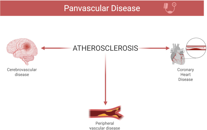
Figure 1: Panvascular disease. Please click here to view a larger version of this figure.
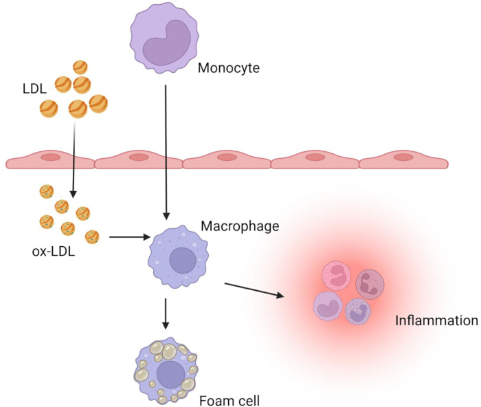
Figure 2: The mechanism of atherosclerosis. Please click here to view a larger version of this figure.
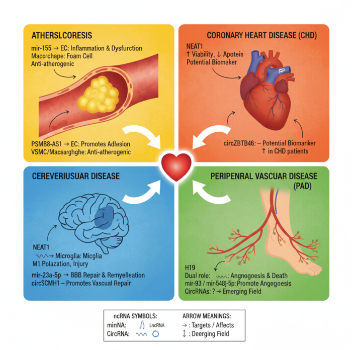
Figure 3: Non-coding RNAs in panvascular diseases. Please click here to view a larger version of this figure.
Table 1: Non-coding RNA and coronary heart disease. Please click here to download this Table.
Table 2: Non-coding RNA and cerebrovascular disease. Please click here to download this Table.
Disclosures
The authors declare that there is no conflict of interest.
Acknowledgements
This work was supported by the Zhanjiang Science and Technology Plan Project (2021A1003-1), the research project of Guangdong Provincial Bureau of Traditional Chinese Medicine (20221441), and the Guangdong Basic and Applied Basic Research Foundation (No.2023A1515010482). Weiyan Li: Conceptualization, Writing - original draft. Lingyan Fang, Fengya Zeng, and Jian Cao: Conceptualization, Writing - review & editing; Can Chen: Writing - review & editing, Supervision, Project administration, Funding acquisition. All authors read and approved the final manuscript.
References
- Zhou, X., Yu, L., Zhao, Y., Ge, J. Panvascular medicine: an emerging discipline focusing on atherosclerotic diseases. Eur Heart J. 43 (43), 4528-4531 (2022).
- Zhu, L., Li, N., Sun, L., Zheng, D., Shao, G. Non-coding RNAs: the key detectors and regulators in cardiovascular disease. Genomics. 113, 1233-1246 (2021).
- Chen, L. L., Kim, V. N. Small and long non-coding RNAs: past, present, and future. Cell. 187 (23), 6451-6485 (2024).
- Diener, C., Keller, A., Meese, E. The miRNA-target interactions: an underestimated intricacy. Nucleic Acids Res. 52 (4), 1544-1557 (2024).
- Saaoud, F., et al. Circular RNAs are a novel type of non-coding RNAs in ROS regulation, cardiovascular metabolic inflammations and cancers. Pharmacol Ther. 220, 107715(2021).
- Liu, C. X., Chen, L. L. Circular RNAs: characterization, cellular roles, and applications. Cell. 185 (12), 2016-2034 (2022).
- Wang, C., et al. Nicotine exacerbates endothelial dysfunction and drives atherosclerosis via extracellular vesicle-miRNA. Cardiovasc Res. 119 (3), 729-742 (2023).
- Guo, B., et al. MiRNA-132/212 encapsulated by adipose tissue-derived exosomes worsen atherosclerosis progression. Cardiovasc Diabetol. 23 (1), 331(2024).
- Rawal, S., et al. miR-369-3p ameliorates diabetes-associated atherosclerosis by regulating macrophage succinate-GPR91 signaling. Cardiovasc Res. 120 (14), 1693-1712 (2024).
- Blaser, M. C., et al. Multiomics of tissue extracellular vesicles identifies unique modulators of atherosclerosis and calcific aortic valve stenosis. Circulation. 148 (8), 661-678 (2023).
- Chen, X., et al. Hepatic steatosis aggravates atherosclerosis via small extracellular vesicle-mediated inhibition of cellular cholesterol efflux. J Hepatol. 79 (6), 1491-1501 (2023).
- Xie, H., et al. Oral pathogen aggravates atherosclerosis by inducing smooth muscle cell apoptosis and repressing macrophage efferocytosis. Int J Oral Sci. 15 (1), 26(2023).
- Cimen, I., et al. Targeting a cell-specific microRNA repressor of CXCR4 ameliorates atherosclerosis in mice. Sci Transl Med. 15 (720), eadf3357(2023).
- Choi, Y. Y., et al. The miR-126-5p and miR-212-3p in the extracellular vesicles activate monocytes in the early stage of radiation-induced vascular inflammation implicated in atherosclerosis. J Extracell Vesicles. 12 (5), e12325(2023).
- Egea, V., et al. Properties and fate of human mesenchymal stem cells upon miRNA let-7f-promoted recruitment to atherosclerotic plaques. Cardiovasc Res. 119 (1), 155-166 (2023).
- Li, S., et al. LncRNA PSMB8-AS1 instigates vascular inflammation to aggravate atherosclerosis. Circ Res. 134 (1), 60-80 (2024).
- Jiang, M., et al. LncRNA NIPA1-SO confers atherosclerotic protection by suppressing the transmembrane protein NIPA1. J Adv Res. 54, 29-42 (2023).
- Winter, H., et al. Targeting long non-coding RNA NUDT6 enhances smooth muscle cell survival and limits vascular disease progression. Mol Ther. 31 (6), 1775-1790 (2023).
- Ding, N., et al. Novel long non-coding lncARF mediated hyperhomocysteinemia-induced atherosclerosis via autophagy inhibition in foam cells. J Adv Res. 73, 311-328 (2024).
- Liang, Y., et al. Linc00657 promoted pyroptosis in THP-1-derived macrophages and exacerbated atherosclerosis via the miR-106b-5p/TXNIP/NLRP3 axis. Int J Biol Macromol. 253 (Pt 4), 126953(2023).
- Qu, W., et al. Long non-coding RNA Gpr137b-ps promotes advanced atherosclerosis via the regulation of autophagy in macrophages. Arterioscler Thromb Vasc Biol. 43 (11), e468-e489 (2023).
- Tong, X., et al. Exosome-derived circ_0001785 delays atherogenesis through the ceRNA network mechanism of miR-513a-5p/TGFBR3. J Nanobiotechnology. 21 (1), 362(2023).
- Chen, Z., et al. A novel circular RNA, circSQSTM1, protects the endothelial function in atherosclerosis. Free Radic Biol Med. 209 (Pt 2), 301-319 (2023).
- Yang, H., et al. Exosomes from IgE-stimulated mast cells aggravate asthma-mediated atherosclerosis through circRNA CDR1as-mediated endothelial cell dysfunction in mice. Arterioscler Thromb Vasc Biol. 44 (3), e99-e115 (2024).
- Zhang, M., et al. circ_0086296 induced atherosclerotic lesions via the IFIT1/STAT1 feedback loop by sponging miR-576-3p. Cell Mol Biol Lett. 27 (1), 576-573 (2022).
- Holdt, L. M., et al. Circular non-coding RNA ANRIL modulates ribosomal RNA maturation and atherosclerosis in humans. Nat Commun. 7, 12429(2016).
- Zhang, Y. H., Pan, X., Zeng, T., Chen, L., Huang, T., Cai, Y. D. Identifying the RNA signatures of coronary artery disease from combined lncRNA and mRNA expression profiles. Genomics. 112 (6), 4945-4958 (2020).
- Zareba, L., et al. MicroRNAs and long non-coding RNAs in coronary artery disease: new and potential therapeutic targets. Cardiol Clin. 38 (4), 601-617 (2020).
- Ebadi, N., Ghafouri-Fard, S., Taheri, M., Arsang-Jang, S., Parsa, S. A., Omrani, M. D. Dysregulation of autophagy-related lncRNAs in peripheral blood of coronary artery disease patients. Eur J Pharmacol. 867, 172852(2020).
- Zhang, H., Ji, N., Gong, X., Ni, S., Wang, Y. NEAT1/miR-140-3p/MAPK1 mediates the viability and survival of coronary endothelial cells and affects coronary atherosclerotic heart disease. Acta Biochim Biophys Sin (Shanghai). 52 (9), 967-974 (2020).
- Xiao, H., et al. CIRKIL exacerbates cardiac ischemia/reperfusion injury by interacting with Ku70. Circ Res. 130 (5), e3-e17 (2022).
- Qi, J., Han, W., Zhong, N., Gou, Q., Sun, C. Integrated analysis of miRNA-mRNA regulatory network and functional verification of miR-338-3p in coronary heart disease. Funct Integr Genomics. 23 (1), 16(2022).
- Flinn, B., Adams, C., Chowdhury, N., Gress, T., Santanam, N. Profiling of non-coding regulators and their targets in epicardial fat from patients with coronary artery disease. Int J Mol Sci. 23 (10), 5297(2022).
- Torres-Paz, Y. E., et al. Overexpression of microRNA-21-5p and microRNA-221-5p in monocytes increases the risk of developing coronary artery disease. Int J Mol Sci. 24 (10), 221-225 (2023).
- Kumar, D., Narang, R., Saluja, D., Srivastava, K. Functional association of miR-133b and miR-21 through novel gene targets ATG5, LRP6 and SGPP1 in coronary artery disease. Mol Diagn Ther. 26 (6), 655-664 (2022).
- Torres-Paz, Y. E., Flores-Rivera, J., Martínez-Aguilar, E., et al. Involvement of expression of miR33-5p and ABCA1 in human peripheral blood mononuclear cells in coronary artery disease. Int J Mol Sci. 25 (16), 8605(2024).
- Shen, M., et al. Prospective findings from the Dongfeng-Tongji cohort: Exposure to various metals, the expression of microRNA-4286, and the incidence of acute coronary syndrome. Environ Res. 250, 118322(2024).
- Liu, Y., et al. MiR-127-3p enhances macrophagic proliferation via disturbing fatty acid profiles and oxidative phosphorylation in atherosclerosis. J Mol Cell Cardiol. 193, 36-52 (2024).
- Mo, P., et al. Decreased plasma miR-140-3p is associated with coronary artery disease. Heliyon. 10 (5), e26960(2024).
- Liu, X., et al. Fast degradation of meccirnas by supv3l1/elac2 provides a novel opportunity to tackle heart failure with exogenous meccirna. Circulation. 151 (17), 1272-1290 (2025).
- Rodríguez-Esparragón, F., de la Rosa, D. A., Santana, A., et al. Analysis of ANRIL isoforms and key genes in patients with severe coronary artery disease. Int J Mol Sci. 24 (22), 16127(2023).
- Fu, Y., et al. Detailed profiling of m6A modified circRNAs and synergistic effects of circRNA and environmental risk factors for coronary artery disease. Eur J Pharmacol. 951, 175761(2023).
- Zhou, H., et al. Identification of circular RNA BTBD7_hsa_circ_0000563 as a novel biomarker for coronary artery disease and the functional discovery of BTBD7_hsa_circ_0000563 based on peripheral blood mononuclear cells: A case control study. Clin Proteomics. 19 (1), 37(2022).
- Pan, Z., et al. CircARCN1 aggravates atherosclerosis by regulating HuR-mediated USP31 mRNA in macrophages. Cardiovasc Res. 120 (13), 1531-1549 (2024).
- Gao, W., et al. Integrated analysis of angiogenesis related lncRNA-miRNA-mRNA in patients with coronary chronic total occlusion disease. Front Genet. 13, 855549(2022).
- Gao, W., Huang, Y., Yuan, J. Circ-MBOAT2 regulates angiogenesis via the miR-495/NOTCH1 axis and associates with myocardial perfusion in patients with coronary chronic total occlusion. Int J Mol Sci. 25 (2), 793(2024).
- Liu, X., Yang, H., Zhang, H. Circulating exosomal circRNAs as diagnostic biomarkers for chronic coronary syndrome. Metabolites. 13 (10), 1066(2023).
- Ye, B., et al. Hsa_circ_0007478 aggravates NLRP3 inflammasome activation and lipid metabolism imbalance in ox-LDL-stimulated macrophage via miR-765/EFNA3 axis. Chem Biol Interact. 368, 110195(2022).
- Holme, F. A., et al. Circular RNA profile in atherosclerotic disease: Regulation during ST-elevated myocardial infarction. Int J Mol Sci. 25 (16), 9014(2024).
- Wang, Z., et al. Role of hsa_circ_0000280 in regulating vascular smooth muscle cell function and attenuating neointimal hyperplasia via ELAVL1. Cell Mol Life Sci. 80 (1), 3(2022).
- Chen, T., Huang, X., Zhao, Y. X., Zhou, Z. W., Zhou, W. S. NEAT1 inhibits the angiogenic activity of cerebral arterial endothelial cells by inducing the M1 polarization of microglia through the AMPK signaling pathway. Cell Mol Biol Lett. 29 (1), 62(2024).
- Wang, J., Cao, B., Gao, Y., Chen, Y. H., Feng, J. Exosome-transported lncRNA H19 regulates insulin-like growth factor-1 via the H19/let-7a/insulin-like growth factor-1 receptor axis in ischemic stroke. Neural Regen Res. 18 (6), 1316-1320 (2023).
- Yu, S., An, J., Sun, R., Feng, J., Yu, M. LncRNA KCNQ1OT1 predicts further cerebral events in patients with transient ischemic attack. Front Pharmacol. 13, 961190(2022).
- Zhou, Y., et al. lncRNA DHFRL1-4 knockdown attenuates cerebral ischemia/reperfusion injury by upregulating the levels of angiogenesis-related genes. Int J Mol Med. 50 (2), 108(2022).
- Xie, X., Cao, Y., Dai, L., Zhou, D. Bone marrow mesenchymal stem cell-derived exosomal lncRNA KLF3-AS1 stabilizes Sirt1 protein to improve cerebral ischemia/reperfusion injury via miR-206/USP22 axis. Mol Med. 29 (1), 3(2023).
- Li, Y., et al. M2 microglia-derived extracellular vesicles promote white matter repair and functional recovery via miR-23a-5p after cerebral ischemia in mice. Theranostics. 12 (7), 3553-3573 (2022).
- Pan, J. J., et al. M2 microglial extracellular vesicles attenuated blood-brain barrier disruption via miR-23a-5p in cerebral ischemic mice. Aging Dis. 15 (3), 1344-1356 (2024).
- Li, Y., et al. A short peptide exerts neuroprotective effects on cerebral ischemia-reperfusion injury by reducing inflammation via the miR-6328/IKKβ/NF-κB axis. J Neuroinflammation. 20 (1), 53(2023).
- Gao, X., et al. Observing extracellular vesicles originating from endothelial cells in vivo demonstrates improved astrocyte function following ischemic stroke via aggregation-induced emission luminogens. ACS Nano. 17 (16), 16174-16191 (2023).
- Liu, Y., et al. CircOGDH is a penumbra biomarker and therapeutic target in acute ischemic stroke. Circ Res. 130 (6), 907-924 (2022).
- Li, B., et al. FTO-dependent m6A modification of Plpp3 in circSCMH1-regulated vascular repair and functional recovery following stroke. Nat Commun. 14 (1), 489(2023).
- Yang, J., Li, Y., Zhou, H. CircUSP36 attenuates ischemic stroke injury through the miR-139-3p/SMAD3/Bcl2 signal axis. Clin Sci (Lond). 136 (12), 953-971 (2022).
- Zhao, Y., et al. CircFndc3b mediates exercise-induced neuroprotection by mitigating microglial/macrophage pyroptosis via the ENO1/KLF2 axis in stroke mice. Adv Sci (Weinh). 12 (1), 2403818(2025).
- Chen, Z., et al. A construction and comprehensive analysis of the immune-related core ceRNA network and infiltrating immune cells in peripheral arterial occlusive disease. Front Genet. 13, 951537(2022).
- Huang, L., et al. LncRNA H19/miR-107 regulates endothelial progenitor cell pyroptosis and promotes flow recovery of lower extremity ischemia through targeting FADD. Biochim Biophys Acta Mol Basis Dis. 1870 (7), 167323(2024).
- Lee, C. Y., Lin, S. J., Wu, T. C. miR-548j-5p regulates angiogenesis in peripheral artery disease. Sci Rep. 12 (1), 838(2022).
- Cheng, H. S., et al. A miRNA/CXCR4 signaling axis impairs monopoiesis and angiogenesis in diabetic critical limb ischemia. JCI Insight. 8 (7), e163360(2023).
- Ismaeel, A., et al. Skeletal muscle miR-210 expression is associated with mitochondrial function in peripheral artery disease patients. Transl Res. 246, 66-77 (2022).
- Cheng, H. S., et al. Impaired angiogenesis in diabetic critical limb ischemia is mediated by a miR-130b/INHBA signaling axis. JCI Insight. 8 (10), e163041(2023).
- Zaied, A. A., et al. Pentose pathway activation is superior to increased glycolysis for therapeutic angiogenesis in peripheral arterial disease. J Am Heart Assoc. 12 (7), e027986(2023).
- McCoy, M. G., et al. MicroRNA-375 repression of Kruppel-like factor 5 improves angiogenesis in diabetic critical limb ischemia. Angiogenesis. 26 (1), 107-127 (2023).