Using a Parkinson's disease (PD)-like mouse model that overexpresses the mutant A53T form of human alpha-synuclein (h-α-Syn) in the dorsal raphe nucleus (DR), we have developed an electrophysiological recording method to investigate the role of the ventromedial prefrontal cortex (vmPFC)-DR circuit in depressive/anxiety disorders in PD.
Method Article
Awake Electrophysiological Profiling of the Ventromedial Prefrontal Cortex in a Mouse Model of Depression and Parkinson's Disease
In This Article
Summary
Abstract
While Parkinson's disease (PD) is primarily known as a motor disorder, non-motor symptoms often emerge before motor deficits and significantly impact disease progression, being crucial at all disease stages. Among these, depression and anxiety are the most prevalent symptoms and cause a higher symptom burden in women than in men. Alterations in the serotonergic (5-HT) system are frequently linked to mood disorders, and accumulations of α-synuclein (α-Syn) have been observed in the 5-HT raphe nuclei (RN) of patients with Parkinson's disease (PD) and depression. Understanding the neural circuits underlying these non-motor symptoms is therefore critical.
Here, we present an improved protocol to investigate the role of the ventromedial prefrontal cortex (vmPFC)-dorsal raphe nucleus (DR) circuit in PD-associated mood disorders using a female mouse model overexpressing the mutant A53T form of human α-syn (h-α-Syn) in the DR. Our method allows high-resolution assessment of neuronal activity in the infralimbic (IL) and prelimbic (PL) cortices under both basal conditions and during aversive conditioning and extinction. Using a tone-light sensory conditioning paradigm in awake, head-fixed mice, we employ multichannel electrophysiological probes to record neuronal responses. Experiments were conducted in virtual reality corridors, combining a cylindrical treadmill with a dual-screen visual display to maintain behavioral engagement while ensuring precise control of sensory stimuli and recording stability.
This protocol builds on the established involvement of the IL and PL cortices in fear conditioning and extinction and provides a robust framework to examine dynamic neural activity in circuits implicated in depressive and anxiety-like behaviors in PD. By enabling simultaneous behavioral and electrophysiological measurements under controlled conditions, this approach offers a powerful tool for elucidating the neurobiological mechanisms of non-motor symptoms in PD and for testing potential interventions.
Introduction
Parkinson's disease (PD) is a motor disorder, but non-motor symptoms are known to precede the motor manifestations and are critical throughout all disease stages1,2,3,4. Among these, depression and anxiety are the most prevalent neuropsychiatric symptoms, severely affecting the quality of life of PD patients and causing a higher symptom burden on women than on men5. Despite this, they are often underdiagnosed and inadequately treated6. For all these reasons, it is essential to understand the neurobiology and the circuits involved in the neuropsychiatric symptoms of PD in order to stratify patients and offer them more personalized care.
Although the etiology of PD remains partially unclear, several key neuropathological features have been identified. The disease is characterized by the progressive degeneration and loss of dopaminergic (DA) neurons and fibers in the nigrostriatal system, leading to the prototypical motor deficits associated with PD7,8. Another hallmark of PD is the presence of Lewy bodies (LB) and Lewy neurites -- intracellular inclusions formed by fibrillar protein aggregates -- composed predominantly of the protein α-synuclein (α-Syn)9,10. Neuroimaging studies also reveal that other neurotransmitter systems are affected -- even before DA involvement -- among them, the serotonergic (5-HT) system11,12,13. Changes in the serotonergic (5-HT) system are commonly associated with mood disorders, and deposits of α-synuclein (α-Syn) have been detected in the 5-HT raphe nuclei (RN) of individuals diagnosed with Parkinson's disease (PD) and depression14.
The dorsal raphe nuclei (DR) is the largest 5-HT nucleus in the RN15, playing a critical role in emotional regulation, perception, reward, aggression, and social interactions. Consequently, dysfunction in the DR is associated with neuropsychiatric disorders such as stress, anxiety, and depression15,16. At this point, it is important to emphasize the strong, bidirectional functional and anatomical connection between the DR and the prefrontal cortex (PFC), a brain region essential for higher-order brain functions. While the more dorsal regions of the PFC are related to cognitive functions, the more ventromedial areas (vmPFC) play a fundamental role in emotional regulation, action control, memory, and decision-making17,18. Therefore, the vmPFC and the DR together significantly influence mood, and altered activity in this circuit is associated with depressive symptoms18. Moreover, the accumulation of aggregated forms of α-Syn in 5-HT neurons in the RN has been shown to induce anxiety and depressive-like phenotypes19,20,21.
Utilizing a PD female mouse model with a depressive phenotype that overexpresses the mutant A53T form of human α-syn (h-α-Syn) in the DR via an adeno-associated viral (AAV) vector, we have developed an electrophysiological recording method. This method aims to investigate the role of the vmPFC-DR circuit in depressive/anxiety disorders in PD. This approach involves assessing the neuronal activity of the infralimbic (IL) and prelimbic (PL) cortices in awake mice through single-unit and local field potentials (LFPs) recordings. This is accomplished using a multichannel probe in conjunction with a virtual reality runner. Recognizing the involvement of the PL and IL cortices in aversive conditioning, we also measure their activity during such a paradigm to evaluate their function in stressful situations. It is crucial to understand that the PL primarily drives conditioned responses, while the IL cortex plays a key role in the extinction of these responses22,23.
The following protocol describes the steps for implanting a head-bar and acutely recording the electrical activity of the PL and IL cortices in vivo. We achieve this using a virtual reality corridor, which combines a cylindrical treadmill for head-fixed mice with a double-screen visual display. The protocol is structured into four main stages: (1) head-bar implantation surgery, (2) habituation, (3) recording and signal acquisition, and (4) data analysis: spike sorting and data preprocessing.
Three weeks after inducing h-α-Syn overexpression in the brain 5-HT system, mice undergo the head-bar implantation surgery, which enables subsequent head-fixation. Following a 1-week recovery period, mice are habituated for 4 days to both the experimenter and the treadmill and head-fixation system. On the recording day, this setup is used to record neural activity from the PL and IL cortices under baseline conditions and during aversive conditioning. The raw neural data is processed via automatic spike sorting with Kilosort4 followed by manual curation in Phy2, in order to extract high-quality single-unit clusters for further analysis (Figure 1).
The proposed awake, head-fixed multichannel vmPFC recording protocol offers several advantages compared with classical alternatives such as anesthetized or ex vivo preparations and wireless recordings in freely moving animals. Unlike anesthetized or ex vivo approaches, this method allows the assessment of vmPFC activity under controlled visual and auditory stimulation within a virtual reality environment, which is essential for implementing aversive conditioning paradigms24,25. In contrast to freely moving recordings, the head-fixed configuration provides precise experimental control over sensory inputs and task contingencies, thereby reducing variability and enabling consistent trial structures. Furthermore, head-fixation reduces the risk of task disengagement observed in freely moving paradigms, particularly under aversive stimulation, while still allowing high-yield, stable multichannel recordings26.
From a practical perspective, this protocol requires animals to be habituated to head-fixation and treadmill running, but it benefits from commercially available, relatively affordable head-fixation systems that incorporate cylindrical treadmills. This makes the approach accessible to smaller laboratories, as probes can be reused multiple times (up to ~10 insertions), substantially lowering costs compared with chronic freely moving implants. Consequently, this method provides a balance between experimental control, behavioral relevance, and cost-effectiveness. Another factor to consider is the implementation of the aversive conditioning paradigm. In this study, we employed an experimental design based on rodents' natural aversion to light: 10 auditory tones (conditioned stimuli) were each followed by a brief exposure to intense light (unconditioned stimulus). Although this pattern is less stressful than other classical conditioning methods, such as those involving electric shocks, it constitutes a more similar real-world stressor.
Protocol
All experiments were performed with age-matched controls, and animal procedures were designed in line with the 3R's rules; conducted in accordance with ethical guidelines outlined in the EU Directive 2010/63 (September 22, 2010), Spanish legislation (RD 1201/2005, L.32/2007, and L.6/2013) and approved by Generalitat Valenciana (ES462500001003; ref. 2023-VSC-PA-0073), and the local Bioethics Committee of the University of Valencia (ref. A20230214153605).
NOTE: To ensure safety and prevent contamination, throughout the entire procedure, wear personal protective equipment, including disposable gloves and booties, a lab coat, mask, and a hair net. Use veterinary drug take-back systems for the disposal of residues resulting from its use, in accordance with local regulations and applicable national take-back programs.
1. Mouse model generation
NOTE: Adult female C57BL/6J mice (9 weeks old) were housed under controlled environmental conditions (22 ± 1 °C; 12-h light/dark cycle) with ad libitum access to food and water.
- Under isoflurane anesthesia (4% for induction, 2% for maintenance) in the mouse anesthesia chamber, randomly assign the mice to receive stereotaxic microinjections into the DR (coordinates relative to bregma: anteroposterior [AP] −4.5 mm, mediolateral [ML] −1.0 mm, dorsoventral [DV] −3.2 mm; injection angle: 20°) using an automated microinjector at a flow rate of 0.4 µL/min, as previously described19,27,28.
- Ensure each mouse receives a total volume of 0.6 µL of either a recombinant adeno-associated virus serotype 1/2 (AAV1/2; concentration: 5.16 × 10¹² gp/mL) encoding the A53T mutant form of human h-α-Syn, to induce overexpression in 5-HT neurons of the DR (n = 4) or an empty AAV1/2 vector containing non-coding stuffer DNA (AAV-EV), used as a control group (n=5).
- To prevent reflux, keep the needle in place for an additional 8 min before slowly withdrawing it at a rate of 320-400 µm/min.
NOTE: For safety, ensure that the scavenging systems are functioning properly to capture anesthetic vapors before any stereotaxic surgery (sections 1 and 2). In case of an isoflurane spill while refilling the vaporizer, immediately clean it up using an inert, absorbent material, such as sawdust. Use veterinary drug take-back systems for the disposal of isoflurane filters and any residues resulting from its use, in accordance with local regulations.
2. Head-bar implantation surgery
NOTE: Perform the head-bar implantation surgery following the steps below, 3 weeks after generating the mouse model.
- Pre-surgical preparation and anesthesia
- Anaesthetize the animal with isoflurane (4% for induction, 1.5-2% for maintenance) delivered via an isoflurane vaporizer.
- Position the mouse in the stereotaxic apparatus on a warming pad to maintain body temperature throughout the procedure.
- Gently open the mouse's mouth to place its teeth on the bite bar and attach the mask connected to a stereotaxic apparatus, to ensure continuous isoflurane delivery for anesthesia maintenance.
- Apply eye drops to prevent ocular ulceration.
- Administer analgesia at least 10 min prior to scalpel incision via subcutaneous injection of butorphanol (5 mg/kg in saline solution).
- Apply regional lidocaine (0.03 mL in each ear, 4 mg/kg, do not exceed maximum doses of 7 mg/kg or lidocaine/prilocaine cream [25 mg/g lidocaine and 25 mg/g prilocaine]) to the inner ear to reduce discomfort from the ear bars.
- Stereotaxic positioning and skull preparation
- Position the ear bars to securely hold the head, ensuring symmetry and minimal movement.
- Adjust the incisor bar to bring bregma and lambda to the same horizontal level, making sure they are on the midline.
- Confirm anesthesia induction by checking for loss of pedal withdrawal, palpebral reflexes, decreased respiratory rate, and absence of whisker movement.
- Ensure the mouse is correctly positioned in the stereotaxic frame with the ear bars carefully adjusted.
- Shave the surgical area and disinfect it using an antiseptic solution (povidone-iodine followed by 70% ethanol) to minimize the risk of infection.
- Make a midline scalp incision and gently retract the underlying tissues to expose the skull while maintaining hemostasis. Ensure that the skin is opened sufficiently to visualize the parietotemporal ridges. To achieve this, place four fine bulldog clamps on the fascia beneath the skin to maximize exposure.
- Clean and thoroughly dry the exposed skull for a clear view of anatomical landmarks (bregma and lambda).
- Level the skull in the anteroposterior axis by aligning the bregma and lambda in the same horizontal plane.
- Level the skull in the mediolateral axis by matching the depth of both parietotemporal ridges.
- Drilling and implant placement
- Drill a 1 mm diameter hole above the vmPFC at coordinates AP: +1.75; ML: −0.4; DV: +3.1 (Figure 2). Use the stereotaxic arm to mark the four vertices with ink to guide silicon probe insertion on the recording day. Additionally, draw a cross by connecting the previously described points with a scalpel and applying an abundance of ink so that it penetrates the incisions, ensuring the marks remain stable. Remove any excess ink with a cotton swab moistened with 70% alcohol.
- Drill two additional holes in the posterior skull region above the cerebellum. Insert one wire manually into each hole for future reference and grounding (Figure 2), respectively.
- Alternatively, use stainless steel screws (A2 stainless steel, M1 × 2 size, or 18-8 stainless steel, 0.061" diameter × 3/16" length) or stainless-steel male jumpers. Regardless of the choice, ensure that the element providing electrical contact remains in direct contact with the brain surface without exerting pressure to avoid tissue damage, compromise animal welfare, or adversely affect recording quality.
- Make shallow grooves on the exposed skull with a scalpel to increase surface area for better implant adhesion.
- Apply an intermediate dental adhesive resin to the cleaned and dried skull in two sequential layers: first with the base-catalyzer mixture, allowing it to dry completely, and then with the powder-containing mixture, ensuring coverage of the entire surface and the skull edges. Verify with tweezers that the adhesive bond between the skull and the future implant cement is fully solidified before proceeding.
- Place a stainless-steel head-bar between the PFC recording hole and the reference/ground holes (Figure 2). Align the head-bar with the stereotaxic arm to ensure it is completely level with the skull, thereby maintaining accurate stereotaxic coordinates during head-fixation and subsequent silicon probe insertion.
- Bond all metal components to the skull using dental cement, carefully avoiding any cement entering the recording hole.
- Cover the craniotomy using a bi-component surgical silicone.
NOTE: If all steps are carefully followed, the implantation and subsequent functionality of the head-bar are expected to proceed without complications, with an estimated success rate greater than 95%.
- Post-operative care
- Carefully monitor mouse recovery following brain surgery, ensuring the animal regains consciousness, maintains body temperature, and experiences pain relief.
- House mice individually in cages and monitor them until full recovery.
- Administer subcutaneous butorphanol (5 mg/kg in saline solution) every 12 h for 3 days post-surgery.
NOTE: (CRITICAL) Throughout the surgical procedure, continuously monitor vital signs (heart and respiratory rate, oxygen saturation, and blood pressure). Apply eye drops as needed to prevent corneal dryness or damage.
3. Habituation
NOTE: Once mice recover their pre-surgical weight (approximately 5 to 7 days post-surgery), begin a 4-day habituation protocol to gradually acclimate the animals to the experimental setup.
- Day 1: Conduct two 10 min handling sessions, with a 1 h rest period between sessions to familiarize the mice with the experimenter. Gently touch the mouse while it is on the forearm to familiarize it with the experimenter.
- Day 2: Perform two 5 min handling sessions. Following each session, gently hold the mice by the head-bar in 10-s intervals for a total of 5 min.
- Day 3: Following a brief handling session, allow the mice to run freely on the treadmill for 7 min. Then, fix the head and maintain the mice in the head-fixed position for 5 min.
- Day 4: Conduct a 15-min head-fixed treadmill running session.
NOTE: (CRITICAL) Animals exhibiting excessive weight loss, barbering, decreased exploratory behavior, or reduced motility, in accordance with established animal welfare guidelines, must be withdrawn from the study. The implant must remain completely stable. Any displacement requires exclusion of the animal from the study, as it may damage the multichannel probe during recording and/or result in unstable, noisy signals.
4. Probe preparation
NOTE: To connect the probe to the adapters (Figure 3A) follow these steps:
- Attach the probe adapter/connector to the stereotaxic arm adapter.
- Couple 2 head-stage adapter (each Om32) with the probe adapter (Figure 3A), ensuring the orientation matches and avoiding excessive force.
- Connect the probe to the adapter (Figure 3B). To enable simultaneous recordings from IL and PL regions at 64 distinct depths, use a one-shank, 64-channel silicon probe (Shank Length: 8 mm; Shank Thickness: 15 µm) (Figure 4A).
- To facilitate the subsequent localization of the probe, coat the probe with a histologically compatible fluorescent dye or ferromagnetic particles, enabling post hoc verification of the recording site.
- Apply the dye by one of the following methods:
- Dip the entire shank into the solution.
- Release a small drop of the solution from a micropipette onto the probe and spread it along the shank.
- Use a fine brush dipped in the solution to paint the back of the probe.
- For steps 4.5.2 and 4.5.3, apply at least three strokes to ensure proper coating of the shank.
NOTE: Steps 4.1 to 4.3 are only required the first time the probe is used, as it can be reused across multiple recording sessions. After recordings, disconnect the serial peripheral interface (SPI) cables while keeping the probe connected to the adapter and the adapters mounted on the stereotaxic arm adapter .
5. Recording and signal acquisition
- Monitor the estrous cycle, using a non-invasive protocol based on cell typology in vaginal smears to ensure that the females are in the diestrus phase29,30.
- Secure the animal in a head-fixed position on a cylindrical treadmill integrated with a stereotaxic frame (Figure 4B). Ensure that the treadmill and frame are properly aligned to provide stability and comparability across subjects.
- To record and acquire signals, follow steps 5.3.1-5.3.15.
- Use a magnifying lens to ensure that the implant remains completely immobile.
- Mount the probe assembly -- comprising the probe, adapters, and headstage adapters -- onto the adapter arm of the stereotaxic frame.
- Connect the SPI cables, previously linked to the acquisition board, to the head-stages (Figure 3B). Keep a record of which SPI cable is attached to each headstage, as this correspondence is essential for preserving the electrode layout during subsequent automatic spike sorting.
- Attach the ground wire from the probe adapter to the male bridge ipsilateral to the recorded hemisphere, and the reference wire to the contralateral side. Apply a small amount of soldering tin to the wire embedded in the brain, then briefly apply heat to secure the connection. Avoid excessive heat to prevent damage to the underlying tissue or to the adapter electronics.
- Position a magnifying lens to visualize the hole made for probe insertion into the PL and IL cortices. Ensure that the ink landmarks remain clearly visible at all times to maintain precise stereotaxic coordinates during the procedure.
- Guide the probe to the center of the vmPFC hole using the stereotaxic arm and the pre-marked landmarks.
- Carefully position the probe at the center of the craniotomy and gently touch the brain surface. Apply warm saline to the exposed tissue to prevent drying.
- Start lowering the probe at a rate of 50 µm/min for 4-5 min to minimize tissue inflammation. Ensure that the probe shank does not bend during this initial insertion.
- Measure the impedance of the recording sites and compare the values with those provided by the manufacturer. This initial assessment ensures that the first channels accessing the brain have not been damaged or blocked during insertion.
NOTE: (CRITICAL) Electrode impedances should be compared against the manufacturer's baseline values. Deviations of up to ~30% are generally acceptable and do not compromise recordings. Increases between 30-100% can still be usable, but warrant caution and closer monitoring. Changes exceeding 100% often indicate damage or blockage, and the affected sites should be considered unreliable. In such cases, the probe should be cleaned to reduce the impedance as described previously. If impedances cannot be restored to an acceptable range, the probe must be discarded. However, it is ultimately the experimenter's consideration how many unreliable channels can be tolerated before discarding the probe; a commonly applied standard is that no more than ~10% of channels should be compromised. - Continue lowering the probe at a rate of 50 µm/min while monitoring the live signal from the channels entering the brain. Verify that these signals resemble those of the previously assessed channels to confirm proper insertion.
- Advance the probe until it reaches a depth of +3.1 mm DV, keeping the craniotomy hydrated by applying warm saline throughout the lowering.
- Remove the magnifying lens and eliminate any other sources of electrical noise from the room.
- Remeasure the impedance to verify that no recording sites became blocked during probe lowering.
- Monitor the signal from 4-5 consecutive channels as a reference and wait 90-120 min for the probe signal stabilization. After this stabilization period, verify that signal drift is minimal to ensure that subsequent automatic spike sorting can reliably isolate single-unit clusters.
- Remeasure the impedance prior to starting data acquisition to confirm proper recording site conditions and establish a baseline impedance measurement. Apply warm saline regularly to prevent tissue from drying. Save the recording as a binary file (e.g., .dat or .bin) to facilitate subsequent preprocessing of the data.
- Once stable, follow steps 5.4.1-5.4.6.
- Record baseline neural activity for 30 min (Figure 4C).
- Initiate the aversive conditioning protocol using Arduino, 30 min after baseline recording. This consists of 10 auditory tones (10 s/tone), delivered once per minute for 10 min. Each tone is immediately followed by a 10-s light stimulus presented via the virtual reality maze screens (Figure 4C).
- Record neural activity for 10 min without any conditioning stimuli.
- Record PL and IL cortical activity during the extinction protocol, in which 10 auditory tones are presented without the subsequent light stimulus.
- Record neural activity for another 10 min in the absence of any stimuli.
- Stop the recording.
- Post-recording procedures: After completing the recording session, follow step 5.5.1.
- Probe withdrawal: Apply warm saline and carefully withdraw the probe at a rate of 100-150 µm/min while continuously monitoring the electrode signals to avoid mechanical damage to the probe and stretching of the tissue, which could compromise subsequent histological reconstruction.
- Once the probe has been removed from the brain, immediately soak the shank in distilled water for 10 min to prevent attached tissue from drying.
- Anesthetize the animal with sodium pentobarbital (100 mg/kg). Then, inside a chemical fume hood, perfuse the mouse transcardially with 80 mL of saline solution (0.9%), followed by 80 mL of paraformaldehyde 4% diluted in PB (0.1 M, pH 7.6).
- To avoid paraformaldehyde, contact and inhalation use eye/face protection equipment, chemically protective gloves, and a dust mask. At the end of the perfusion, deposit the remaining paraformaldehyde in a container for non-halogenated solvents.
- In case of a spill, moisten the material to prevent dust dispersion and place it in sealed containers. Clean contaminated areas and work surfaces with bleach to neutralize the paraformaldehyde.
- Remove the brain, post-fix overnight at 4 °C, and store in 30% sucrose in PB (0.1 M, pH 7.6) at 4 °C for cryoprotection. Obtain 30 µm coronal sections using a freezing microtome, and mount the slices onto gelatinized glass slides.
NOTE: These sections will be used to confirm DR α-Syn overexpression by immunohistochemistry as previously19,20,21and probe location. - Probe cleaning: Submerge the implantable region of the probe in a 1% enzymatic detergent solution in distilled water for 1 h.
- Soak the probe shank in distilled water for another 10 min to remove detergent residues.
- Dry for 20-30 min in an upright position. After drying, inspect the probe under a microscope to confirm that no tissue or debris remains on the recording sites. If the probe is completely clean, dip it in high-grade isopropyl alcohol for 1 min and store it in its original antistatic box.
6. Data analysis: Spike sorting and data preprocessing
NOTE: To analyze extracellular neural recordings and attribute individual spikes to specific neurons, use the Kilosort431 spike-sorting algorithm.
- Perform automatic spike sorting using Kilosort431. In the Kilosort4 GUI, select the binary recording file and specify the probe layout according to the manufacturer's configuration sheet. Run the spike sorting using the default parameters, which are ideal to capture the waveforms recorded in the IL and PL cortices without further adjustments.
- Inspect the Kilosort output using Phy2 (kwikteam/phy) for additional manual curation. Incorporate community-developed plugins (petersenpeter/phy2-plugins; jiumao2/PhyWaveformPlugin) to add extra functionalities to Phy2 and improve the quality of curated data.
- Label as noise and discard any clusters with a frequency <0.05 Hz or containing fewer than 500 spikes.
- Also, mark the following clusters as noise:
- Mark those that display triangular-shaped auto-correlograms as noise.
- Mark those with waveform shapes that do not resemble spikes as noise.
- Mark those detected on a single recording channel or across all channels with varying amplitude as noise.
- Mark those exhibiting strong periodicity in their auto-correlograms as noise.
- Select only clusters that exhibit physiologically appropriate waveforms, evidence of a refractory valley in the auto-correlogram, plausible amplitude variations, and clearly separated principal components.
- Manually clean each cluster by applying Mahalanobis distance-based outlier removal.
- Continue reviewing waveform shapes and amplitude changes as key indicators of the validity of the manual cleaning.
NOTE: Once spike sorting is completed, the neuronal activity patterns of the vmPFC can be analyzed using the routines implemented in Python.
Results
The recent recordings of PL and IL cortical activity, utilizing the detailed experimental protocol, reveal significant findings: h-α-Syn overexpression in the DR, which induces an anxiety-like phenotype in female mice, also alters ventromedial vmPFC activity. Through Python-based analysis, we demonstrated that α-Syn overexpression in 5-HT neurons of the DR nucleus altered the firing dynamics and functional organization of the vmPFC-DR circuit, reducing the electrophysiological distinction between neuronal subtypes. In control animals, neuronal subtypes were clearly separable based on electrophysiological properties, allowing accurate classification of SST+, pyramidal, and PV+ neurons. In contrast, A53T α-Syn overexpression led to a marked loss of separability, with overlapping feature spaces and extensive misclassification between neuronal populations (Figure 5). This disruption reflects a collapse of firing-rate distributions and indicates that α-Syn pathology compromises the distinct electrophysiological identities that sustain circuit-level organization under normal conditions.
Figure 6 shows the clustering of neurons from control animals and those overexpressing the mutated A53T form of h-α-Syn using our own phyton routines (https://github.com/NeuronalCircuitsLab-UV/Papers-Projects/tree/main/ParkinsonModel). Neurons expressing A53T-h-α-syn exhibited higher and more variable firing rates compared with controls, resulting in segregation between clusters. These findings indicate that α-Syn overexpression reshapes basal firing dynamics, promoting sustained hyperactivity and a reorganization of the functional identity of neuronal populations within the circuit. The presence of different clusters in both groups demonstrates that h-α-Syn overexpression alters vmPFC activity (data not shown).
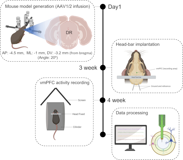
Figure 1: Timeline diagram of the experimental design. Generation of the Parkinson's disease animal model based on the overexpression of the A53T mutant form of human α-synuclein (h-α-Syn) in the dorsal raphe nucleus (DR). Three weeks after viral injection, animals undergo a second surgical procedure to implant a head-bar onto the skull. Following habituation, this allows head-fixation for the recording of neural activity in the prelimbic (PL) and infralimbic (IL) cortices, performed four weeks after model generation. After recording, the raw data is processed for spike sorting and signal cleaning using Kilosort4 and Phy2. Please click here to view a larger version of this figure.
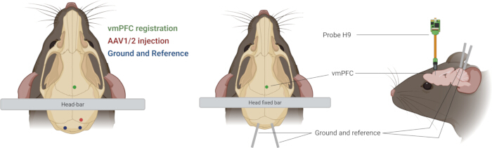
Figure 2: Injection and implantation coordinates used in the stereotaxic surgical procedure. Schematic representation of the craniotomy showing the implantation of a 3D-printed head-bar for positioning in the activity wheel using a head-fixation system, along with a ground screw, a reference electrode, and a hole for introduce multichannel probe and record neuronal activity in the prelimbic (PL) and infralimbic (IL) cortices. Please click here to view a larger version of this figure.
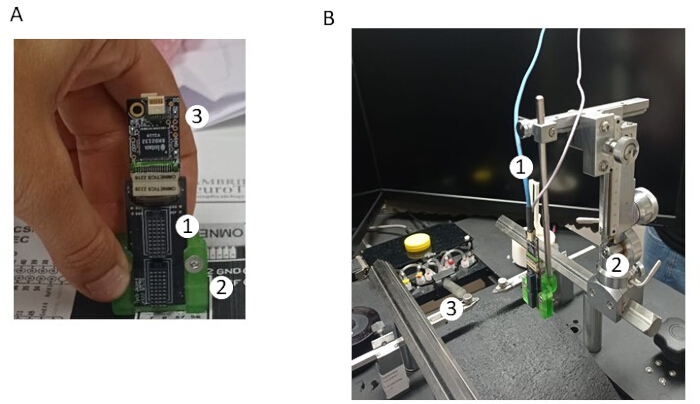
Figure 3: Components and setup of electrophysiology recording system. (A) Probe adapter/connector (1) mounted on the stereotaxic arm adapter (2). Two headstage adapters (3) are connected to the probe adapter. (B) SPI cables (1) from the Open Ephys acquisition system connected to the headstage adapters. Probe connected to the adapters and placed in the stereotaxic (2) of the head-fix system (3). Please click here to view a larger version of this figure.
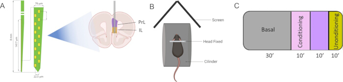
Figure 4: Equipment and experimental design. (A) Probe characteristics and localization of the recording sites within the PL and IL cortices. (B) Virtual reality system and activity wheel. (C) Diagram of the experimental paradigm. Please click here to view a larger version of this figure.
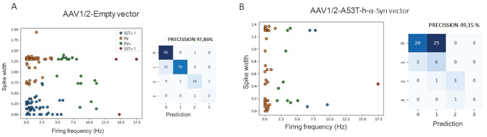
Figure 5: Overexpression of the mutant A53T form of human α-Synuclein (h-α-Syn) in serotonergic (5-HT) neurons of the dorsal raphe nucleus (DR) induces functional alterations in the ventromedial prefrontal cortex (vmPFC)-DR circuit. (A) Scatter plot illustrating spike width versus firing frequency and the corresponding confusion matrix for neuronal classification in control animals. (B) Equivalent analysis in AAV1/2-A53T-h-α-Syn-injected mice showing disrupted separability of neuronal populations and altered firing dynamics within the circuit. Please click here to view a larger version of this figure.
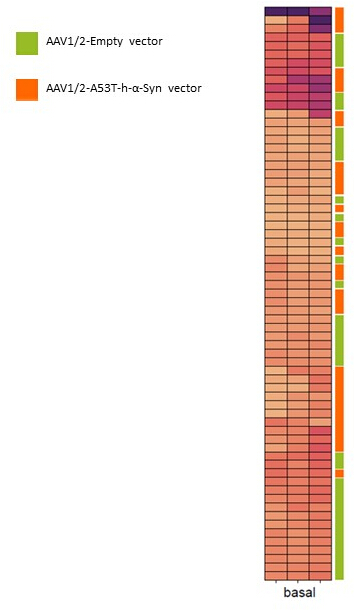
Figure 6: Heatmap of mean firing rates recorded in control and A53T-h-α-synuclein. Each row represents a single neuron and each column a consecutive temporal window during the basal recording period. Color intensity indicates the mean firing rate, and neurons are organized according to unsupervised clustering. Color bars (green) on the right denote the resulting clusters for each experimental condition. Please click here to view a larger version of this figure.
Discussion
This protocol presents a robust methodology for in vivo electrophysiological assessment of PL and IL cortices in awake, head-fixed mice, utilizing a virtual reality corridor system. The significance of this method lies in its ability to investigate the vmPFC-DR circuit directly in awake, behaving mice within a PD model. Even if head-fixation restricts animal movements and can be stressful, it offers precise stimulus control, recording stability, and is appropriate for studying perception and cognitive processes, though it may not reflect natural behavior24,25. This approach offers significant advantages over ex vivo or anesthetized preparations by allowing for the study of neural activity in a more physiologically relevant state, particularly during aversive conditioning, which models stress-related situations32,33. Indeed, during general anesthesia, behavioral state and global brain activity, in particular cortical responses, are profoundly altered34,35,36. Indeed, during general anesthesia, behavioral state and global brain activity, in particular cortical responses, are profoundly altered34,35,36. Existing methods often rely on post-mortem analysis or in vitro techniques, which cannot capture the dynamic neural responses to behavioral paradigms32,37,38.
This protocol provides a powerful tool to understand the neurobiology of neuropsychiatric symptoms in PD, especially depression and anxiety, which are highly prevalent and impact patient quality of life but are often underdiagnosed and inadequately treated. By enabling detailed analysis of vmPFC neuronal firing patterns, including pyramidal neurons, SST+ and PV+ interneurons, this method can reveal circuit-level dysfunctions underlying these non-motor symptoms. Ultimately, this detailed understanding is essential for patient stratification and developing more personalized therapeutic approaches for PD. The findings from this protocol, such as the observed disruption of vmPFC neuronal firing patterns due to h-α-Syn overexpression in the DR, directly support its utility in advancing our knowledge of PD-related mood disorders.
There are some limitations and points to consider regarding the technique. The use of head-fixation, while necessary for stable electrophysiological recordings, restricts the mouse's natural movement25. This, combined with the virtual reality environment, may not fully replicate the complexity of natural behaviors and could potentially influence stress levels and neural activity in ways that differ from a freely moving context. To allow for more natural behavior, wireless electrophysiology technologies could be employed. These systems enable the recording of neural activity in freely moving animals within their home cages or more complex behavioral environments. While this can introduce motion artifacts into the signal, advances in hardware and analysis software can help correct for them. This would eliminate the stress associated with head-fixation39 and allow for the exploration of a broader behavioral repertoire. Additionally, the technique is highly invasive as it requires viral injections, craniotomy, and probe insertion directly into the brain tissue. These interventions can lead to tissue damage, inflammation, and gliosis, which might alter the physiological function of the circuits under investigation. To reduce tissue damage, smaller and more flexible probes, such as ultrafine polymer probes that conform better to the brain tissue, could be used. Surgical techniques can also be refined to minimize inflammation40,41. Alternatively, less invasive functional imaging techniques like two-photon calcium imaging could be employed42,43. This allows for the visualization of activity in hundreds of neurons simultaneously at cellular resolution in awake animals, although it is generally limited to more superficial cortical layers. Furthermore, the entire experimental process, including surgery, recovery, handling, and head-fixation, is inherently stressful for the animals. While a habituation period is included, it may not eliminate all stress-induced effects, which could act as a confounding variable when interpreting the results of the aversive conditioning paradigm. To reduce animal stress, the habituation protocol could be extended and refined. Positive reinforcement training could be incorporated, where mice receive a small reward for voluntarily entering and remaining in the head-fixation apparatus. Monitoring corticosterone levels (a stress hormone) could serve as an objective biomarker to ensure that stress is effectively minimized before experimental recordings begin.
Finally, while the protocol uses established tools for spike sorting, the process requires significant manual curation and subjective decisions to distinguish true neuronal spikes from noise. Furthermore, the classification of neuronal subtypes (e.g., pyramidal, PV+, SST+) is based on electrophysiological properties (firing patterns) and not on direct molecular identification, which represents an indirect method of cell-type classification. For a more precise identification of neuron types, electrophysiological recordings could be combined with optogenetic techniques. This would involve using transgenic mice in which specific cell types (e.g., PV+ or SST+ interneurons) express light-sensitive ion channels. By applying a pulse of light during the recording, these neurons can be selectively activated, allowing for their unambiguous identification in the electrophysiological data. Post-experiment histology with specific markers can then confirm the identity of the recorded neural populations.
Finally, it should be emphasized that this technique comprises a series of critical steps in which strict adherence to the methodology is required in order to ensure successful recordings and maximize the number of neurons recorded. Among the most relevant are: the proper infection of the model to achieve α-Syn overexpression and the careful manipulation of the probe, given its fragility and susceptibility to breakage. In this regard, the probe must be introduced with precision and lowered at the prescribed rate to prevent structural damage and to ensure optimal signal acquisition. Another highly critical step is the stabilization of the response, during which tissue adaptation may result in the loss of activity in certain neurons. Therefore, it is recommended to allow a stabilization period of 90-120 min to ensure reliable signal acquisition.
Disclosures
The authors declare no competing interests.
Acknowledgements
This work has been funded by the 2023 BBRF Young Investigator Grant 31547 (MSA), MCIU/AEI/FEDER EU grant PID2022-141700OB-I00, MCIN/AEI/10. 13039/501100011033 (AB), AGAUR 2021-SGR-01358, Government of Catalonia (AB), and PID2022-141733NB-I00 (VT). We would also like to thank the Spanish Network for Research on Stress, MCIN/AEI/10.13039/501100011033 and CB/07/09/0034 Centre for Biomedical Research in Mental Health Network (CIBERSAM). MSA has a Margarita Salas Scholarship (MS21-132) from the University of Valencia (reclassification of the Spanish University System of the Ministry of Universities of the Government of Spain, financed by the European Union, Next Generation EU).
Materials
| Name | Company | Catalog Number | Comments |
|---|---|---|---|
| AAV1/2-CMV/CBA-Human A53T aSyn-WPRE-BGH-polyA | Charles River | GD1001-RV | Human A53T aSyn |
| AAV1/2-CMV/CBA-Null/Empty-WPRE-BGH-polyA | Charles River | GD1004-RV | Empty Vector Control |
| ADPT A64-Om32x2 | Cambridge NeuroTech | Omnetics 2228 | Adapter |
| bi-component surgical silicone | World Precision Instruments | KWIK-SIL | |
| Butorphanol | Torbugesic vet / cymit química | 67753-31-5 | |
| DuraLay | Reliance Dental Manufacturing / Kalma | 43640 | dental cement |
| H9 Multichannel probe | Cambridge NeuroTech | ASSY-77 H9 | |
| EMLA cream (Lidocaine / pricolaine) | Lirios Vañó Sempere Pharmacy | 679290 | |
| microinjector | World Precision Instrument | KDS-310-PLUS | |
| MSS Isoflurane Vaporizer | Medical Supplies and Services, UK | MSSVAP02 | |
| Open Ephys Acquisition Board | Open Ephys | OEPS-9029 | Acquisition board; The Open Ephys Acquisition Board can stream up to 512 channels of neural data to a computer via USB |
| C&B MetaBond | Parkell | S380-RD | Quick adhesive cement |
| Pentobarbital | Dolethal Vetoquinol /Merck Life Science, S.L.U. | P-010-1ML | Barbiturate |
| RHD 32-channel headstage | Intan Technologies | #C3314 | head-stage adapter |
| Siccafluid | Thea S.A. Laboratories / Lirios Vañó Sempere Pharmacy | 6515167 | Ophthalmic Gel |
| stereotaxic apparatus | Stoelting | 51730U | |
| stereotaxic frame | Narishige | SR-6N/SM-15R | |
| Terg-a-zyme | Sigma Aldrich | Z273287 | Probe cleaning solution |
References
- Cooney, J. W., Stacy, M. Neuropsychiatric issues in Parkinson's disease. Curr Neurol Neurosci Rep. 16 (5), 49(2016).
- Schapira, A. H. V., Chaudhuri, K. R., Jenner, P. Non-motor features of Parkinson disease. Nat Rev Neurosci. 18 (7), 435-450 (2017).
- Ahmad, M. H., Rizvi, M. A., Ali, M., Mondal, A. C. Neurobiology of depression in Parkinson's disease: Insights into epidemiology, molecular mechanisms and treatment strategies. Ageing Res Rev. 85 (1), 101840(2023).
- Absalyamova, M., Traktirov, D., Burdinskaya, V., Artemova, V., Muruzheva, Z., Karpenko, M. Molecular basis of the development of Parkinson's disease. Neuroscience. 565 (1), 292-300 (2024).
- Crispino, P., et al. Gender differences and quality of life in Parkinson's disease. Int J Environ Res Public Health. 18 (1), 198(2020).
- Chuquilín-Arista, F., Álvarez-Avellón, T., Menéndez-González, M. Prevalence of depression and anxiety in Parkinson's disease and impact on quality of life:A community-based study in Spain. J Geriatr Psychiatry Neurol. 33 (1), 207-213 (2020).
- Greffard, S., et al. Motor score of the Unified Parkinson Disease Rating Scale as a good predictor of Lewy body-associated neuronal loss in the substantia nigra. Arch Neurol. 63 (1), 584-588 (2006).
- Mamelak, M. Parkinson's disease, the dopaminergic neuron and gammahydroxybutyrate. Neurol Ther. 7 (1), 5-11 (2018).
- Wakabayashi, K., Tanji, K., Mori, F., Takahashi, H. The Lewy body in Parkinson's disease:Molecules implicated in the formation and degradation of alpha-synuclein aggregates. Neuropathology. 27 (5), 494-506 (2007).
- Sezgin, M., Bilgic, B., Tinaz, S., Emre, M. Parkinson's disease dementia and Lewy body disease. Semin Neurol. 39 (2), 274-282 (2019).
- Jellinger, K. A. The pathobiological basis of depression in Parkinson disease:Challenges and outlooks. J Neural Transm. 129 (12), 1397-1418 (2022).
- Wang, J., Sun, J., Gao, L., Zhang, D., Chen, L., Wu, T. Common and unique dysconnectivity profiles of dorsal and median raphe in Parkinson's disease. Hum Brain Mapp. 44 (1), 1070-1078 (2023).
- Miquel-Rio, L., Sarriés-Serrano, U., Pavia-Collado, R., Meana, J. J., Bortolozzi, A. The role of α-synuclein in the regulation of serotonin system: Physiological and pathological features. Biomedicines. 11 (2), 541(2023).
- Halliday, G. M., Blumbergs, P. C., Cotton, R. G., Blessing, W. W., Geffen, L. B. Loss of brainstem serotonin- and substance P-containing neurons in Parkinson's disease. Brain Res. 510 (1), 104-107 (1990).
- Ren, J., et al. Single-cell transcriptomes and whole-brain projections of serotonin neurons in the mouse dorsal and median raphe nuclei. Elife. 8 (1), e49424(2019).
- Huang, K. W., et al. Molecular and anatomical organization of the dorsal raphe nucleus. Elife. 8 (1), e46464(2018).
- Hiser, J., Koenigs, M. The multifaceted role of the ventromedial prefrontal cortex in emotion, decision making, social cognition, and psychopathology. Biol Psychiatry. 83 (1), 638-647 (2018).
- Alexander, L., Wood, C. M., Roberts, A. C. The ventromedial prefrontal cortex and emotion regulation: Lost in translation. J Physiol. 601 (1), 37-50 (2023).
- Miquel-Rio, L., et al. Human α-synuclein overexpression in mouse serotonin neurons triggers a depressive-like phenotype. Rescue by oligonucleotide therapy. Transl Psychiatry. 12 (1), 79(2022).
- Sarries-Serrano, U., Miquel-Rio, L., Surroca-Lopez, A., Paz, V., Meana, J. J., Bortolozzi, A. Sex-based modulation of endoplasmic reticulum stress and antidepressant response in a mouse model of α-synucleinopathy. Neuroscience Appl. 2 (1), 101028(2023).
- Miquel-Rio, L., et al. Early synaptic changes and reduced brain connectivity in PD-like mice with depressive phenotype. NPJ Parkinsons Dis. 11 (1), 242(2025).
- Vidal-Gonzalez, I., Vidal-Gonzalez, B., Rauch, S. L., Quirk, G. J. Microstimulation reveals opposing influences of prelimbic and infralimbic cortex on the expression of conditioned fear. Learn Mem. 13 (6), 728-733 (2006).
- Giustino, T. F., Maren, S. The role of the medial prefrontal cortex in the conditioning and extinction of fear. Front Behav Neurosci. 9 (1), 298(2015).
- Guo, Z. V., et al. Procedures for behavioral experiments in head-fixed mice. PLoS One. 9 (2), e88678(2014).
- Nasr, A., Dominiak, S. E., Sehara, K., Nashaat, M. A., Sachdev, R. N. S., Larkum, M. E. Efficient training approaches for optimizing behavioral performance and reducing head fixation time. PLoS One. 17 (11), e0276531(2022).
- Bjerre, A. S., Palmer, L. M. Probing cortical activity during head-fixed behavior. Front Mol Neurosci. 13 (1), 30(2020).
- Alarcón-Arís, D., et al. Anti-α-synuclein ASO delivered to monoamine neurons prevents α-synuclein accumulation in a Parkinson's disease-like mouse model and in monkeys. EBioMedicine. 59 (1), 102944(2020).
- Pavia-Collado, R., et al. Intracerebral administration of a ligand-ASO conjugate selectively reduces α-synuclein accumulation in monoamine neurons of double mutant human A30PA53Tα-synuclein transgenic mice. Int J Mol Sci. 22 (6), 2939(2021).
- McLean, A. C., Valenzuela, N., Fai, S., Bennett, S. A. Performing vaginal lavage, crystal violet staining, and vaginal cytological evaluation for mouse estrous cycle staging identification. J Vis Exp. (67), e4389(2012).
- Byers, S. L., Wiles, M. V., Dunn, S. L., Taft, R. A. Mouse estrous cycle identification tool and images. PLoS One. 7 (4), e35538(2012).
- Pachitariu, M., Sridhar, S., Pennington, J., Stringer, C. Spike sorting with Kilosort4. Nat Methods. 21 (5), 914-921 (2024).
- Opitz, A., Falchier, A., Linn, G. S., Milham, M. P., Schroeder, C. E. Limitations of ex vivo measurements for in vivo neuroscience. Proc Natl Acad Sci U S A. 114 (20), 5243-5246 (2017).
- Sorrenti, V., Cecchetto, C., Maschietto, M., Fortinguerra, S., Buriani, A., Vassanelli, S. Understanding the effects of anesthesia on cortical electrophysiological recordings: A scoping review. Int J Mol Sci. 22 (3), 1286(2021).
- Brown, E. N., Purdon, P. L., Van Dort, C. J. General anesthesia and altered states of arousal :A systems neuroscience analysis. Annu Rev Neurosci. 34 (1), 601-628 (2011).
- Sellers, K. K., Bennett, D. V., Hutt, A., Williams, J. H., Fröhlich, F. Awake vs. anesthetized: Layer-specific sensory processing in visual cortex and functional connectivity between cortical areas. J Neurophysiol. 113 (10), 3798-3815 (2015).
- Qiu, J., et al. The volatile anesthetic isoflurane differentially inhibits voltage-gated sodium channel currents between pyramidal and parvalbumin neurons in the prefrontal cortex. Front Neural Circuits. 17 (1), 1185095(2023).
- Windels, F. Neuronal activity: From in vitro preparation to behaving animals. Mol Neurobiol. 34 (1), 1-26 (2006).
- Belle, A. M., et al. Evaluation of in vitro neuronal platforms as surrogates for in vivo whole brain systems. Sci Rep. 8 (1), 10820(2018).
- Juczewski, K., et al. Stress and behavioral correlates in the head-fixed method:Stress measurements, habituation dynamics, locomotion, and motor-skill learning in mice. Sci Rep. 10 (1), 12245(2020).
- Chen, P. C., Young, C. G., Schaffer, C. B., Lal, A. Ultrasonically actuated neural probes for reduced trauma and inflammation in mouse brain. Microsyst Nanoeng. 8 (1), 117(2022).
- Wang, S., et al. Mechanically adaptive and deployable intracortical probes enable long-term neural electrophysiological recordings. Proc Natl Acad Sci U S A. 121 (40), e2403380121(2024).
- Andermann, M. L., et al. Chronic cellular imaging of entire cortical columns in awake mice using microprisms. Neuron. 80 (4), 900-913 (2013).
- Kondo, M., Kobayashi, K., Ohkura, M., Nakai, J., Matsuzaki, M. Two-photon calcium imaging of the medial prefrontal cortex and hippocampus without cortical invasion. Elife. 6 (1), e26839(2017).