Case Report

Allogeneic HSCT For Pediatric Extranodal NK/T-Cell Lymphoma Transformed From Chronic Active Epstein–Barr Virus Infection: A Case Report

DOI:

10.3791/69867

⸱

June 16th, 2026

In This Article

Summary

Loading...
$$\rightleftharpoonup{xx}$$ $$\longleftharp{xx}$$, $$\longrightharp{xx}$$,

Allo-HSCT achieved complete remission and full donor chimerism in a child with CAEBV-transformed ENKTL. No EBV reactivation, GVHD, or complications occurred during the 1-year follow-up. Adjuvant microecological preparation helped reduce transplant-related risks.

Abstract

Loading...
$$\rightleftharpoonup{xx}$$ $$\longleftharp{xx}$$, $$\longrightharp{xx}$$,

This study aimed to explore the clinical characteristics, diagnostic criteria, therapeutic regimens, and prognostic features of pediatric extranodal natural killer/T-cell lymphoma (ENKTL) transformed from chronic active Epstein-Barr virus infection (CAEBV), to provide evidence-based references for standardized clinical diagnosis and treatment of these refractory diseases. A pediatric patient with CAEBV-transformed ENKTL admitted to Nanshan Hospital Affiliated to Shenzhen University was retrospectively enrolled in this study, who received allogeneic hematopoietic stem cell transplantation (allo-HSCT). The patient achieved complete remission after multiple cycles of preoperative chemotherapy. Triple probiotic preparations containing Bifidobacterium, Bacillus licheniformis, and Lactobacillus were administered throughout the preconditioning phase and post-transplant period. Persistent complete remission was achieved after transplantation with full donor chimerism of 100%. Long-term follow-up over 1 year post-transplantation showed no disease recurrence, EBV reactivation, severe infectious complications, or acute and chronic transplantation-related complications. This case study confirmed that allo-HSCT is safe and effective for the treatment of pediatric CAEBV-transformed ENKTL, with a significantly superior long-term prognosis compared with chemotherapy alone. Peri-transplant adjuvant application of microecological preparations may reduce the risks of respiratory tract infection and graft-versus-host disease in children. Given that this is a single-case study with limited follow-up duration, there are certain limitations in the clinical generalization of the conclusions.

Introduction

Loading...
$$\rightleftharpoonup{xx}$$ $$\longleftharp{xx}$$, $$\longrightharp{xx}$$,

Epstein-Barr virus (EBV) is a double-stranded DNA herpesvirus with a high infection rate, affecting more than 90% of the global population. Following EBV infection, the virus may remain latent in the host or enter a lytic phase. EBV can lead to a wide range of diseases, varying in severity from mild infectious mononucleosis to more severe conditions such as chronic active EBV infection (CAEBV), EBV-related malignancies, lymphoproliferative disorders, and autoimmune diseases1,2. CAEBV is a lymphoproliferative disorder caused by EBV infection in T or natural killer (NK) cells, characterized by both systemic inflammatory and neoplastic features. This condition predominantly affects children and adolescents, with adults typically experiencing a worse prognosis. Clinical manifestations include recurrent fever, lymphadenopathy, hepatosplenomegaly, EBV-associated hepatitis, liver dysfunction, and thrombocytopenia3,4. Unlike some viral infections, CAEBV is not self-limiting; without treatment, it can progress to hemophagocytic lymphohistiocytosis (HLH), leukemia, lymphoma, and multiple organ failure, making it both aggressive and fatal. In 2016, the World Health Organization classified CAEBV as an EBV-associated T or NK cell lymphoproliferative disease5.

Extranodal NK/T-cell lymphoma (ENKTL) is another EBV-associated extranodal malignancy, primarily originating from NK cells and, less commonly, from T cells. It is characterized by local tumor necrosis, vascular destruction, a cytotoxic phenotype, and EBV infection. ENKTL most commonly occurs in the nasal cavity, while extranasal lesions (such as skin, subcutaneous tissue, and multiple organ involvement) tend to be highly malignant and invasive, with rapid progression, a poor prognosis, and a high recurrence rate6. The 5-year overall survival rate for ENKTL with extranasal lesions is approximately 34%, and the median survival time for patients with extracutaneous involvement is only 4 months6. ENKTL typically shows poor responsiveness to anthracycline-based chemotherapy, and due to the high rate of relapse following chemotherapy, allo-HSCT remains the only curative option for patients with advanced or relapsed/refractory ENKTL6,7.

CAEBV transformed into ENKTL is an extremely rare clinical scenario with a worse prognosis than either disease alone, as it combines the persistent inflammatory damage of CAEBV and the aggressive tumorigenic characteristics of ENKTL. Currently, there is no standardized treatment protocol for this condition. Existing therapeutic options include chemotherapy, targeted therapy, and allo-HSCT. Chemotherapy alone often fails to achieve long-term disease control due to the drug resistance of ENKTL cells and the persistent EBV infection in CAEBV8,9. Targeted therapies such as HDAC inhibitors (cedamide), JAK/STAT pathway inhibitors (ruxolitinib), and PD-1 monoclonal antibodies have shown certain efficacy in small-scale clinical trials, but their long-term therapeutic effects and safety require further verification10,11. In contrast, allo-HSCT can achieve radical cure of tumors and EBV infection through immune reconstitution, making it the most effective treatment option for eligible patients5,7. However, post-transplant infections and GVHD remain significant challenges affecting prognosis5,12.

Research and expert consensus in China suggest that microecological interventions can reduce respiratory tract infections in tumor patients, and fecal microbiota transplantation is recommended as a second-line therapy for acute gastrointestinal GVHD following allo-HSCT12,13. Pretreatment prior to allo-HSCT (including high-dose chemotherapy) can disrupt the patient's intestinal microecological balance, damage the intestinal epithelium, and compromise the intestinal barrier. The use of broad-spectrum antibiotics further alters the intestinal flora, increasing the risk of infections and GVHD14,15,16. Microecological preparations (probiotics) can regulate intestinal flora balance, enhance mucosal immune barriers, and inhibit donor T cell activation, thereby reducing the risks of infection and GVHD12,13,14.

Case presentation:

We present a case of a 15-year-old female patient with ENKTL transformed from CAEBV who underwent allo-HSCT combined with adjuvant triple microecological preparations. This case report details the clinical diagnosis, treatment process, and follow-up outcomes, aiming to provide practical clinical references for the management of similar rare cases.

Protocol

Loading...
$$\rightleftharpoonup{xx}$$ $$\longleftharp{xx}$$, $$\longrightharp{xx}$$,

This study was approved by the Ethics Committee of Affiliated Nanshan Hospital of Shenzhen University (No.: ky-2026-0570701; Date: May 9, 2026). Written informed consent was signed and obtained from the patient’s legal guardians for the publication of this case report and related clinical data. All procedures followed the Declaration of Helsinki. The reagents and the equipment used are listed in the Table of Materials.

Inclusion and exclusion criteria
Patients included in the present study were aged ≤18 years and had a definitive diagnosis of pathologically confirmed EBER-positive extranodal natural killer/T-cell lymphoma (ENKTL) complicated by chronic active Epstein–Barr virus (CAEBV) infection, in accordance with standard diagnostic criteria. All enrolled patients had no contraindications to chemotherapy or allogeneic hematopoietic stem cell transplantation (allo-HSCT) and had an available 10/10 human leukocyte antigen (HLA)-matched donor, with voluntary written informed consent obtained from their legal guardians for study participation and long-term follow-up. Patients were excluded if they presented with concomitant malignant tumors or severe autoimmune diseases, had a documented hypersensitivity to any chemotherapeutic agents or microecological preparations used in the treatment protocol, were pregnant or lactating, or were unable to complete scheduled follow-up due to geographical constraints or other unavoidable factors.

1. Diagnostic methods

  1. Clinical symptom collection
    1. The patient's main symptoms (fever, sore throat, cough, chest pain, etc.), onset time, duration, and changes during treatment were recorded.
  2. Laboratory examinations
    1. Routine blood tests: Peripheral blood smear analysis was performed to detect white blood cell count, lymphocyte ratio, platelet count, etc., using a hematology analyzer.
    2. EBV-DNA detection: Plasma and blood cell EBV-DNA copy numbers were detected using real-time PCR; if blood cell EBV-DNA was positive, lymphocyte sorting was performed to identify the infected cell subset.
    3. Immunological indicators: Soluble CD25 receptor level and NK cell activity were detected using flow cytometry.
    4. Biochemical examinations: Liver function, kidney function, coagulation function, etc., were detected using an automatic biochemical analyzer.
  3. Imaging studies
    1. PET-CT scans were performed to evaluate the extent of lesion involvement, treatment response, and recurrence during follow-up.
  4. Pathological and immunohistochemical examinations
    1. A biopsy of the scalp mass was performed, pathological sections were prepared, and hematoxylin-eosin staining and immunohistochemical staining (detection of CK, CD3, CD5, CD10, CD4, CD8, CD2, CD7, CD43, CD20, CD56, Granzyme B, TIA-1, Ki-67, CD79a, CD34, CD117, TdT, EBER, etc.) were conducted to confirm the pathological type and EBV infection status.
  5. Bone marrow examination
    1. Bone marrow smear, pathological examination, and flow cytometry were performed to evaluate whether the bone marrow was involved.

2. Treatment procedure

  1. Chemotherapy regimens
    1. VDLP regimen: Vincristine 2 mg was administered on days 1, 8, 15, and 22; doxorubicin liposome 20 mg was administered on days 1, 8, 15, and 22; pegaspargase was administered on days 2 and 16; prednisone 80 mg was administered on days 1–21.
    2. MEAD regimen: Mitoxantrone liposome 30 mg was administered on day 1; etoposide 100 mg was administered on days 2–4 and 15–16; dexamethasone 10 mg was administered on days 2–4 and 15–16; cytarabine 100 mg was administered on days 2–5.
    3. Gemox + pegaspargase regimen: Gemcitabine 1.5 g was administered on days 1 and 8; pegaspargase 3750 IU was administered on day 2; oxaliplatin 150 mg was administered on day 1.
    4. HLH04 regimen (short-term): Etoposide 100 mg was administered on days 1–2; dexamethasone 10 mg was administered on days 1–7.
    5. Consolidation chemotherapy: Gemcitabine 1.5 g was administered on day 1; oxaliplatin 150 mg was administered on day 1; pegaspargase 3750 IU was administered on day 2; VP-16 (etoposide) 100 mg was administered on days 1–2; dexamethasone 10 mg was administered on days 1–7; intrathecal injection of cytarabine and methotrexate via lumbar puncture was performed to prevent central nervous system involvement.
  2. Stem cell mobilization and collection
    1. The donor (patient's 25-year-old sister, 10/10 HLA-matched, blood type A+, EBV-seronegative: EBV-VCA-IgG(−), IgM(−), EA-IgG(−), NA-IgG(−); EBV-DNA undetectable) was subcutaneously injected with granulocyte colony-stimulating factor 5 µg/kg twice daily for 4 days to mobilize stem cells.
    2. On the morning of the 5th day, 200 mL of peripheral blood stem cell suspension was collected using a blood cell separator; three 10 mL bags of stem cells were reserved for backup.

3. Pre-transplant preparation

  1. One week before conditioning therapy, the patient began taking oral levofloxacin to clear intestinal bacteria.
  2. NOTE: Levofloxacin is indicated for adults aged 18 years and above; the patient was over 16 years old before transplantation, with height and weight meeting adult standards, classified under the adult hematological malignancy category; the guardian was fully informed of potential risks and signed an informed consent form.
  3. Pre-transplantation assessments, including organ function tests, infection screenings, and physical fitness evaluations, were performed.
  4. After a medicated bath, the patient was transferred into a Class 100 laminar flow ward for strict aseptic isolation.

4. Transplant conditioning regimen

  1. A mitoxantrone liposome-enhanced BUCY protocol was adopted: mitoxantrone liposome 40 mg was administered on day -8; busulfan 3.2 mg/kg was administered on days -7, -6, and -5 (busulfan blood concentration was monitored, and the therapeutic range was maintained at 600–900 ng/mL); cyclophosphamide 50 mg/kg was administered on days -4 and -3; antithymocyte globulin 1.5 mg/kg was administered on days -5, -4, and -3.

5. Stem cell infusion

  1. A total of 170 mL of donor hematopoietic stem cell suspension (CD34+ cell count: 2.94 × 10⁶/kg, nucleated cell count: 5.7 × 10⁸/kg) was transfused into the patient at an infusion rate of 5–10 mL/min.

6. Post-transplant supportive care and prophylaxis

  1. Anti-infection prophylaxis: Fluconazole was administered for fungal prophylaxis, valacyclovir was administered for viral prevention, and trimethoprim-sulfamethoxazole was administered for Pneumocystis jiroveci pneumonia; compound chlorhexidine was used as a mouthwash, and levofloxacin eye drops were used for local prophylaxis.
  2. Organ protection: Antiemetics, hydration, and alkalinization were administered during chemotherapy to protect liver and kidney function; sodium valproate and phenytoin were administered alongside busulfan to prevent central nervous system toxicity.
  3. GVHD prophylaxis: Cyclosporine combined with short-course methotrexate was used.
  4. Hematopoietic recovery promotion: Granulocyte colony-stimulating factor and thrombopoietin were administered post-transplantation.
  5. Microecological preparation administration: A triple probiotic regimen of Bifidobacterium, Bacillus licheniformis, and Lactobacillus was administered prior to conditioning therapy and continued during the post-transplant period, 3 times a day, 1 sachet each time.

7. Post-transplant monitoring and follow-up

  1. Hematopoietic reconstitution monitoring: Routine blood tests were performed daily until neutrophil and platelet engraftment (neutrophil count ≥0.5 × 10⁹/L for 3 consecutive days, platelet count ≥20 × 109/L without transfusion support).
  2. Chimerism monitoring: PCR-based DNA fingerprinting was used to detect the chimerism rate at 1, 2, 3, 6, and 12 months post-transplant.
  3. EBV-DNA monitoring: Plasma and blood cell EBV-DNA copy numbers were detected monthly; if blood cell EBV-DNA was positive, lymphocyte sorting was performed to identify the infected cell subset.
  4. Imaging follow-up: PET-CT scans were performed at 3 months and 12 months post-transplant to evaluate treatment response.
  5. Complications monitoring: The occurrence of acute and chronic GVHD, infections, and other adverse events was recorded.

Results

Loading...
$$\rightleftharpoonup{xx}$$ $$\longleftharp{xx}$$, $$\longrightharp{xx}$$,

General information

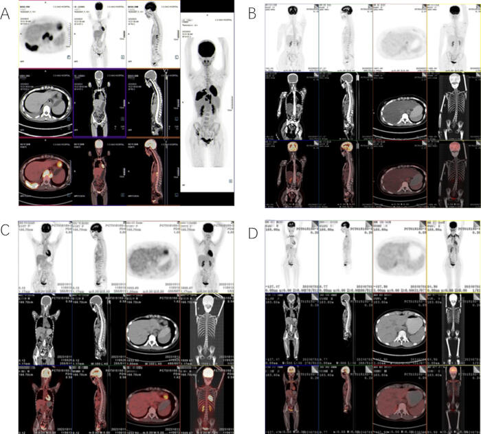
The patient, a 15-year-old female, presented with a fever of unknown origin in October 2022. The fever episodes primarily occurred at night, with a maximum temperature of 38.3 °C, and were accompanied by sore throat, chills, and fatigue. Initial blood tests revealed a mildly decreased white blood cell count, normal C-reactive protein levels, elevated lymphocytes, and an EBV-DNA level of 1.50 × 10³ copies/mL, leading to a diagnosis of infectious mononucleosis. After receiving symptomatic and supportive treatment, her symptoms improved. However, three months later, she experienced recurrent fever, with temperatures peaking at 38.5 °C, along with a dry cough and the appearance of a 1 cm × 1 cm mass in the left frontal and temporal regions, as well as multiple enlarged bilateral cervical lymph nodes. Repeat EBV-DNA testing showed a level of 1.99 × 10³ copies/mL, and a peripheral blood smear revealed 38% lymphocytes, consistent with a diagnosis of CAEBV (meeting diagnostic criteria: persistent infectious symptoms for more than 6 months, sustained elevation of EBV-DNA). Despite treatment with ganciclovir, antipyretics, and fluid replacement, her symptoms persisted. By February 2023, the patient experienced persistent high fever, with temperatures consistently exceeding 40°C, along with right-sided chest pain and chest tightness. Positron emission tomography-computed tomography (PET-CT) revealed thickening of the subcutaneous soft tissue with increased metabolic activity in the left frontal, temporal, and parietal regions. Additionally, diffuse thickening of the right pleura with increased metabolic activity, multiple hypermetabolic and enlarged lymph nodes deep to the bilateral diaphragms, and hypermetabolic foci in the right costal arch and left pubic bone were observed, suggesting a high likelihood of multisystem involvement by a malignant lymphohematopoietic tumor (Figure 1A).

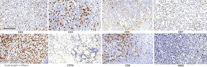
A biopsy of the scalp mass revealed pathology consistent with ENKTL. Most of the biopsy tissue showed necrotic changes, including necrosis of the striated muscle tissue. Atypical cells with large, dark-staining nuclei were observed infiltrating the areas surrounding residual blood vessels, with abundant nuclear debris in the background. Immunohistochemistry findings were as follows: CK (-), CD3 (+), CD5 (-), CD10 (-), CD4 (-), CD8 (+), CD2 (+), CD7 (-), CD43 (+), CD20 (-), CD56 (+), Granzyme B (+), TIA-1 (+), Ki-67 (high proliferation index, ~90%), CD79a (-), CD34 (-), CD117 (-), TdT (-)and EBER (+) (Figure 2). Molecular pathology confirmed Epstein-Barr virus-encoded RNA positivity. Bone marrow smear, pathology, and flow cytometry findings were unremarkable. The patient had lymphadenopathy in the early stage, but no lymph node biopsy was performed, making it impossible to determine the initial onset time of ENKTL. Final Diagnosis: ENKTL transformed from CAEBV.

Chemotherapy cycle

The patient began chemotherapy with the VDLP regimen on March 8, 2023, which included vincristine (2 mg on days 1, 8, 15, and 22), doxorubicin liposome (20 mg on days 1, 8, 15, and 22), pegaspargase (on days 2 and 16), and prednisone (80 mg on days 1–21). Concurrently, symptomatic and supportive treatments were enhanced. Following chemotherapy, the patient developed neutropenia with fever and abnormal coagulation function. These complications were managed with anti-infective therapy using cefoperazone-sulbactam, meropenem, and caspofungin. Transfusions of blood products, including cryoprecipitate, plasma, and platelets, were also administered. After the first cycle of chemotherapy, there was a slight shrinkage of the patient's lymph nodes, but fever recurred intermittently. The patient fulfilled 6 out of 8 diagnostic criteria for HLH according to the HLH-2004 guidelines: (1) persistent fever; (2) elevated soluble CD25 (4213 U/mL); (3) decreased NK cell activity (13.8%); (4) serum ferritin 742.5 ng/mL (normal: 13–150 ng/mL); (5) splenomegaly; (6) hemophagocytosis in bone marrow smears, meeting the HLH diagnostic criteria17. On April 7, 2023, the chemotherapy regimen was switched to MEAD (reason: The MEAD regimen contains more potent antitumor and anti-inflammatory agents such as mitoxantrone liposome and etoposide, which are more suitable for lymphoma complicated by HLH), comprising mitoxantrone liposome (30 mg on day 1), etoposide (100 mg on days 2–4 and 15–16), dexamethasone (10 mg on days 2–4 and 15–16), and cytarabine (100 mg on days 2–5). On April 10, 2023, the patient’s fever improved (max 37.5 °C), indicating an initial response. Fever recurred on April 17, 2023, complicated by infection. Immune checkpoint inhibitors were added on April 26, 2023, after which the fever resolved completely. On May 4, 2023, the patient received the Gemox chemotherapy regimen combined with pegaspargase  (reason: The Gemox regimen is a first-line chemotherapy regimen for ENKTL, and the addition of pegaspargase can further enhance antitumor efficacy to consolidate CR). This included gemcitabine (1.5 g on days 1 and 8), pegaspargase (3750 IU on day 2), and oxaliplatin (150 mg on day 1). Additionally, a short-term HLH04 regimen (etoposide 100 mg on days 1–2 and dexamethasone 10 mg on days 1–7) was implemented to address HLH. During the subsequent bone marrow suppression phase, the patient developed an abdominal infection, which was gradually controlled with treatment, including tigecycline, cefoperazone-sulbactam, and caspofungin for antifungal therapy. A repeat PET-CT scan on May 27, 2023, showed complete resolution of the lesions, and the patient was evaluated as having achieved complete remission (Figure 1B). On June 5, 2023, consolidation chemotherapy was initiated again with gemcitabine (1.5 g on day 1), oxaliplatin (150 mg on day 1), pegaspargase (3750 IU on day 2), VP-16 (etoposide, 100 mg on days 1–2), and dexamethasone (10 mg on days 1–7). Intrathecal injections of cytarabine and methotrexate were also administered via lumbar puncture to prevent central nervous system involvement by lymphoma. With a fully HLA-matched donor identified, the patient proceeded to undergo allo-HSCT.

Stem cell collection and implantation testing

The patient had a fully matched sibling donor, with a 10/10 HLA match. The donor was the patient's 25-year-old sister, both sharing blood type A+. To mobilize stem cells, the donor was subcutaneously injected with granulocyte colony-stimulating factor at 5 µg/kg twice daily for four days. On the morning of the 5th day, 200 mL of peripheral blood stem cell suspension was collected using a blood cell separator. On the day of transplantation, 170 mL of the collected suspension was transfused back into the patient. The infused cells included a CD34+ cell count of 2.94 × 10⁶/kg and a nucleated cell count of 5.7 × 10⁸/kg. Additionally, three bags of stem cells were reserved, each containing 10 mL, with CD34+ cell counts of 0.17 × 10⁶/kg and nucleated cell counts of 0.34 × 10⁸/kg. Following transplantation, donor lymphocyte infusion was performed as a preventive measure against recurrence. The patient's engraftment status will be monitored at 1, 2, 3, 6, and 12 months post-transplant. Since the donor and recipient are of the same sex, DNA fingerprinting via PCR technology will be used to monitor chimerism. The follow-up period will continue until September 2024, allowing analysis of hematopoietic reconstitution, the occurrence of acute and chronic GVHD or other complications, as well as tracking imaging results, EBV-DNA levels, and the chimerism rate.

Transplantation process and outcomes

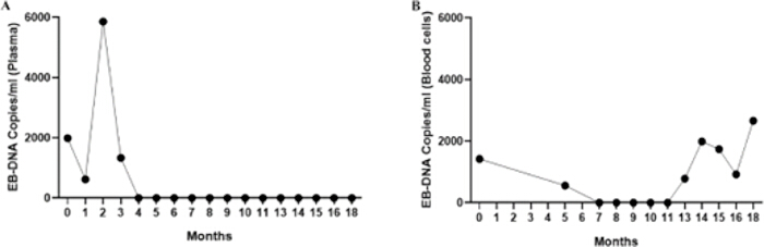
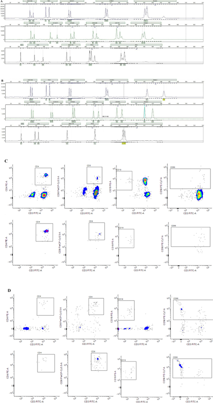
The patient completed pre-transplant preparation and received the mitoxantrone liposome-enhanced BUCY conditioning regimen (rationale for adding mitoxantrone liposome: It has strong antitumor activity, can enhance the killing of residual tumor cells and EBV-infected cells, and synergize with busulfan and cyclophosphamide to improve transplant efficacy; administered on day -8, with an 8-day interval before stem cell infusion, allowing sufficient time for drug metabolism (half-life of approximately 40 h) without affecting donor stem cell engraftment). On July 7, 2023, stem cell infusion was successfully completed, followed by supportive care such as anti-infection, GVHD prophylaxis, and hematopoietic recovery promotion. On the 4th day after transplantation, the patient experienced abdominal pain, fever, and neutropenia, which was complicated by an abdominal infection. Following treatment with meropenem and tigecycline for bacterial infection and caspofungin for antifungal therapy, the patient’s symptoms improved, and her body temperature normalized. Neutrophil and platelet engraftment occurred on day 12 post-transplant, and one month later, her complete blood count normalized. Bone marrow biopsy showed active nucleated cell proliferation, and DNA fingerprinting revealed a 100% donor cell engraftment rate. The patient was monitored for over 1 year post-transplant, and no complications such as acute or chronic GVHD or infections were observed. Follow-up is ongoing until September 2024. PET-CT scans conducted at 3 months and 12 months post-transplant showed the patient remained in complete remission. EBV-DNA in plasma was undetectable post-transplant, and occasional low-level EBV-DNA in blood cells was considered normal (Figure 3). DNA fingerprinting at 1 month and 6 months post-transplant confirmed 100% donor engraftment, demonstrating full donor hematopoiesis (Figure 4). Lymphocyte subset analysis showed that CD3+CD8+ cells and EBV-infected CD56+ cells were undetectable after transplantation (Figure 4).

figure-results-1

Figure 1: PET-CT imaging results during treatment and follow-up. (A) Pre-induction chemotherapy PET-CT image showing lymphoma involvement in the subcutaneous soft tissue of the left frontotemporal-parietal region, right pleura, and lymph nodes below the bilateral diaphragms with increased metabolic activity. (B) Post-induction chemotherapy PET-CT image showing complete resolution of all lesions, consistent with complete remission. (C,D) PET-CT follow-up images obtained at 3 months (C) and 12 months (D) after transplantation, demonstrating sustained complete remission. Please click here to view a larger version of this figure.

figure-results-2
Figure 2: Pathological and immunohistochemical findings of scalp mass. Biopsy tissue showed necrotic changes; atypical large hyperchromatic cells infiltrated around residual blood vessels with abundant nuclear debris in the background. Immunohistochemistry: CK(-), CD3(+), CD5(-), CD10(-), CD4(-), CD8(+), CD2(+), CD7(-), CD43(+), CD20(-), CD56(+), Granzyme B(+), TIA-1(+), Ki-67 (proliferation index ~90%), CD79a(-), CD34(-), CD117(-), TdT(-), EBER(+), consistent with EBV-associated extranodal NK/T-cell lymphoma. Scale bars: 100 µm. Please click here to view a larger version of this figure.

figure-results-3
Figure 3: Dynamic monitoring of Epstein–Barr virus DNA levels. (A) Plasma Epstein–Barr virus (EBV)-DNA levels over time. Plasma EBV-DNA remained undetectable at all post-transplantation time points, indicating the absence of EBV reactivation. (B) Cellular EBV-DNA levels over time. Occasional low-level cellular EBV-DNA was detected after transplantation without apparent clinical significance.Please click here to view a larger version of this figure.

figure-results-4
Figure 4: Post-transplant chimerism analysis and detection of Epstein–Barr virus-infected lymphocyte subsets. (A) Pre-transplant DNA fingerprinting profile of the recipient. (B) DNA fingerprinting analysis at 1 month after transplantation, demonstrating 100% donor chimerism and complete donor-derived hematopoiesis. (C) Pre-transplant lymphocyte subset analysis showing Epstein–Barr virus (EBV) infection in CD3+CD8+ and CD56+ cells. (D) Post-transplant lymphocyte subset analysis showing no detectable EBV-infected CD3+CD8+ or CD56+ cells, indicating complete clearance of EBV infection. Please click here to view a larger version of this figure.

Discussion

Loading...
$$\rightleftharpoonup{xx}$$ $$\longleftharp{xx}$$, $$\longrightharp{xx}$$,

CAEBV transformed into ENKTL is an extremely rare and aggressive clinical entity, with a worse prognosis than either disease alone, as it involves both persistent EBV-driven lymphoproliferation and malignant transformation7,17,18. Early diagnosis and timely intervention are crucial to improving patient outcomes. In this case, the patient initially presented with infectious mononucleosis-like symptoms, progressed to CAEBV, and eventually developed ENKTL with multisystem involvement. The diagnosis was confirmed through clinical manifestations, EBV-DNA detection, pathological and immunohistochemical examinations (EBER positivity is a key marker for EBV-associated tumors)18,19. A limitation of this case is the lack of an early lymph node biopsy, which prevented the determination of the exact timing of ENKTL transformation; therefore, for CAEBV patients with lymphadenopathy or mass lesions, a timely biopsy is recommended to avoid missed diagnosis or delayed treatment.

The treatment principles for CAEBV primarily focus on controlling inflammation, antitumor chemotherapy, and immunotherapy. Recent advances have introduced new therapeutic options. The HDAC inhibitor cedamide, for instance, can eliminate EBV-infected T cells. Although immunochemotherapy and targeted therapies may provide some benefit, more extensive laboratory and clinical research is required to refine treatment strategies and improve outcomes for CAEBV patients. For those who meet the criteria for transplantation, allo-HSCT remains the most effective treatment option. Treatment typically begins by controlling the inflammatory response through immunosuppression, followed by chemotherapy to eliminate EBV-infected lymphocytes. Finally, allo-HSCT is performed to restore the immune system and, ultimately, achieve a cure. It is essential to perform allo-HSCT as soon as possible once CAEBV enters the inactive stage after immunosuppression and chemotherapy, to maximize survival.

Following the diagnosis of CAEBV transformed into ENKTL, the patient achieved rapid complete remission after receiving four cycles of a multi-drug regimen, including etoposide, liposomal doxorubicin, mitoxantrone liposome, pegaspargase, immune checkpoint inhibitors, cedamide, and hormones. Once all symptoms and signs subsided, allo-HSCT was promptly performed. The effectiveness of the treatment was monitored using EBV-DNA quantification and PET/CT scans. A gradual decline in EBV-DNA levels during the transplantation process indicated good disease control. Because EBV-infected memory B cells can persist in the body, EBV-DNA measurements in blood cells may be prone to inaccuracies. Therefore, it is recommended to assess plasma EBV-DNA levels instead. Clinically, many laboratories monitor both plasma and blood cell EBV-DNA simultaneously. If the primary disease remains in remission, plasma EBV-DNA should continue to test negative. When blood cell EBV-DNA remains positive, further analysis by lymphocyte sorting is needed to identify which lymphocyte subset is infected. If pre-transplant NK or T cells are infected, the possibility of disease recurrence should be considered. Conversely, if B cells are infected, close monitoring of EBV-DNA copy number changes is necessary to prevent the development of post-transplant lymphoproliferative disorders. This suggests that simultaneous monitoring of plasma and blood cell EBV-DNA, combined with lymphocyte sorting for positive blood cell EBV-DNA, can avoid misdiagnosis or missed diagnosis.

Moreover, allo-HSCT-related complications significantly impact the prognosis and survival of patients with CAEBV and ENKTL. The role of specific bacteria in regulating GVHD presents an important avenue for exploration. Pretreatment prior to allo-HSCT, which often includes high-dose chemotherapy and radiotherapy, can disrupt the patient's intestinal microecological balance, leading to damage of the intestinal epithelium and compromising the intestinal barrier. The use of broad-spectrum antibiotics for prophylaxis and therapy during allo-HSCT can further alter the patient's intestinal flora, thereby increasing the risk of infections. It should be noted that short-term probiotic administration does not increase the risk of infection, as pre-transplant intestinal decontamination with levofloxacin and strict aseptic isolation in a laminar-flow ward can reduce pathogenic bacterial invasion, while probiotics can rapidly colonize the intestines to form a protective barrier20,21,22. To improve intestinal flora after allo-HSCT, strategies such as adjusting antibiotic regimens, administering probiotics and prebiotics, performing fecal microbiota transplantation, and using other microbiome-enhancing preparations can significantly improve prognosis14,15,16. There are differences in treatment regimens between ENKTL patients with a history of CAEBV and primary ENKTL: (1) Strengthened control of EBV infection (long-term antiviral prophylaxis post-transplant) to prevent EBV reactivation-induced disease recurrence; (2) Vigilance against HLH (close monitoring of soluble CD25 and NK cell activity) as CAEBV patients are prone to secondary HLH; (3) Enhanced infection prevention (prolonged antibiotic prophylaxis and intensified intestinal decontamination) due to pre-existing immune dysfunction in CAEBV patients.

This case also has limitations: it is a single-case report with a small sample size and a follow-up duration of only 1 year; the long-term prognosis and the generalizability of the treatment regimen require verification in large-sample, multicenter studies. Future research directions include: (1) Exploring the mechanism of action of microecological preparations in preventing post-transplant infections and GVHD; (2) Developing more personalized conditioning regimens and targeted therapies for ENKTL transformed from CAEBV; (3) Establishing standardized diagnostic and treatment protocols for this rare disease through multicenter collaboration.

In conclusion, allo-HSCT is an effective curative option for pediatric patients with ENKTL transformed from CAEBV. Timely achievement of CR through multi-cycle chemotherapy, followed by allo-HSCT with a fully matched donor, and adjuvant microecological preparations to prevent complications can significantly improve patient outcomes. This case provides valuable clinical experience for diagnosing and treating similar rare cases.

Disclosures

Loading...
$$\rightleftharpoonup{xx}$$ $$\longleftharp{xx}$$, $$\longrightharp{xx}$$,

The authors have nothing to disclose.

Acknowledgements

Loading...
$$\rightleftharpoonup{xx}$$ $$\longleftharp{xx}$$, $$\longrightharp{xx}$$,

This study was supported by Municipal Financial Subsidy of Nanshan District Medical Key Discipline Construction, Shenzhen Nanshan District Health System Science and Technology Major Project (No. NSZD2023018), NHC Key Laboratory of Nuclear Technology Medical Transformation (Mianyang Central Hospital) (Grant No.2023HYX033).

Materials

List of materials used in this article
NameCompanyCatalog NumberComments
Anthracycline LiposomeCSPC Ouyi Pharmaceutical Co., Ltd.National Drug Approval No. H20113320Doxorubicin Hydrochloride Liposome Injection, 20mg/10mL, used for solid tumors and hematological malignancies
Anticoagulant (Heparin Sodium)Changzhou Qianhong Biochemical Pharmaceutical Co., Ltd.National Drug Approval No. H32022088Unfractionated heparin, injection 10000 USP units/mL, used for anticoagulant therapy
Blood Cell SeparatorTerumo BCT, USAModel: COBE SpectraBlood cell separation system, used for platelet separation and stem cell collection
Carbapenem (Meropenem)Jiangsu Ruishi Biotechnology Co., Ltd.National Drug Approval No. H20243624Broad-spectrum antibiotic, lyophilized powder for injection 1g/vial, used for severe bacterial infections
Cephalosporin (Ceftriaxone)Shenzhen Lijian Pharmaceutical Co., Ltd.National Drug Approval No. H20058024Third-generation cephalosporin, lyophilized powder for injection 1g/vial, used for bacterial infections
Compound Sulfonamide (Sulfamethoxazole-Trimethoprim)Teiyi Pharmaceutical Group Co., Ltd.National Drug Approval No. H44023640Compound antibiotic, 800mg/160mg/tablet, used for bacterial and protozoal infections
CytarabinePfizer Pharmaceuticals Ltd.National Drug Approval No. H20205028Antimetabolite, lyophilized powder for injection 100mg/vial, used for acute myeloid leukemia and lymphoma
DexamethasoneHenan Runhong Pharmaceutical Co., Ltd.National Drug Approval No. H41020330Glucocorticoid, 4mg/tablet, anti-inflammatory and immunosuppressive in chemotherapy
EBER ProbeDako (Agilent), DenmarkY5200In situ hybridization probe, biotin-labeled, used for detection of EBV-encoded small RNAs
EBV-related Phenotypic Antibody (EBNA-1)Abcam, UKab40794Polyclonal antibody, rabbit anti-EBNA-1, used for EBV infection detection
EtoposideQilu Pharmaceutical Co., Ltd.National Drug Approval No. H20143143Topoisomerase II inhibitor, injection 100mg/5mL, used for lymphoma and solid tumors
Flow CytometerBD Biosciences, USAModel: FACSCanto IISix-color flow cytometer, used for cell phenotypic analysis and immunophenotyping
GemcitabineJiangsu Hausen Pharmaceutical Group Co., Ltd.National Drug Approval No. H20030104Antimetabolite, lyophilized powder for injection 200mg/vial, used for pancreatic cancer and hematological malignancies
Hematology AnalyzerSysmex, JapanModel: XN-9000Automatic hematology analyzer, 5-part differential, used for complete blood count analysis
Hematopoietic Growth Factor (G-CSF)Kexing Biopharmaceutical Co., Ltd.National Drug Approval No. S20103004Granulocyte colony-stimulating factor, injection 300mcg/mL, used for prevention of neutropenia
Immunosuppressant (Cyclosporine)Livzon Group Pharmaceutical FactoryNational Drug Approval No. H10950335Calcineurin inhibitor, 100mg/capsule, used for post-transplant immunosuppression
LevofloxacinChangchun Haiyue Pharmaceutical Co., Ltd.National Drug Approval No. H20203307Fluoroquinolone, 750mg/tablet, used for respiratory and urinary tract infections
Lymphoma-related Phenotypic Antibody (CD20)Dako (Agilent), DenmarkM0755Monoclonal antibody, mouse anti-human CD20, used for B-cell lymphoma detection
Nucleoside Antiviral (Acyclovir)Shandong Qidu Pharmaceutical Co., Ltd.National Drug Approval No. H20056584Antiviral drug, 800mg/tablet, used for herpes simplex and varicella-zoster virus infections
Optical MicroscopeOlympus, JapanModel: BX53Brightfield microscope, magnification 40-1000x, used for pathological slide observation
OxaliplatinJiangsu Hengrui Medicine Co., Ltd.National Drug Approval No. H20213312Platinum-based drug, lyophilized powder for injection 50mg/vial, used for colorectal cancer and lymphoma
Pathological Slide Staining MachineLeica Biosystems, GermanyModel: Bond-MaxAutomatic immunohistochemistry/in situ hybridization staining system, used for pathological slide processing
PegaspargaseJiangsu Hengrui Medicine Co., Ltd.National Drug Approval No. H20090015Pegylated Asparaginase, 3750 IU/vial, used for acute lymphoblastic leukemia
PET-CT ScannerSiemens Healthineers, GermanyModel: Biograph mCT128-slice PET-CT, used for tumor functional and anatomical imaging
PrednisoneXinxiang Changle Pharmaceutical Co., Ltd.National Drug Approval No. H41020214Glucocorticoid, 5mg/tablet, used in chemotherapy regimens and anti-inflammatory treatment
Real-time Quantitative PCR InstrumentThermo Fisher Scientific, USAModel: Applied Biosystems 7500Real-time quantitative PCR system, 96-well plate, used for nucleic acid detection
TigecyclineZhengda Tianqing Pharmaceutical Group Co., Ltd.National Drug Approval No. H20133044Glycylcycline antibiotic, lyophilized powder for injection 50mg/vial, used for multidrug-resistant bacterial infections
Topical Preparation (Mupirocin Ointment)Sino-US Tianjin SmithKline Pharmaceutical Co., Ltd.National Drug Approval No. H10930064Topical antibiotic, 2% ointment, used for skin and soft tissue infections
ToripalimabSuzhou Zhonghe Biomedical Technology Co., Ltd.National Drug Approval No. S20191003PD-1 monoclonal antibody, widely used for anti-tumor therapy
Triazole Antifungal (Fluconazole)Sichuan Kelun Pharmaceutical Co., Ltd.National Drug Approval No. H20094047Antifungal drug, injection 200mg/100mL, used for candidiasis and cryptococcosis
Triple Probiotics (Bifidobacterium + Bacillus licheniformis + Lactobacillus)Jincheng Haisi Pharmaceutical Co., Ltd./Zhejiang Jingxin Pharmaceutical Co., Ltd.National Drug Approval No. S19993065/National Drug Approval No. S20083112Probiotic preparation, used for intestinal flora regulation
VincristineShenzhen Wanle Pharmaceutical Co., Ltd.National Drug Approval No. H44021772Chemotherapeutic drug, vinca alkaloid, used for lymphoma/leukemia treatment, injection 1mg/mL

Reprints and Permissions

Request permission to reuse the text or figures of this JoVE article

Request Permission

Tags

Allogeneic HSCTPediatric ENKTLChronic Active EBVHematopoietic Stem CellExtranodal NK T Cell LymphomaPreoperative ChemotherapyComplete RemissionDonor ChimerismProbiotic PreparationsGraft Versus Host Disease

Related Articles