This protocol describes a mouse subcutaneous infection model for Streptococcus pyogenes that enables independent quantification of bacterial burden and lesion progression. The method provides a standardized and reproducible platform for evaluating the roles of bacterial virulence factors, host immune responses, or therapeutic interventions aimed at reducing tissue damage.
Method Article
A Mouse Model of Streptococcus pyogenes Necrotizing Skin Infection
In This Article
Summary
Abstract
Streptococcus pyogenes is a human pathogen that causes severe necrotizing soft tissue infections characterized by rapid and extensive tissue destruction. A key challenge in studying these infections is the lack of experimental models that allow independent assessment of bacterial replication and host-mediated tissue damage. This article presents a standardized and reproducible murine subcutaneous model of S. pyogenes necrotizing skin infection designed to overcome this limitation. The protocol describes the preparation of a log-phase bacterial inoculum, subcutaneous infection of mice, and longitudinal monitoring of lesion development. Key steps include sonication of bacterial cultures to standardize chain length, digital imaging of lesions for quantitative analysis of ulcer area, and determination of bacterial burden through tissue homogenization and plating. This versatile model can be readily adapted to evaluate the effects of specific bacterial genes, host factors, or therapeutic interventions on infection progression and tissue pathology. By enabling reproducible and quantitative assessment of both bacterial and host parameters, this method provides a robust platform for studying pathogenesis and testing strategies to limit tissue damage during invasive streptococcal infection.
Introduction
Streptococcus pyogenes (Group A Streptococcus, GAS) is a significant human pathogen that causes necrotizing soft tissue infections (NSTIs)1, resulting in rapid destruction of skin and underlying tissues2,3,4. Studying the pathogenesis of these infections requires animal models that recapitulate localized tissue injury while allowing independent assessment of bacterial replication and host-mediated pathology5. Many traditional infection models, such as systemic inoculation or in vitro cell culture systems, are limited in their ability to separate bacterial burden from tissue damage, making it challenging to evaluate the specific contributions of virulence factors or host responses6.
The overall goal of this method is to provide a standardized and reproducible mouse subcutaneous model of S. pyogenes necrotizing skin infection. The rationale for this technique is to establish a tractable in vivo system that decouples pathogen replication from host-mediated tissue injury. This model is highly adaptable to common laboratory mouse strains. While the SKH1 (hairless) strain is often used for ease of lesion monitoring7,8, the protocol works effectively on haired strains (e.g., C57BL/6J) following depilation. Published reports typically use either sex or a single sex (often females) and often find similar lesion area/bacterial burden under the specific experimental conditions reported; however, host genetic background and sex can influence GAS disease severity in some settings and should be considered when designing experiments and analyzing results9. A key advantage of this model over alternatives is its capacity for longitudinal monitoring of a localized lesion, enabling precise, independent quantification of ulcer area and bacterial burden from the same animal over time. With an appropriately chosen inoculum and strain, subcutaneous infections are often localized and permit study of lesion progression and resolution. That said, whether infection remains localized is strain-, inoculum-, and host-dependent: certain clinical isolates (or strains that acquire covRS mutations) and sufficiently large inocula can disseminate and cause bacteremia or lethal disease in immunocompetent mice10. Authors should report strain, inoculum, route, and any host modifications and titrate inocula when adapting the model to a new isolate. This approach is highly reproducible, accessible, and compatible with diverse downstream analyses, including histology and immune profiling.
The protocol has been applied to multiple GAS strains (including M1-type isolates such as HSC511 and other M1T110,12) and to clinical NSTI isolates. Because virulence phenotypes differ among emm types and individual isolates13, investigators should empirically optimize inoculum and monitor for systemic spread when translating the protocol to new strains.
This protocol is appropriate for researchers in microbial pathogenesis, immunology, and translational research who wish to investigate bacterial virulence, host immune responses, or novel therapeutic strategies targeting tissue preservation during invasive infection. Its standardized, quantitative design allows for rigorous evaluation of interventions and provides a versatile platform for studying the interplay between pathogen replication and host-mediated tissue injury.
Protocol
All animal experiments were performed in compliance with the Marshall University School of Medicine Institutional Animal Care and Use Committee (IACUC) and Animal Resource Facility. All procedures involving Streptococcus pyogenes were conducted in the PHS-assured, AAALAC-accredited animal facility under Biosafety Level 2 (BSL-2) containment in accordance with institutional biosafety protocols. The experimental workflow is illustrated in Figure 1.
NOTE: Specific details for reagents, materials, and equipment are provided in the Table of Materials.
1. Preparation of bacteria for inoculum
- Prepare an overnight culture by inoculating Streptococcus pyogenes HSC5 from a frozen stock into 10.5 mL of C medium (0.5% peptone, 1.5% yeast extract, 10 mM K2HPO4, 0.4 mM MgSO4, and 17 mM NaCl). Incubate statically at 37 °C for 16-20 h.
- Measure the optical density at 600 nm (OD600) of the overnight culture. Centrifuge 10 mL of culture at 6,000 x g for 5 min at room temperature.
- Resuspend the bacterial pellet in 0.5 mL of fresh, pre-warmed Todd Hewitt broth supplemented with 1% yeast extract.
- Inoculate 50 mL of pre-warmed Todd Hewitt broth supplemented with 1% yeast extract in a conical tube with the resuspended bacteria to a starting OD600 of 0.05.
- Incubate the tube at 37 °C without shaking. Monitor the OD600 every 30 min.
- Harvest the bacteria when the culture reaches an OD600 of ~0.200 by centrifugation at 6,000 x g for 5 min at 4 °C.
- Discard the supernatant and resuspend the pellet in 10 mL of ice-cold, sterile phosphate-buffered saline (PBS) solution.
- Centrifuge the suspension again at 6,000 x g for 5 min at 4 °C. Resuspend the final pellet in 1 mL of ice-cold, sterile PBS solution. Keep the suspension on ice.
NOTE: This concentrated bacterial suspension can be held on ice for up to 30 min before proceeding to sonication.
2. Standardization of bacterial concentration
- Transfer the 1 mL bacterial suspension to a microcentrifuge tube. Sonicate the suspension on ice using a cup-horn sonicator to break long chains of S. pyogenes.
CAUTION: Wear appropriate hearing protection during sonication. The procedure generates high-frequency noise that can cause hearing damage. - Set the sonicator to deliver 5 cycles of 1-min pulses (dependent on sonicators). To confirm effective chain disruption, place a 10 µL drop of the sonicated suspension on a microscope slide, cover with a coverslip, and visualize under 40X.
NOTE: The goal of sonication is to reduce long chains to predominantly short chains (ideally 1-4 cells per chain) and single cocci. If long chains (>10 cells) are still prevalent, repeat the sonication cycle. - Prepare a 1:100 dilution of the sonicated bacteria in sterile PBS solution.
- Load 10 µL of the diluted bacteria onto a counting chamber/bacterial hemocytometer.
- Count the number of bacterial chains in 10 large squares of the chamber under a microscope.
- Calculate the bacterial concentration in colony-forming units per mL (CFU/mL) using the formula: Concentration = (Total Count / 10) × Dilution Factor × 1.25 × 10⁶.
NOTE: Each chain of bacterial cells is counted as a single CFU. - Dilute the sonicated bacterial stock with ice-cold PBS to a final working concentration of 1 × 10⁸ CFU/mL.
3. Subcutaneous infection of mice
- One day prior to infection, shave the left hind flank of each mouse. Use electric clippers to remove all hair completely from an area of approximately 2 cm x 2 cm.
- Anesthetize a mouse in an induction chamber (4% induction, 1.5% maintenance isoflurane). To maintain synchronization of the infection, process mice in small, manageable batches (e.g., 5-10 mice per batch).
- Using a 1 mL syringe fitted with a 27-G needle, draw up 100 µL of the bacterial inoculum.
- Remove the mouse from the isoflurane chamber and immediately insert the needle subcutaneously into the shaved hind flank of the anesthetized mouse with the bevel facing up.
- Inject the entire 100 µL volume slowly to create a localized bleb. Withdraw the needle.
- Return the mouse to its cage and monitor until fully recovered from anesthesia.
- Repeat steps 3.2-3.5 for all mice in the experimental cohort.
- Prepare serial 10-fold dilutions of the remaining bacterial inoculum in PBS.
- Spot-plate 5 µL of each dilution onto agar plates. Incubate plates anaerobically at 37 °C overnight to confirm the delivered dose.
4. Longitudinal monitoring of infection progression
- Monitor all infected mice at least once daily for any signs of discomfort. Adhere to established humane endpoints, which are particularly important when using hypervirulent strains or immunocompromised hosts.
- Anesthetize each mouse following the procedure described in step 3.2.
- Remove the mouse from the isoflurane chamber and position it on its side next to an identifier label. To enable longitudinal tracking of individual mice, mark (e.g., ear punch, ear tag) each animal prior to the start of the experiment.
- Capture a digital photograph of the lesion area using a camera mounted directly above the mouse.
- Open the digital image in image analysis software (ImageJ/Fiji). Select the Freehand Selections tool from the toolbar.
- Manually trace the perimeter of the visible ulcer in the image.
- Open the Analyze menu and select Measure to calculate the area within the traced perimeter.
- Record the measured area in square millimeters for each mouse at each time point.
NOTE: The experiment can be paused at this point. Mice are returned to their cages for continued monitoring until the next observation time point or endpoint.
5. Tissue collection and bacterial burden quantification
- Euthanize mice at the experimental endpoint using carbon dioxide inhalation followed by cervical dislocation, a method approved by the Marshall University IACUC.
- Using sterile surgical scissors and forceps, aseptically excise the entire lesion and a minimal amount of surrounding tissue. Ensure the excision includes the full thickness of the skin and the underlying subcutaneous tissue, typically extending to but not including the muscle layer, unless the infection has visibly invaded the muscle fascia.
- Transfer the tissue to a pre-weighed, sterile 2 mL screw-cap microcentrifuge tube containing 1 mL of sterile PBS and two sterile 2.8 mm stainless steel beads.
- Record the weight of the tissue.
- Homogenize the tissue using a mechanical homogenizer set to 6.5 m/s for 60 s.
- Place the tube on ice for 5 min to prevent overheating.
- Homogenize the tissue for a second cycle of 60 s at 6.5 m/s.
- Prepare serial 10-fold dilutions of the tissue homogenate in a 96-well plate using sterile PBS as diluent. Spot-plate 5 µL of each dilution onto agar plates.
- Incubate the plates anaerobically at 37 °C for 24-48 h. For example, place plates inside a sealed jar with an anaerobic gas generator sachet and an anaerobic indicator.
- Count the colonies on plates with 30-300 colonies. Calculate the colony-forming units per mL (CFU/mL) of tissue homogenate.
Results
The mouse subcutaneous infection model for S. pyogenes consistently generates a localized, progressive necrotic ulcer, enabling simultaneous quantification of bacterial burden and tissue pathology. Representative results from this standardized protocol are as follows.
Experimental groups typically consist of 6-10 mice to provide sufficient statistical power (e.g., 80-90%) for detecting significant differences in lesion size and bacterial burden, based on prior studies8,14,15. Data are analyzed using appropriate statistical tests (e.g., unpaired t-test for two groups, one-way ANOVA with post-hoc test for multiple groups). Control groups should include mice injected with vehicle (e.g., sterile PBS) alone, and when testing mutant strains, a parallel group infected with the wild-type (WT) parent strain.
Following subcutaneous inoculation with 1 × 10⁷ CFU of wild-type S. pyogenes strain HSC5, a well-defined area of induration develops at the injection site within 24 h post-infection (hpi), which progresses to a necrotic ulcer by 48-72 hpi (Figure 2A). The lesion area, measured daily from digital images using ImageJ software, typically increases from approximately 5-10 mm2 at 1 day post-infection (dpi) to a peak of 30-45 mm2 by 5-6 dpi, during which eschar formation and tissue contraction indicate the onset of healing. Representative quantitative data (Figure 2B) show the mean lesion area ± SD peaks at approximately 40 mm² by day 6 post-infection before resolution begins. A successful infection is characterized by this predictable progression and low inter-animal variability (typically <20% coefficient of variation in lesion area at peak severity).
Bacterial burden, determined by homogenizing excised lesions and enumerating CFUs, typically ranges from 1 × 10⁷ to 1 × 10⁹ CFU per mL of tissue homogenate at 3 dpi. As shown in a representative experiment (Figure 2C), the mean bacterial burden at 3 dpi is 1.1 × 10⁸ ± 0.4 × 10⁸ CFU/mL (mean ± SD). A key strength of this model is its ability to decouple bacterial growth from tissue damage. For instance, infection with a ΔPdh mutant yields a bacterial load comparable to wild-type at 3 dpi but results in a significantly smaller lesion area, demonstrating a specific alteration in disease pathology independent of bacterial replication15,16. Suboptimal results, such as failure to form an ulcer or CFUs below 1 × 10⁵ CFU/mL, often indicate technical error, potentially inoculum delivered as an intramuscular injection instead of the intended subcutaneous route, or a non-viable inoculum.
Histological analysis of lesions by H&E staining reveals progressive tissue destruction over the course of infection (Figure 2D). Uninfected skin shows normal architecture with intact epidermis, dermis, and underlying muscle. By 1 dpi, a developing subcutaneous lesion is visible, characterized by early necrosis and an emerging inflammatory infiltrate. At 4 dpi, the lesion reaches maximal size with a central necrotic core, surrounded by a dense peripheral infiltrate of immune cells. Immunofluorescence staining of tissue sections15 can further delineate host-pathogen interactions, such as the predominantly extracellular localization of wild-type bacteria versus the increased intracellular presence of specific mutants within phagocytes15,16. For immunofluorescence, bacteria can be labeled using a Group A carbohydrate-specific antibody15. High-quality sections show clear tissue architecture and specific staining with minimal background fluorescence17.
The model is highly adaptable for downstream applications. Flow cytometric analysis of single-cell suspensions from lesions provides quantitative data on immune cell recruitment, typically showing a predominance of these cells: CD11b+ Ly6G+ neutrophils, and CD11b+ F4/80+ macrophages at 3 dpi15. Furthermore, the protocol supports cytokine quantification from tissue homogenates15. These integrated readouts validate the model's utility for comprehensive analysis of the host immune response during a localized, tissue-destructive infection.
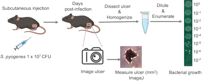
Figure 1: Schematic of the murine subcutaneous Streptococcus pyogenes infection model. The procedure begins with subcutaneous injection of a defined bacterial inoculum (typically 1 × 10⁷ CFU in 100 µL PBS) into the shaved left flank of anesthetized mice. Lesion development is monitored daily, and ulcer images are captured using a calibrated digital camera. Ulcer area (mm2) is quantified using ImageJ based on pixel-to-millimeter calibration. At designated time points, the entire lesion and surrounding tissue are aseptically excised, weighed, and homogenized in sterile PBS using a bead mill homogenizer. Homogenates are serially diluted and plated on Todd Hewitt agar for enumeration of viable S. pyogenes colony-forming units (CFU). Figure partially created with Biorender.com Please click here to view a larger version of this figure.
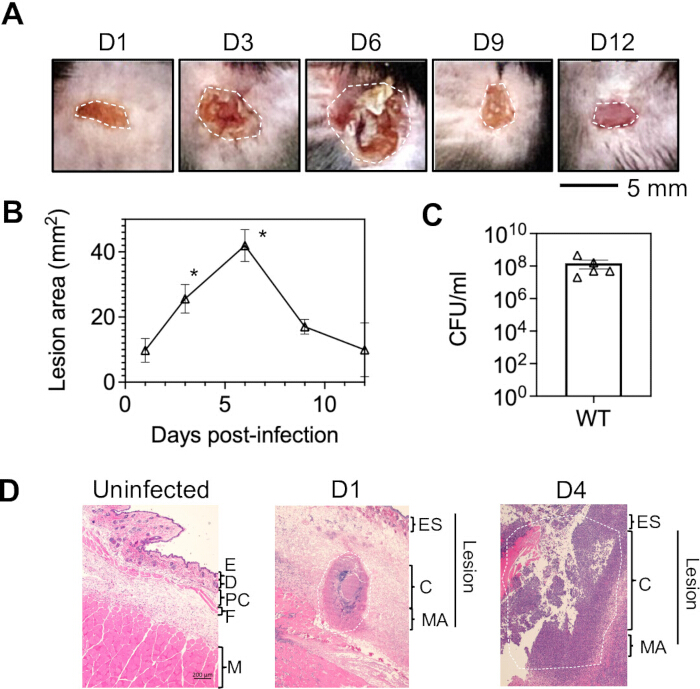
Figure 2: Subcutaneous infection and lesion progression in mice. (A) C57BL/6J mice were subcutaneously infected with 1 × 10⁷ CFU wild-type S. pyogenes HSC5 and monitored over 12 days for tissue damage. Representative ulcers are shown, with lesion areas outlined by a dashed white line. (B) Lesion area was measured daily from digital images; data are presented as mean ± SD (n = 5 mice per time point). Statistical significance between consecutive days was determined by one-way ANOVA with Tukey's post-hoc test (*p < 0.05). (C) Bacterial loads were determined from homogenized lesions at 3 dpi. Each symbol represents an individual mouse. (D) Histopathology of S. pyogenes HSC5 infection following subcutaneous inoculation of 1 × 10⁷ CFU into the flank skin of C57BL/6J mice. Hematoxylin and eosin (H&E)-stained sections of infected tissue are compared with uninfected controls. By Day 1, a well-defined subcutaneous lesion forms with a necrotic core, surrounded by an inflammatory margin rich in polymorphonuclear leukocytes. By Day 4, the lesion reaches maximal size and forms an open ulcer with eschar development. Streptococci remain confined to the core by inflammatory cells in the margin. In most strains, including HSC5, the lesion resolves within 14 days. Abbreviations: E, epidermis; D, dermis; PC, panniculus carnosus; F, fascia; M, muscle; Es, eschar; C, Core; MA, margin. Please click here to view a larger version of this figure.
Discussion
The mouse subcutaneous S. pyogenes necrotizing skin infection model described here provides a robust and reproducible system for dissecting the mechanisms underlying severe soft tissue infection. Its primary value lies in the ability to decouple bacterial burden from tissue pathology, enabling mechanistic studies of how host and pathogen factors independently contribute to disease progression. This distinction is particularly important in S. pyogenes infection, where necrotizing fasciitis severity often reflects an excessive host inflammatory response to virulence factors such as streptolysins rather than bacterial proliferation alone18.
Several methodological steps are critical for consistent and interpretable results. Standardization of the bacterial inoculum is essential. Cultures should be grown overnight in C medium, subcultured into Todd-Hewitt (THY) broth, and harvested at mid-log phase (OD600 ≈ 0.200) to ensure uniform expression of virulence factors. The composition of C medium (containing casein hydrolysate, yeast extract, and mineral salts) supports robust overnight growth while maintaining GAS viability. Before infection, bacteria must be thoroughly sonicated for approximately 5 min to disrupt the characteristic streptococcal chains. This step ensures an even inoculum and prevents the formation of clumps that can obstruct capillaries or produce variable lesion sizes. The injection technique is another major determinant of success. The needle should be inserted at a shallow angle to achieve a true subcutaneous bleb. Intramuscular injection alters disease kinetics and lesion morphology, while puncturing through the skin can lead to leakage and failure to establish infection. Consistency in injection depth and site is especially important when comparing strains or treatment conditions.
The model is easily adapted to specific experimental goals. Inoculum size can be titrated to modulate infection severity, and the system is compatible with host-targeted interventions such as immune cell depletion or cytokine neutralization. Variability in lesion size is most often due to inconsistent bacterial growth phase or inadequate sonication. Low bacterial recovery can result from the use of hypotonic or acidic buffers during tissue homogenization; sterile PBS (pH 7.4) should be used to preserve bacterial viability.
A key limitation of this model is its localized nature. It does not fully recapitulate systemic dissemination or multiorgan involvement characteristic of invasive S. pyogenes disease, such as streptococcal toxic shock syndrome19,20,21. Additionally, murine and human skin differ in structure and immune cell composition, particularly in T-cell subsets and antimicrobial peptide profiles22. Furthermore, several critical GAS virulence factors, including superantigens23 and streptokinase24, exhibit human-specific activity and do not function equivalently in mice, which must be considered when interpreting results. Therefore, findings derived from this system should be validated in complementary models or human tissue when possible.
Compared to alternative infection routes, this subcutaneous approach offers distinct advantages. Intraperitoneal infection models primarily assess survival and systemic bacterial load, limiting insight into localized pathology13,14. The subcutaneous method described here reliably produces a well-defined, expanding ulcer that can be monitored longitudinally to assess both bacterial growth and tissue destruction within the same animal.
This model has broad applications in both microbiology and immunology. It is ideally suited for functional studies of bacterial virulence determinants -- ranging from secreted toxins25 such as NAD-glycohydrolase26,27 and streptolysin O28,29,30 to regulatory systems like CovRS31 -- that modulate tissue damage. For immunological research, it provides a controlled platform to examine innate immune dynamics, such as neutrophil recruitment, macrophage polarization, or cytokine signaling32, including the roles of IL-1β, IL-10, and inflammasome pathways in modulating infection outcomes. Moreover, the model is highly amenable to therapeutic testing, allowing evaluation of antimicrobial agents17, toxin inhibitors, and host-directed therapies that attenuate tissue injury without affecting bacterial clearance.
Overall, this standardized subcutaneous infection model serves as a versatile and reproducible platform to study the interplay between bacterial virulence and host response in necrotizing skin infection and to accelerate translational efforts toward more effective treatments.
Disclosures
The authors declare no competing interests.
Acknowledgements
This work was supported by the Marshall University Startup Fund and the WV Clinical and Translational Science Institute Bench-to-Bedside grant (U54GM104942) to WX. National Institutes of Health Grants R21 AI163825 (to MGC). We thank Dr. Jill Khan (MA, DVM, MPH, DACLAM), Director of the Marshall University Animal Resource Facility, and Dr. Monica Valentovic (PhD), Chair of the IACUC, for their critical review of this manuscript.
Materials
| Name | Company | Catalog Number | Comments |
|---|---|---|---|
| 1 mL Syringe | BD | 309628 | For subcutaneous injection |
| 28-G Needle | BD | 305109 | For subcutaneous injection |
| Bacterial Hemocytometer / Counting Chamber | Hausser Scientific | 02-671-6 | For bacterial counting pre-inoculation |
| BD BBL GasPak EZ Anaerobic Container System | Becton, Dickinson and Company (BD) | 260001 | For anaerobic incubation |
| C57BL/6J mice | The Jackson Laboratory | 664 | 8-12 weeks old |
| Carbon Dioxide (CO2) Tank | Local supplier | N/A | For euthanasia |
| Cup-Horn Sonicator (e.g., Q700) | Qsonica | 431C2 | For standardizing bacterial chain length |
| Digital Camera | N/A | N/A | For lesion imaging |
| Electric Clippers | Wahl | e.g., 8655-200 | For shaving mouse flank |
| FastPrep-24 Homogenizer | MP Biomedicals | 116005500 | For tissue homogenize |
| ImageJ Software | National Institutes of Health (NIH) | N/A | https://imagej.nih.gov/ij/ |
| Isoflurane | Baxter | 1001936060 | For anesthetic |
| Isoflurane Anesthesia System (Chamber & Vaporizer) | VetEquip or similar | N/A | For mouse anesthesia |
| Peptone | Becton, Dickinson and Company (BD) | 211677 | For preparation of C medium |
| Phosphate-Buffered Saline (PBS) | Gibco | 10010023 | Sterile, pH 7.4 |
| S. pyogenes strain HSC5 | Caparon lab | M1T1 clinical isolate | |
| SKH1 mice | Charles River Laboratories | 477 | 8-12 weeks old, hairless |
| Stainless steel beads, 2.8 mm | Universal Medical | D1133-28 | Sterile |
| Sterile Surgical Scissors and Forceps | Fine Science Tools or similar | N/A | For aseptic tissue collection |
| Todd Hewitt Broth | Becton, Dickinson and Company (BD) | 249240 | For bacterial subculture |
| Yeast Extract (for C medium) | Becton, Dickinson and Company (BD) | 212750 | For preparation of C medium (same as for broth supplement) |
References
- Hakkarainen, T. W., Kopari, N. M., Pham, T. N., Evans, H. L. Necrotizing soft tissue infections: review and current concepts in treatment, systems of care, and outcomes. Curr Probl Surg. 51 (8), 344-362 (2014).
- Brouwer, S., et al. Pathogenesis, epidemiology and control of Group A Streptococcus infection. Nat Rev Microbiol. 21 (7), 431-447 (2023).
- Cunningham, M. W. Pathogenesis of group A streptococcal infections and their sequelae. Adv Exp Med Biol. 609, 29-42 (2008).
- Henningham, A., Barnett, T. C., Maamary, P. G., Walker, M. J. Pathogenesis of group A streptococcal infections. Discov Med. 13 (72), 329-342 (2012).
- Pancholi, V., Caparon, M. Streptococcus pyogenes Metabolism. Streptococcus pyogenes: Basic Biology to Clinical Manifestations. , University of Oklahoma Health Sciences Center. Oklahoma City. (2022).
- Ravins, M., et al. Murine soft tissue infection model to study Group A Streptococcus (GAS) pathogenesis in necrotizing fasciitis. Methods Mol Biol. 2427, 185-200 (2022).
- Benavides, F., Oberyszyn, T. M., VanBuskirk, A. M., Reeve, V. E., Kusewitt, D. F. The hairless mouse in skin research. J Dermatol Sci. 53 (1), 10-18 (2009).
- Cusumano, Z. T., Watson, M. E. Jr, Caparon, M. G. Streptococcus pyogenes arginine and citrulline catabolism promotes infection and modulates innate immunity. Infect Immun. 82 (1), 233-242 (2014).
- Chella Krishnan, K., et al. Genetic architecture of Group A streptococcal necrotizing soft tissue infections in the mouse. PLoS Pathog. 12 (10), e1005732(2016).
- Pato, C., Melo-Cristino, J., Ramirez, M., Friaes, A. Portuguese Group for the Study of Streptococcal, I. Streptococcus pyogenes causing skin and soft tissue infections are enriched in the recently emerged emm89 clade 3 and are not associated with abrogation of CovRS. Front Microbiol. 9, 2372(2018).
- Port, G. C., Paluscio, E., Caparon, M. G. Complete genome sequence of emm type 14 Streptococcus pyogenes strain HSC5. Genome Announc. 1 (6), e00612-e00613 (2013).
- Le Breton, Y., et al. Genome-wide discovery of novel M1T1 group A streptococcal determinants important for fitness and virulence during soft-tissue infection. PLoS Pathog. 13 (8), e1006584(2017).
- Bergsten, H., Nizet, V. The intricate pathogenicity of Group A Streptococcus: a comprehensive update. Virulence. 15 (1), 2412745(2024).
- Watson, M. E. Jr, Neely, M. N., Caparon, M. G. Animal Models of Streptococcus pyogenes Infection. Streptococcus pyogenes: Basic Biology to Clinical Manifestations. , University of Oklahoma Health Sciences Center. Oklahoma City. (2022).
- Xu, W., et al. Reprogramming aerobic metabolism mitigates Streptococcus pyogenes tissue damage in a mouse necrotizing skin infection model. Nat Commun. 16, 2559(2025).
- Merriman, J. A., Xu, W., Caparon, M. G. Central carbon flux controls growth/damage balance for Streptococcus pyogenes. PLoS Pathog. 19 (5), e1011481(2023).
- Zou, Z., et al. Dihydrothiazolo ring-fused 2-pyridone antimicrobial compounds treat Streptococcus pyogenes skin and soft tissue infection. Sci Adv. 10 (13), eadn7979(2024).
- Stevens, D. L., Bryant, A. E. Impetigo, Erysipelas and Cellulitis. Streptococcus pyogenes: Basic Biology to Clinical Manifestations. , University of Oklahoma Health Sciences Center. Oklahoma City. (2022).
- Mania, A., et al. Invasive group A streptococcal infections as a consequence of coexisting or previous viral infection in the post-COVID-19 pandemic period. J Infect Public Health. 18, 102622(2025).
- Stevens, D. L., Bryant, A. E. Severe Group A Streptococcal Infections. Streptococcus pyogenes: Basic Biology to Clinical Manifestations. , University of Oklahoma Health Sciences Center. Oklahoma City. (2016).
- Thacharodi, A., et al. The burden of group A Streptococcus (GAS) infections: the challenge continues in the twenty-first century. iScience. 28, 111677(2025).
- Qu, G., et al. Comparing mouse and human tissue-resident γδ T cells. Front Immunol. 13, 891687(2022).
- Proft, T., Fraser, J. D. Streptococcal Superantigens: Biological Properties and Potential Role in Disease. treptococcus pyogenes: Basic Biology to Clinical Manifestations. , University of Oklahoma Health Sciences Center. Oklahoma City. (2022).
- McArthur, J. D., Cook, S. M., Venturini, C., Walker, M. J. The role of streptokinase as a virulence determinant of Streptococcus pyogenes-potential for therapeutic targeting. Curr Drug Targets. 13 (3), 297-307 (2012).
- Barnett, T. C., et al. Streptococcal toxins: role in pathogenesis and disease. Cell Microbiol. 17 (12), 1721-1741 (2015).
- Ghosh, J., Anderson, P. J., Chandrasekaran, S., Caparon, M. G. Characterization of Streptococcus pyogenes beta-NAD+ glycohydrolase: re-evaluation of enzymatic properties associated with pathogenesis. J Biol Chem. 285 (8), 5683-5694 (2010).
- Ghosh, J., Caparon, M. G. Specificity of Streptococcus pyogenes NAD(+) glycohydrolase in cytolysin-mediated translocation. Mol Microbiol. 62 (5), 1203-1214 (2006).
- Vega, L. A., Caparon, M. G. Cationic antimicrobial peptides disrupt the Streptococcus pyogenes ExPortal. Mol Microbiol. 85 (6), 1119-1132 (2012).
- Mozola, C. C., Magassa, N., Caparon, M. G. A novel cholesterol-insensitive mode of membrane binding promotes cytolysin-mediated translocation by Streptolysin O. Mol Microbiol. 94 (3), 675-687 (2014).
- Meehl, M. A., Caparon, M. G. Specificity of streptolysin O in cytolysin-mediated translocation. Mol Microbiol. 52 (6), 1665-1676 (2004).
- Hollands, A., et al. Genetic switch to hypervirulence reduces colonization phenotypes of the globally disseminated Group A Streptococcus M1T1 clone. J Infect Dis. 202 (1), 11-19 (2010).
- Xu, W., et al. Host cysteine proteases promote the severity of catheter-associated urinary tract infection and kidney fibrosis. mBio. 16 (11), e0216125(2025).
Reprints and Permissions
This article has been published
Video Coming Soon