This protocol describes a retrospective single-center method for quantifying postoperative length of stay after minimally invasive transforaminal lumbar interbody fusion and testing whether patient-level perioperative variables independently predict extended postoperative stay, using a clinically grounded cutoff, multivariable logistic regression, bootstrap validation, and pre-specified sensitivity analyses.
Research Article
Postoperative Length of Stay After Minimally Invasive Transforaminal Lumbar Interbody Fusion: Analysis of Perioperative Predictors
In This Article
Summary
Abstract
Length of stay is a key quality and cost metric after minimally invasive transforaminal lumbar interbody fusion (MIS-TLIF). Published benchmarks vary widely between healthcare systems, and in tertiary referral settings total hospital stay is confounded by preoperative workup time. Whether patient-level perioperative factors independently predict extended postoperative length of stay in this procedure remains unclear. This retrospective cohort analysis included 101 consecutive patients who underwent MIS-TLIF at a single tertiary spine center between January 2019 and October 2024. To isolate surgical recovery from preoperative workup time, the primary outcome was postoperative length of stay, measured from the day of surgery to the day of discharge. Extended postoperative length of stay was pre-specified as ≥ 7 days (cohort 75th percentile). Candidate predictors—age, body mass index (BMI), preoperative C-reactive protein, number of fused levels, operative time, estimated blood loss, preoperative pain scores, Oswestry Disability Index (ODI), and perioperative complications—were screened by univariate logistic regression and entered into a multivariable model, validated by 1000-iteration bootstrap resampling. Pre-specified sensitivity analyses used cutoffs of ≥ 6 days and ≥ 5 days.
Median postoperative length of stay was 4 days (interquartile range 3–6), consistent with international benchmarks. Eighteen patients (17.8%) met the primary extended-stay definition. BMI showed a marginal inverse association at the primary threshold (adjusted odds ratio (OR) 0.83, p = 0.039), but this signal did not persist at either ≥ 6-day (p = 0.12) or ≥ 5-day (p = 0.11) cutoffs, indicating that it is not robust. No patient-level perioperative factor was robustly and independently associated with extended postoperative stay. Once surgical recovery was isolated from preoperative workup time, the drivers of extended postoperative stay appeared to lie outside traditionally targeted patient-level variables, supporting a shift in focus toward system-level factors such as standardized discharge pathways and enhanced recovery protocols.
Introduction
Degenerative lumbar spine disease is a leading cause of disability globally, affecting over 266 million individuals worldwide and imposing a substantial socioeconomic burden1. When conservative management fails to provide adequate symptom relief, lumbar interbody fusion remains the gold standard surgical intervention to achieve biomechanical stability, restore disc height, and decompress neural elements2,3,4.
MIS-TLIF, first described by Foley et al. in 20035, has emerged as the predominant minimally invasive fusion technique. By utilizing tubular retractors and muscle-splitting approaches, MIS-TLIF significantly reduces iatrogenic tissue trauma while maintaining fusion rates comparable to those of open procedures6,7. Multiple systematic reviews and meta-analyses8,9,10 have demonstrated superior perioperative outcomes, including reduced blood loss (150–300 mL vs 400–800 mL), shorter hospitalization (3–5 vs 5–7 days), and lower infection rates (1–2% vs 3–5%) compared to traditional open TLIF.
The recent evolution of full-endoscopic lumbar fusion represents the frontier of ultra-minimally invasive spinal surgery11. This technique, which employs a uniportal working channel of 6.9–10 mm diameter under continuous saline irrigation, enables complete neural decompression and interbody fusion through a single portal. Early adopters have reported remarkable outcomes, with a mean blood loss below 100 mL, ambulation within 6–12 h, and hospital discharge within 24–72 h12. However, the steep learning curve (estimated at 30–50 cases) and technical demands have limited its widespread adoption13.
Despite these technological advances, considerable variability exists in postoperative recovery trajectories and hospital length of stay (LOS)14. Extended hospitalization not only increases healthcare costs by approximately $2,000–3,000 per additional day, but also elevates the risk of nosocomial complications, including healthcare-associated infections (1% increased risk per day), venous thromboembolism, and deconditioning15. Furthermore, prolonged LOS correlates with decreased patient satisfaction scores and delayed return to work16,17.
This study aimed to (1) identify independent predictors of extended hospital stay following MIS-TLIF and full-endoscopic lumbar fusion, (2) develop and validate a predictive model for risk stratification, and (3) provide evidence-based recommendations for perioperative optimization strategies.
To date, this is the first single-center analysis of MIS-TLIF in a Chinese tertiary referral setting to (i) pre-specify extended postoperative LOS using a clinically grounded threshold rather than a cohort-median split, (ii) evaluate the robustness of identified predictors through pre-specified sensitivity analyses at alternative thresholds, and (iii) explicitly isolate postoperative recovery from preoperative workup time, thereby permitting direct comparison with published international benchmarks. This methodological framework yields a clinically important negative finding that would not have emerged under conventional single-threshold analyses: in a program whose postoperative LOS is already close to international benchmarks, routine patient-level perioperative variables do not robustly and independently predict extended postoperative stay. By redirecting attention from patient-level optimization toward system-level factors such as standardized discharge pathways and ERAS protocols, the present study contributes a cautionary and hypothesis-reshaping perspective to the literature on length-of-stay prediction after minimally invasive lumbar fusion.
Protocol
This study was reviewed and approved by the Institutional Review Board of Hechuan District People's Hospital, Chongqing, China (IRB approval number: CQZR-2025008). All study procedures were performed in accordance with the principles of the Declaration of Helsinki and the local research ethics regulations applicable at the institution. Given the retrospective design of the study and the exclusive use of de-identified clinical and radiographic data extracted from the institutional electronic medical record, the requirement for individual informed consent was formally waived by the Institutional Review Board.
Study design and patient selection
This retrospective cohort study included 101 consecutive patients who underwent MIS-TLIF between January 2019 and October 2024 at Hechuan District People's Hospital, Chongqing, China. An additional 10 patients who underwent full-endoscopic lumbar interbody fusion (Endo-LIF) during the same period were analyzed as an exploratory descriptive subgroup only, because the event count in this subgroup was insufficient to support inferential statistics.
The inclusion criteria were: age 18–80 years; degenerative lumbar disease confirmed by magnetic resonance imaging or computed tomography (spinal stenosis, spondylolisthesis Meyerding grade I–II, or degenerative disc disease); failure of structured conservative treatment as defined below; and complete medical records with a minimum of six months of radiographic follow-up. Exclusion criteria were previous surgery at the index level, acute trauma, active infection, malignancy, severe osteoporosis (T-score below -3.0), and intraoperative conversion to open surgery.
Failed conservative treatment was defined as persistent disabling lumbar or radicular symptoms lasting at least six continuous weeks despite a structured non-operative program (oral pharmacological therapy, supervised physical therapy or rehabilitation, activity modification, and—where documented—adjunctive non-pharmacological therapies), meeting at least one of the following endpoints: persistent or worsening pain (visual analog scale, VAS, ≥ 5/10 for back or leg pain), persistent or worsening ODI consistent with moderate-to-severe disability (typically ≥ 40%), progressive neurological deficit, functional impairment incompatible with daily or occupational activities, or patient-reported intolerance of further conservative management. Image-guided epidural or transforaminal corticosteroid injection was not routinely offered at the institution and was therefore not a component of the conservative pathway in this cohort. Full itemized criteria are provided in Supplementary Material 1.
Surgical techniques
An overview of the procedural workflow is provided in Figure 1, which illustrates the eight sequential steps of the procedure together with the six pre-specified fluoroscopy checkpoints.
Materials and equipment
The following equipment was used uniformly across all cases in the cohort. The specifications are listed by procedural phase in the order in which each item is encountered during the operation, and the reader is referred to the corresponding Protocol subsection for the operational parameters and decision rules governing use of each item. Full manufacturer details, catalog information, and generic descriptions for all commercial products used in this protocol are provided in the Table of Materials at the end of the manuscript.
Operating table and positioning equipment. All procedures were performed on a radiolucent operating table fitted with a Wilson frame. Patient warming during the procedure was provided by a standard forced-air warming system, and sequential compression devices for venous thromboembolism prophylaxis were applied bilaterally before induction of anesthesia.
Intraoperative fluoroscopy. Intraoperative imaging was performed using a mobile C-arm fluoroscopy unit operated in pulsed fluoroscopy mode throughout each procedure. Cumulative fluoroscopy time was recorded by the C-arm system and averaged approximately 30 s per case across the study cohort. The six pre-specified fluoroscopy checkpoints applied during each procedure are described in the Intraoperative imaging subsection.
Tubular retractor system for MIS-TLIF. Minimally invasive exposure was achieved using a commercially available tubular retractor system comprising serial muscle dilators and a final working channel of 22–24 millimeters in diameter. The retractor was anchored to the operating table rail using the manufacturer-supplied articulated arm.
Endoscopic system for full-endoscopic transforaminal lumbar interbody fusion. The endoscopic procedures in the exploratory subgroup were performed using a rigid working-channel lumbar endoscope paired with a commercial high-definition camera and light-source platform. Continuous saline irrigation was delivered via a dedicated irrigation pump as described in the Endoscopic surgical technique subsection, at a target working-channel pressure within the manufacturer-recommended range of 30–45 millimeters of mercury.
High-speed surgical drill. Facetectomy and bony decompression were performed using a commercial high-speed surgical drill. Diamond and matchstick burrs were selected by the operating surgeon according to the specific bony work required at each step.
Interbody cage system. Interbody fusion was performed using a commercial polyetheretherketone (PEEK) interbody cage system, available at the institution in heights of 8–14 millimeters and widths of 22–30 millimeters. The specific cage selected for each patient followed the four-step decision algorithm described in the Cage selection and graft preparation subsection, and was not determined by the stocked size range. For osteoporotic patients and revision cases, a three-dimensionally printed titanium cage was used in accordance with the same decision algorithm.
Pedicle screw and rod system. Posterior fixation was achieved using a commercial percutaneous pedicle screw and rod system, with screw diameters stocked at the institution in the range of 6.0–7.5 millimeters. Screw diameter and length for each level were selected by the operating surgeon based on intraoperative pedicle morphology as assessed on true anteroposterior (AP) and lateral fluoroscopy, and on the patient's preoperative computed tomography images.
Hemostatic and supporting agents. Bipolar electrocautery was used for coagulation of soft tissue and epidural venous bleeding. Topical hemostatic agents available on the surgical field included flowable gelatin matrix and oxidized regenerated cellulose, applied as described in the Troubleshooting subsection. Fibrin sealant was available for use in the management of incidental durotomy. Antiseptic skin preparation was performed with a 10 percent povidone-iodine solution.
Patient positioning and preparation. After induction of general anesthesia, the patient was positioned prone on a radiolucent operating table fitted with a Wilson frame (full product details provided in the Table of Materials). The Wilson frame was adjusted to produce moderate lumbar flexion in order to open the posterior interlaminar space and facilitate access to the target disc space, while avoiding excessive kyphosis that would be restored during cage insertion. The abdomen was kept free of compression to minimize intra-abdominal pressure, reduce epidural venous engorgement, and limit intraoperative blood loss. The head was supported in a neutral position on a padded prone headrest, with the cervical spine in neutral alignment and without rotation, and the eyes were checked to ensure that no direct pressure was applied to the globes. The arms were positioned with both shoulders abducted to no more than 90°, and the elbows flexed to approximately 90°, with the forearms resting on padded arm boards; shoulder hyperabduction beyond 90° was deliberately avoided in order to prevent brachial plexus traction injury. Pressure points were padded with gel or foam cushions, with specific attention to the knees, anterior iliac crests, chest, elbows, and forearms. The knees were flexed to approximately 30° with a pillow supporting the ankles to reduce lower-extremity venous stasis. A sequential compression device was applied to the lower extremities for venous thromboembolism prophylaxis before the induction of anesthesia. Intraoperative normothermia was maintained using a forced-air warming blanket applied to the non-surgical body surface. After positioning was complete, overall spinal alignment was verified visually, and a preliminary lateral fluoroscopic image was obtained to confirm that the target lumbar segment was clearly visualized in the lateral plane before skin preparation and draping. Skin was prepared with a chlorhexidine–alcohol or povidone–iodine solution according to institutional antiseptic protocol, and the surgical field was draped using a standard sterile technique.
All procedures were performed under general anesthesia by board-certified spine surgeons with more than five years of post-fellowship experience, using minimally invasive techniques.
All 101 MIS-TLIF procedures and the 10 full-endoscopic procedures were performed by single board-certified spine surgeons meeting the training requirements described in Supplementary Material 1. Full operator qualifications, recommended training pathway for new operators, and general teaching principles are provided in Supplementary Material 1.
MIS-TLIF was performed using a standard tubular retractor approach with sequential dilators creating a 22–24 mm muscle-splitting corridor (Figure 2A). After facetectomy using a high-speed burr and ligamentum flavum removal with Kerrison rongeurs, complete neural decompression and discectomy were performed under direct tubular or endoscopic visualization (Figure 2B), followed by endplate preparation with pituitary rongeurs and curettes. the construct was completed with bilateral percutaneous pedicle screw–rod fixation verified on AP fluoroscopy (Figure 2C), and an interbody cage packed with morselized local autologous bone was then inserted and its position confirmed on intraoperative lateral fluoroscopy (Figure 2D).
Endo-LIF was performed through a uniportal transforaminal approach with the patient positioned prone on a Wilson frame. After percutaneous guidewire placement, a 7.5–8.5 mm working cannula was advanced to the facet–transverse process junction. Through an endoscope with continuous saline irrigation, progressive foraminoplasty, discectomy, and endplate preparation were performed using specialized endoscopic instruments. Continuous saline irrigation was delivered via a dedicated irrigation pump. The pump was operated within the manufacturer-recommended pressure range of 30–45 mmHg, with a typical working setting of approximately 35 mm Hg. The inflow rate was adjusted intraoperatively by the assisting scrub nurse to maintain visual clarity of the surgical field, while deliberately avoiding sustained pressure elevation that could compromise epidural venous drainage or promote fluid extravasation into the epidural or retroperitoneal space. A standardized sterile isotonic saline solution warmed to body temperature was used throughout. An endoscopic-specific cage was inserted through the working channel, followed by percutaneous screw–rod fixation. All endoscopic procedures were performed by a single surgeon with fellowship training in endoscopic spine surgery to minimize interoperate variability.
Procedural endpoints
To ensure consistent interpretation of each critical step across operators, the following procedural endpoints were pre-specified and applied uniformly to all cases in this cohort. These endpoints define the point at which each step was considered complete and formed the basis for all intraoperative decisions regarding progression to the next step.
Complete discectomy. A discectomy was considered complete when all four of the following were achieved: (i) removal of the nucleus pulposus and mobile annular fragments from the target disc space under tubular or endoscopic visualization; (ii) direct visualization of the contralateral annulus and the posterior longitudinal ligament through the working channel, confirming the absence of residual bulging disc material at the midline; (iii) free passage of a blunt probe or angled curette across the full width of the disc space from the ipsilateral to the contralateral pedicle, without obstruction by residual disc material; and (iv) absence of residual soft disc fragments on a final inspection using angled curettes and pituitary rongeurs.
Adequate endplate preparation. Endplate preparation was considered adequate when all three of the following were achieved: (i) removal of the cartilaginous endplate using serial curettes and shavers until the subchondral bone was exposed across the planned cage footprint; (ii) the appearance of punctate bleeding from the subchondral bone, confirming vascular access to the fusion bed; and (iii) preservation of the cortical bony endplate as structural support for the cage, verified by tactile feedback of the curette meeting firm bone rather than cancellous give-way. The guiding principle of this step was removal of the cartilage while preservation of the cortical endplate, in order to minimize the risk of subsequent cage subsidence.
Adequate neural decompression. Neural decompression was considered adequate when (i) the traversing nerve root was freely mobile medially using a blunt nerve hook, (ii) the exiting nerve root at the foraminal zone was visualized or palpated without residual compression, and (iii) pulsation of the thecal sac was restored and visible under direct tubular or endoscopic visualization.
Adequate cage positioning. The interbody cage was considered adequately positioned when (i) the anterior margin of the cage lay at or just posterior to the anterior vertebral cortex on lateral fluoroscopy, (ii) the cage crossed the midline on AP fluoroscopy, and (iii) there was no cage overhang at the posterior vertebral margin that could impinge on the thecal sac or traversing nerve root.
Cage selection and graft preparation
Interbody cage selection followed a pre-specified four-step decision algorithm applied uniformly to all cases in the cohort. The algorithm was designed to ensure reproducibility across operators and to separate pre-specified criteria from intraoperative judgment at each step.
Step 1 — Material. A polyetheretherketone cage was used as the default material for all primary MIS-TLIF cases. A three-dimensionally printed titanium cage was used in two specific situations: (i) patients with osteoporosis, and (ii) revision cases at the index level. Osteoporosis was defined a priori as a T-score of −2.5 or below on preoperative dual-energy X-ray absorptiometry when available. In patients in whom preoperative dual-energy X-ray absorptiometry had not been performed, a diagnosis of osteoporosis was made on the basis of clinical and radiographic criteria, including (a) vertebral body cortical thinning, loss of trabecular density, or prior low-energy vertebral fracture on preoperative computed tomography, and (b) the judgment of the operating surgeon at the time of surgical planning. Outside these two indications, polyetheretherketone was used.
Step 2 — Footprint (width × length). After complete discectomy and adequate endplate preparation as defined in Section procedural endpoints, the disc space was trialed using sequential cage sizers of progressively larger footprint. The largest footprint that could be seated fully within the prepared disc space without generating excessive insertion resistance, iatrogenic endplate injury, or cortical rim fracture was selected. Trialing proceeded in a step-wise fashion from the smallest size upward, and trialing was terminated when either a snug fit was achieved or the next sizer could not be seated without undue impaction force.
Step 3 — Height. Cage height was selected to restore foraminal height to that of the adjacent unaffected motion segment, as assessed by intraoperative trial insertion combined with lateral fluoroscopic confirmation. Trial spacers of incrementally greater height were inserted until foraminal dimensions on lateral fluoroscopy approximated those of the reference segment. The final cage was of the same height as the accepted trial. Over-distraction, defined as fluoroscopic foraminal height exceeding the reference segment or requiring excessive impaction force, was explicitly avoided because of the associated risk of iatrogenic nerve root irritation and cage subsidence.
Step 4 — Shape and orientation. A curved (banana-type) interbody cage was used in all cases. The cage was inserted unilaterally through the working corridor under direct tubular visualization and rotated transversely to span the midline of the disc space, thereby maximizing anterior column support across the full width of the vertebral body while requiring only a single-side approach.
Graft preparation and packing. The interbody cage was packed with morselized local autologous bone harvested from the laminectomy and facetectomy bone removed during the exposure and decompression steps. The bone was morselized manually using a rongeur and a graft-packing funnel, and was packed into the cage immediately before insertion. No allograft, synthetic bone substitute, or bone morphogenetic protein was used.
Approximate procedural timing
To provide an orientation for operators learning this technique, the following typical phase durations are provided for an uncomplicated single-level MIS-TLIF performed by an experienced operator. These values represent the time expected for each phase in an efficient single-level case and are intended as a learning reference rather than a report of timestamped intraoperative data.
In the present retrospective cohort, observed total operative time across all cases was longer than the typical single-level reference range above, reflecting the mixture of single-level and two-level cases and the expected real-world variation associated with patient anatomy, case complexity, and intraoperative decision-making. The mean total operative time across the entire MIS-TLIF cohort (n = 101) was 153.6 min ± 41.9 min (median 145 min, range 80–340 min). When stratified by the number of fused levels, mean operative time was 148.0 ± 37.1 min for single-level procedures (n = 90) and 199.1 min ± 52.5 min for two-level procedures (n = 11), reflecting the approximately 50 min increment typically added by a second operative level.
Troubleshooting and management of intraoperative challenges
Successful reproduction of this technique requires preparedness for a small number of recurring intraoperative challenges. The following resolutions were applied consistently in the program and are provided here as a practical reference for operators learning the technique. Where a corresponding event occurred in the present cohort, the case-level outcome is also reported.
Management protocols for seven recurring intraoperative challenges encountered during reproduction of this technique — incidental durotomy, pedicle breach during percutaneous screw placement, inadequate working corridor, endplate violation during trialing or cage insertion, intraoperative epidural venous bleeding, nerve-root irritation, and postoperative wound complications — are provided in detail in Supplementary Material 1. Where a corresponding event occurred in the present cohort, the case-level outcome is also reported.
Overall, the three complications recorded in the present cohort (one incidental durotomy, one transient L5 motor weakness, and one superficial wound infection) corresponded to an overall complication rate of 3.0 percent, and all three resolved without long-term sequelae.
Operators new to the technique should anticipate that early cases may exceed the reference range above while proficiency is being established, and should prioritize adherence to the pre-specified procedural endpoints defined in the Procedural Workflow subsection over adherence to any specific time target. Phase durations were not individually timestamped in the electronic medical record for each case in this cohort; the phase-by-phase estimates above represent the consensus of the operating surgeons in the program.
Intraoperative imaging
Intraoperative fluoroscopic imaging followed a pre-specified sequence of six checkpoints applied uniformly across all cases in the cohort. All images were acquired using pulsed fluoroscopy to optimize image quality while minimizing cumulative radiation exposure. Cumulative fluoroscopy time was routinely recorded by the C-arm system at the end of each procedure, and averaged approximately 30 s per case across the study cohort. Full product details for the mobile C-arm fluoroscopy unit are provided in the Table of Materials.
Safety considerations and intraoperative precautions
Safe replication of this protocol depends on adherence to a small number of core safety considerations that apply across the full operative workflow. These are distributed throughout the preceding subsections of the Protocol so that each safety point is presented at the procedural step to which it applies, and are consolidated here as a reference summary for operators learning the technique.
Outcome definition
Primary outcome — postoperative LOS. The primary outcome of this study was postoperative LOS, measured in whole days from the day of surgery to the day of hospital discharge. Postoperative length of stay, rather than total length of stay (admission to discharge), was chosen as the primary outcome for three reasons. First, in the Chinese tertiary referral setting, total length of stay is heavily influenced by preoperative workup time — including imaging, multidisciplinary consultation, and preoperative optimization — which is not a function of surgical recovery and is therefore not modifiable by any intraoperative or early postoperative intervention. Second, postoperative length of stay is the metric most directly comparable to published international benchmarks for MIS-TLIF, which typically report values of 2–5 days measured from the day of surgery. Third, isolating the postoperative interval reduces distortion from a small number of outliers whose total length of stay was prolonged by non-surgical factors.
Primary extended-stay definition. extended postoperative LOS was pre-specified as a postoperative stay of 7 days or longer. This threshold was selected a priori on three converging grounds: (i) it corresponds to the 75th percentile of the observed postoperative length-of-stay distribution in the study cohort, (ii) it represents a clinically meaningful one-week threshold commonly used in spine surgery quality reporting, and (iii) it is consistent with prolonged-stay cutoffs cited in the international minimally invasive lumbar fusion literature. Patients with a postoperative LOS of 7 days or longer were classified as having extended postoperative LOS; all other patients were classified as non-extended.
Sensitivity definitions. To assess the robustness of the primary analysis to the choice of threshold, two alternative pre-specified cutoffs were used in sensitivity analyses: a postoperative LOS of 6 days or longer, and 5 days or longer, corresponding to the upper bound of the published international benchmark range. Predictors identified in the primary analysis were re-evaluated under each alternative definition, and concordance across thresholds was used as an indicator of robustness.
Data collection
The following data were collected for all patients from the institutional electronic medical record and from prospectively maintained outpatient follow-up records, using a pre-specified data collection schedule applied uniformly across the cohort.
Demographics and anthropometrics. Age, sex, height, weight, and BMI were recorded at the time of admission. Body mass index (BMI) was categorized using Asian-specific cut-off points20: underweight below 18.5, normal 18.5–22.9, overweight 23.0–27.4, and obese 27.5 kg/m2 or above.
VAS pain scores. Back pain and leg pain were each rated on a 0–10 visual analog scale at the following pre-specified timepoints: at the outpatient spine clinic visit at which surgery was indicated (within two weeks before admission), on postoperative day 1, on postoperative day 3, on the day of hospital discharge, and at the six-week outpatient follow-up visit. The preoperative outpatient assessment was used as the baseline preoperative score for analysis.
ODI21. The ODI was administered at the outpatient spine clinic visit at which surgery was indicated (within two weeks before admission, used as the baseline preoperative score), and again at the six-week, three-month, and six-month outpatient follow-up visits.
CRP. Serum C-reactive protein (CRP) concentration was measured by a commercial high-sensitivity turbidimetric immunoassay (institutional normal range below 10 mg/L) at two timepoints: on the morning of the day of surgery, used as the baseline preoperative value, and on postoperative day 3, used as the postoperative value.
Operative variables. Operative time was recorded in min from skin incision to wound closure. Estimated intraoperative blood loss was recorded in milliliters by the circulating nurse using a combined assessment of suction canister volume and weighed sponges. Both variables were extracted from the operative record.
LOS (primary outcome). postoperative LOS was measured in whole days from the day of surgery to the day of hospital discharge, using day-zero counting in which the day of surgery was counted as day zero. A patient discharged on the calendar day following surgery was therefore recorded as having a postoperative length of stay of one day.
Perioperative complications.
Any complication occurring during the index hospital admission or during the postoperative wound surveillance period was identified by chart review. Complications were classified according to the Clavien–Dindo classification system22. Grade assignment for each case was performed by chart review against the original operative notes, nursing records, and follow-up documentation. The type and management of each individual complication observed in the present cohort are described in the Troubleshooting subsection of the Protocol and in the Results.
Radiographic fusion. Radiographic fusion was assessed at the first scheduled outpatient follow-up visit at or after six months postoperatively. Fusion was defined as the presence of continuous trabecular bridging across the interbody disc space on computed tomography, or as less than 3 mm of translation on dynamic flexion–extension lumbar radiographs. Fusion was assessed by the operating surgeon and confirmed by an independent reviewer where possible.
Follow-up adherence. All follow-up visits were conducted in person at the institutional outpatient spine clinic. The six-week visit was attended by the great majority of patients; approximately 85% attended the six-month visit. Patients missing a scheduled visit were classified as lost to follow-up for that timepoint, with outcome data treated as missing under the complete-case approach described in the Statistical Analysis subsection.
Statistical analysis
Continuous variables are reported as mean ± standard deviation or median with interquartile range, as appropriate to distribution, and categorical variables as counts and percentages. Between-group comparisons used Student's t-test or the Mann–Whitney U test for continuous variables, and the chi-square test or Fisher's exact test for categorical variables, as appropriate.
Candidate predictors—age, sex, BMI, preoperative CRP, number of fused levels, operative time, estimated blood loss, preoperative back/leg pain scores, preoperative ODI, and perioperative complications—were screened by univariate logistic regression. Variables reaching p < 0.10, together with age and BMI as essential covariates, were entered into a multivariable logistic regression model for extended postoperative LOS, respecting the approximately ten-events-per-variable rule.
Model coefficient stability was assessed by 1000-iteration nonparametric bootstrap resampling, with 95% CIs for adjusted ORs computed from the 2.5th and 97.5th percentiles of the bootstrap distribution. Pre-specified sensitivity analyses were performed at the two alternative extended-stay thresholds defined in the Outcome Definition subsection. Tests were two-sided with p < 0.05 considered significant.
Complete-case analysis was used; missing rates were below 3% for the primary outcome and predictors (approximately 15% for six-month fusion/ODI due to incomplete follow-up). Little's test did not reject missing-completely-at-random for the primary variables (χ2 = 8.4, p = 0.59); no imputation was performed.
Results
The results are presented in an order that mirrors the structure of the Protocol, beginning with cohort characteristics and baseline data, followed by postoperative LOS, predictors of extended postoperative LOS, pre-specified sensitivity analyses, the exploratory endoscopic cohort, and a summary of findings.
Cohort characteristics and baseline data
The flow of patients through eligibility, allocation, and analysis is summarized in Figure 3. A total of 101 consecutive patients who underwent MIS-TLIF at a single tertiary spine center between January 2019 and October 2024 were included in the primary cohort, and an additional 10 patients who underwent Endo-LIF during the same period were analyzed as an exploratory descriptive subgroup only. Mean age was 60.0 years ± 9.0 years, and 48 patients (47.5%) were male. Mean BMI was within the overweight range by Chinese classification criteria, and the distribution across underweight, normal, overweight, and obese categories is summarized in Table 1. Preoperative disability and pain scores reflected symptomatic lumbar degenerative disease appropriate for surgical indication, and surgical characteristics (single- versus two-level fusion, operative time, estimated blood loss) are reported in Table 1.
Radiographic fusion at the first follow-up visit at or after six months postoperatively was achieved in all patients with available imaging at that timepoint (representing approximately 85 percent of the cohort due to incomplete follow-up attendance). This 100 percent fusion rate among assessed patients should be interpreted cautiously given the retrospective design, the substantial proportion of patients without six-month imaging, and the potential for assessment bias in non-blinded radiographic review.
Three perioperative complications (3.0 percent of the cohort) were recorded during the study period and were classified using the Clavien–Dindo system. The first was an incidental durotomy identified during ligamentum flavum removal, managed by primary suture repair under tubular visualization reinforced by fibrin sealant, with no postoperative cerebrospinal fluid leakage; this was classified as Clavien–Dindo Grade I on the basis that the repair was performed at the index operation and did not require any pharmacological intervention beyond routine perioperative care, although some classifiers may consider the application of fibrin sealant a Grade II intervention. The second was a case of transient L5 motor weakness graded 4 out of 5 on the immediate postoperative examination, managed conservatively with physical therapy and fully resolved by the six-week follow-up visit; this was classified as Clavien–Dindo Grade I. The third was a superficial wound infection presenting on postoperative day 12, managed successfully with oral antibiotics and local wound care without return to the operating room; this was classified as Clavien–Dindo Grade II on the basis that it required pharmacological treatment. No Clavien–Dindo Grade III, IV, or V complications occurred in the present cohort, and all three complications resolved without long-term sequelae. Detailed management protocols for these and other potential intraoperative challenges are described in the Troubleshooting subsection of the Protocol.
Core safety considerations spanning patient positioning, radiation, neurological, vascular, and infection control are integrated throughout the Protocol at each procedural step and are summarized together, with the conversion-to-open threshold, in Supplementary Material 1.
Intraoperative blood loss is minimized through a combination of meticulous bipolar hemostasis at each step, abdominal decompression from the Wilson frame positioning, and the stepwise management of epidural venous bleeding described in the Troubleshooting subsection. Estimated intraoperative blood loss is recorded for every case, and postoperative blood loss is monitored daily through serial hemoglobin measurement and wound drainage output until both stabilize. No formal volume threshold is applied for intraoperative re-assessment; instead, a sustained or escalating rate of bleeding that cannot be controlled by routine hemostatic measures prompts immediate reassessment of patient positioning, hemostasis technique, and the surgical corridor, independently of absolute blood loss volume.
Postoperative LOS
Median postoperative LOS, measured from the day of surgery to the day of discharge, was 4 days (interquartile range 3–6 days; range 2–13 days). This value is consistent with published international benchmarks of 2–5 days for MIS-TLIF. Under the pre-specified primary definition of extended postoperative LOS (≥ 7 days, corresponding to the cohort 75th percentile), 18 of 101 patients (17.8%) were classified as having extended postoperative LOS, and 83 (82.2%) as non-extended.
Predictors of extended postoperative LOS
Baseline characteristics of patients with and without extended postoperative LOS are compared in Table 1. Patients classified as having extended postoperative LOS had a significantly lower BMI than those with non-extended stay (p = 0.039), whereas age, sex, preoperative CRP, preoperative back and leg pain VAS scores, preoperative ODI, number of fused levels, operative time, estimated intraoperative blood loss, and perioperative complication rate did not differ significantly between groups.
Univariate and multivariable logistic regression results are presented in Table 2. In univariate analysis, BMI was the only candidate predictor associated with extended postoperative LOS at the pre-specified screening threshold of p < 0.10 (OR 0.83 per kg/m2, 95% CI 0.70–0.99, p = 0.039). No other candidate variable — including age, sex, preoperative CRP, number of fused levels, operative time, estimated intraoperative blood loss, preoperative pain scores, preoperative disability, or perioperative complications — reached this threshold.
In the multivariable logistic regression model adjusted for age, BMI retained marginal significance (adjusted OR 0.83 per kg/m2, 95% CI 0.70–0.99, p = 0.039). Nonparametric bootstrap resampling with 1000 iterations yielded a 95% confidence interval for the adjusted body mass index odds ratio of 0.68–0.99, indicating that the point estimate was stable across resamples but the lower bound of the confidence interval approached the null. Age was not associated with extended postoperative LOS in the multivariable model (adjusted odds ratio 1.00, 95% confidence interval 0.94–1.06, p = 0.94). The direction of the body mass index association was inverse — that is, higher body mass index was associated with lower odds of extended postoperative stay — which is opposite to the direction expected if body mass index acted as a direct risk factor for prolonged surgical recovery.
The adjusted ORs and 95% CIs from the multivariable logistic regression, together with the corresponding estimates from the pre-specified sensitivity analyses, are presented graphically in Figure 4.
Sensitivity analyses
Pre-specified sensitivity analyses at two alternative thresholds for extended postoperative LOS are summarized in Supplementary Table 1. At a cutoff of ≥ 6 days (28 events), BMI was no longer significantly associated with extended postoperative stay (adjusted OR 0.89, 95% CI 0.77–1.03, p = 0.12). At a cutoff of ≥ 5 days (32 events), the association was similarly non-significant (adjusted odds ratio 0.89, 95% confidence interval 0.78–1.02, p = 0.11). The non-persistence of the body mass index association across alternative pre-specified thresholds indicates that the marginal signal observed at the primary cutoff is not robust to the choice of threshold and should not be interpreted as a stable independent association.
Exploratory endoscopic cohort
An exploratory cohort of 10 patients who underwent endoscopic transforaminal lumbar interbody fusion during the same study period is summarized descriptively in Table 3. Because the event count in this cohort (3 events) is insufficient to support inferential statistics, no hypothesis tests or regression models were performed on this subgroup. These data are presented for hypothesis generation only and should be interpreted accordingly.
Summary of findings
In summary, postoperative LOS in this single-center MIS-TLIF cohort was short and comparable to international benchmarks. BMI showed a marginal inverse association with extended postoperative LOS at the primary threshold but did not retain this association under pre-specified sensitivity analyses. No other patient-level perioperative factor was independently associated with extended postoperative LOS.
DATA AVAILABILITY:
The de-identified individual-level dataset supporting the findings of this study, together with the analysis code used to generate the reported results, has been deposited in the Zenodo repository and is publicly available at https://doi.org/10.5281/zenodo.19656395. The deposited archive includes the anonymized patient-level data table (age in years, sex, BMI, preoperative CRP, number of fused levels, operative time, estimated blood loss, preoperative pain and ODI scores, perioperative complications by Clavien–Dindo grade, and postoperative LOS), a data dictionary, and the R and SPSS scripts used for univariate and multivariable logistic regression, bootstrap validation, and pre-specified sensitivity analyses. All directly identifying variables (name, medical record number, exact dates of admission and surgery) have been removed in accordance with the approval granted by the Institutional Review Board of Hechuan District People’s Hospital (CQZR-2025008). Where any portion of the raw data cannot be released because of residual re-identification risk, those variables have been excluded from the public archive and noted in the repository README.
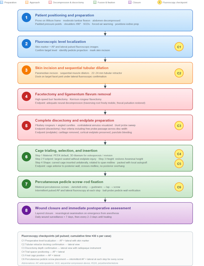
Figure 1: Procedural workflow for minimally invasive transforaminal lumbar interbody fusion. The eight sequential steps of the procedure are shown from top to bottom, color-coded by phase: preparation (steps 1 and 2), surgical approach (step 3), neural decompression and discectomy (steps 4 and 5), interbody fusion and pedicle screw fixation (steps 6 and 7), and wound closure and postoperative assessment (step 8). Yellow circular markers (C1–C6) indicate the six pre-specified intraoperative fluoroscopy checkpoints, all performed using pulsed fluoroscopy with a cumulative average of approximately 30 s per case across the present cohort. Key procedural endpoints — for complete discectomy, endplate preparation, neural decompression, and cage positioning — are summarized within their corresponding step boxes. Cage selection follows a four-step decision algorithm (material, footprint, height, shape) detailed in step 6. Abbreviations: AP, anteroposterior; SCD, sequential compression device; PEEK, polyetheretherketone. Please click here to view a larger version of this figure.
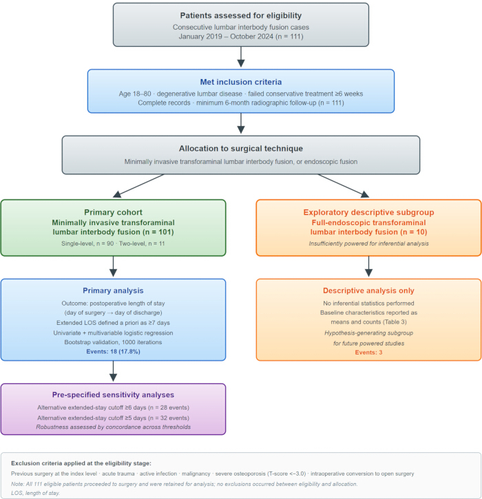
Figure 2: Representative intraoperative and fluoroscopic images from a single-level minimally invasive transforaminal lumbar interbody fusion case in the present cohort. (A) Tubular retractor docking. Paired tubular working channels (22–24 mm) are anchored to the operating table rail and the mobile C-arm is positioned for lateral fluoroscopy (Checkpoint C2 in Figure 1). (B) Endoscopic view during neural decompression. The traversing nerve root is exposed after ligamentum flavum removal; cottonoid patties control epidural venous bleeding (Checkpoint C3–C4 in Figure 1). (C) Final anteroposterior fluoroscopy (Checkpoint C6). Four percutaneous pedicle screws with bilateral rods are shown; screw trajectories are contained within the pedicle margins and parallel to the superior endplates. (D) Final lateral fluoroscopy (Checkpoint C6). The interbody cage is centrally positioned within the anterior two-thirds of the disc space, restoring disc height, with bilateral pedicle screws and rods forming a stable construct. Together, panels A–D illustrate the four pre-specified procedural endpoints used in this protocol (tubular docking, neural decompression, cage positioning, and instrumentation). Please click here to view a larger version of this figure.
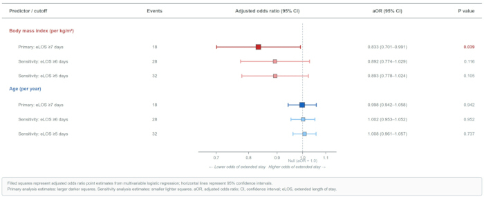
Figure 3: Cohort selection and allocation. The flow diagram summarizes patient eligibility, inclusion, and allocation to the primary minimally invasive transforaminal lumbar interbody fusion cohort and to the exploratory endoscopic subgroup. The primary cohort (n = 101) comprised 90 single-level and 11 two-level cases and was analyzed by univariate and multivariable logistic regression with bootstrap validation and two pre-specified sensitivity analyses at alternative extended length-of-stay thresholds. The exploratory endoscopic subgroup (n = 10, 3 events) was analyzed descriptively only and is presented for hypothesis generation; no inferential statistics were performed on this subgroup. Exclusion criteria are listed in the footer panel. No exclusions occurred between eligibility and allocation; all 111 eligible patients proceeded to surgery and were retained for analysis. LOS, length of stay. Please click here to view a larger version of this figure.
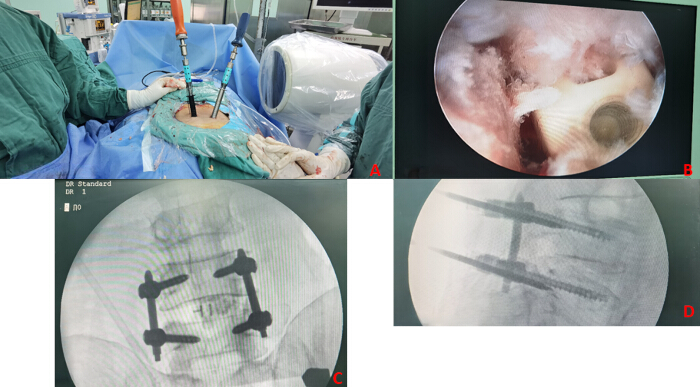
Figure 4: Forest plot of multivariable logistic regression results for extended postoperative length of stay. Adjusted odds ratios and 95% confidence intervals are shown for the two predictors retained in the multivariable model (body mass index, red; age, blue). For each predictor, results are shown for the primary analysis (pre-specified cutoff of postoperative length of stay ≥7 days) and for both pre-specified sensitivity analyses (cutoffs of ≥6 days and ≥5 days). Primary analysis estimates are shown as larger darker squares, and sensitivity analysis estimates as smaller lighter squares. The dashed vertical line represents the null value of adjusted odds ratio = 1.0. Body mass index showed a marginally significant inverse association with extended postoperative length of stay at the primary threshold (adjusted odds ratio 0.833, 95% confidence interval 0.701–0.991, p = 0.039), but this association did not persist at either sensitivity threshold (p = 0.116 at ≥6 days; p = 0.105 at ≥5 days), indicating that the finding is not robust to the choice of threshold. Age was not associated with extended postoperative length of stay at any threshold. aOR, adjusted odds ratio; CI, confidence interval; eLOS, extended length of stay. Please click here to view a larger version of this figure.
| Characteristic | All (n=101) | Non-eLOS (n=83) | eLOS ≥7 days (n=18) | p-value |
| DEMOGRAPHICS | ||||
| Age, years, mean ± SD | 60.0 ± 9.0 | 60.0 ± 9.2 | 60.1 ± 8.4 | 0.956 |
| Male sex, n (%) | 48 (47.5) | 40 (48.2) | 8 (44.4) | 0.977 |
| Height, cm, mean ± SD | 1.6 ± 0.1 | 1.6 ± 0.1 | 1.6 ± 0.1 | 0.879 |
| Weight, kg, mean ± SD | 60.6 ± 8.5 | 61.4 ± 8.5 | 56.8 ± 7.9 | 0.036 |
| Body mass index, kg/m², mean ± SD | 24.8 ± 3.2 | 25.2 ± 3.2 | 23.4 ± 3.1 | 0.039 |
| Underweight (<18.5), n (%) | 3 (3.0) | 2 (2.4) | 1 (5.6) | 0.449 |
| Normal (18.5–23.9), n (%) | 32 (31.7) | 25 (30.1) | 7 (38.9) | 0.656 |
| Overweight (24–27.9), n (%) | 50 (49.5) | 41 (49.4) | 9 (50.0) | 1.000 |
| Obese (≥28), n (%) | 16 (15.8) | 15 (18.1) | 1 (5.6) | 0.292 |
| PREOPERATIVE CLINICAL STATUS | ||||
| Preoperative CRP, mg/L, median (IQR) | 1.7 (1.0–6.5) | 1.4 (1.0–6.5) | 6.5 (1.1–6.5) | 0.106 |
| Preoperative VAS back pain, mean ± SD | 5.9 ± 0.4 | 5.9 ± 0.4 | 5.8 ± 0.5 | 0.753 |
| Preoperative VAS leg pain, mean ± SD | 5.0 ± 0.7 | 4.9 ± 0.7 | 5.1 ± 0.6 | 0.411 |
| Preoperative ODI, mean ± SD | 59.1 ± 3.3 | 59.0 ± 3.4 | 59.2 ± 2.8 | 0.797 |
| SURGICAL CHARACTERISTICS | ||||
| Single-level fusion, n (%) | 90 (89.1) | 74 (89.2) | 16 (88.9) | 1.000 |
| Two-level fusion, n (%) | 11 (10.9) | 9 (10.8) | 2 (11.1) | 1.000 |
| Operative time, min, mean ± SD | 153.6 ± 41.9 | 151.6 ± 36.9 | 162.8 ± 60.5 | 0.458 |
| Estimated blood loss, mL, median (IQR) | 200.0 (200.0–300.0) | 200.0 (200.0–300.0) | 200.0 (100.0–387.5) | 0.519 |
| POSTOPERATIVE OUTCOMES | ||||
| Postoperative LOS, days, median (IQR) | 4.0 (3.0–6.0) | 3.0 (3.0–4.0) | 9.0 (7.2–10.0) | — |
| Any complication, n (%) | 3 (3.0) | 2 (2.4) | 1 (5.6) | 0.449 |
| Fusion achieved, n (%) | 101 (100.0) | 83 (100.0) | 18 (100.0) | 1.000 |
Table 1: Baseline characteristics and perioperative variables comparing patients with and without extended postoperative length of stay in the MIS-TLIF cohort (n = 101). Extended postoperative length of stay was pre-specified as ≥ 7 days (cohort 75th percentile). Data are presented as mean ± standard deviation for continuous variables and n (%) for categorical variables. P-values were calculated using Student's t-test or the Mann–Whitney U test for continuous variables, and Fisher's exact test or chi-square test for categorical variables, as appropriate. Bold values indicate statistical significance (p < 0.05). BMI, body mass index; CRP, C-reactive protein; eLOS, extended length of stay; ODI, Oswestry Disability Index; VAS, visual analog scale.
| Variable | Univariate OR (95% CI) | Univariate p | Multivariable aOR (95% CI) | Multivariable p | Bootstrap 95% CI | ||
| Age (per year) | 1.002 (0.946–1.060) | 0.958 | — | — | — | ||
| Male sex | 0.860 (0.309–2.396) | 0.773 | — | — | — | ||
| Body mass index (per kg/m²) | 0.834 (0.701–0.991) | 0.039 | — | — | — | ||
| Preoperative CRP (per mg/L) | 1.003 (0.969–1.039) | 0.858 | — | — | — | ||
| Two-level fusion (vs single) | 1.028 (0.202–5.218) | 0.974 | — | — | — | ||
| Operative time (per min) | 1.006 (0.995–1.018) | 0.307 | — | — | — | ||
| Estimated blood loss (per mL) | 1.000 (0.997–1.004) | 0.820 | — | — | — | ||
| Preoperative VAS back pain (per point) | 0.826 (0.261–2.617) | 0.745 | — | — | — | ||
| Preoperative VAS leg pain (per point) | 1.308 (0.635–2.691) | 0.466 | — | — | — | ||
| Preoperative ODI (per point) | 1.019 (0.871–1.192) | 0.816 | — | — | — | ||
| Any perioperative complication | 2.382 (0.204–27.794) | 0.489 | — | — | — | ||
| MULTIVARIABLE MODEL (primary, eLOS ≥7 days) | |||||||
| Body mass index (per kg/m²) | — | — | 0.833 (0.701–0.991) | 0.039 | 0.678–0.991 | ||
| Age (per year) | — | — | 0.998 (0.942–1.058) | 0.942 | 0.935–1.056 | ||
Table 2: Univariate and multivariable logistic regression analyses identifying predictors of extended postoperative length of stay (≥ 7 days) in the MIS-TLIF cohort (n = 101). Values represent odds ratios (OR) and adjusted odds ratios (aOR) with 95% confidence intervals (CI) and p-values from Wald tests. The multivariable model was adjusted for age (clinically essential covariate) and entered variables reaching p < 0.10 in univariate analysis, in accordance with the approximately 10-events-per-variable rule given 18 extended-stay events. Bootstrap 95% confidence intervals were derived from 1000 iterations of nonparametric resampling. Bold values indicate statistical significance (p < 0.05). BMI, body mass index; CI, confidence interval; CRP, C-reactive protein; ODI, Oswestry Disability Index; OR, odds ratio; VAS, visual analog scale.
| Characteristic | All Patients (n=10) | Non-eLOS (n=7, 70%) | eLOS (n=3, 30%) |
| CAUTION: Small sample size (n=10, 3 events) precludes formal statistical inference. Minimum recommended n≥80. | |||
| DEMOGRAPHICS | |||
| Age (years) | 59.2 ± 7.1 | 58.6 ± 7.8 | 60.7 ± 6.2 |
| Male sex, n (%) | 6 (60.0) | 4 (57.1) | 2 (66.7) |
| ANTHROPOMETRIC MEASURES | |||
| Height (cm) | 165.3 ± 8.4 | 164.8 ± 9.2 | 166.7 ± 7.1 |
| Weight (kg) | 74.8 ± 13.2 | 73.2 ± 14.6 | 78.7 ± 9.8 |
| Body Mass Index (kg/m²) | 27.3 ± 3.9 | 26.8 ± 4.2 | 28.6 ± 3.1 |
| Comparison to MIS-TLIF: BMI +4.0 kg/m² higher (p=0.02) | |||
| PREOPERATIVE CLINICAL STATUS | |||
| VAS Back Pain (0-10) | 6.9 ± 1.3 | 6.7 ± 1.4 | 7.3 ± 1.2 |
| VAS Leg Pain (0-10) | 7.4 ± 1.5 | 7.3 ± 1.6 | 7.7 ± 1.5 |
| ODI Score (0-100) | 59.8 ± 11.4 | 58.2 ± 12.1 | 63.7 ± 9.6 |
| SURGICAL VARIABLES | |||
| Time admission to surgery (days) | 4.8 ± 2.3 | 4.4 ± 2.1 | 5.7 ± 2.9 |
| Operative time (minutes) | 235.0 ± 78.4 | 227.1 ± 82.3 | 257.3 ± 72.1 |
| Estimated blood loss (mL) | 130.0 ± 78.9 | 118.6 ± 72.4 | 160.0 ± 98.6 |
| Multi-level surgery, n (%) | 3 (30.0) | 2 (28.6) | 1 (33.3) |
| Comparison to MIS-TLIF: Operative time +76.4 min longer (p<0.001) | |||
| Comparison to MIS-TLIF: Blood loss -113.8 mL less (p=0.005) | |||
| POSTOPERATIVE OUTCOMES | |||
| Postoperative CRP Day 3 (mg/L) | 38.2 ± 15.6 | 36.8 ± 16.8 | 41.7 ± 13.2 |
| Complications, n (%) | 0 (0) | 0 (0) | 0 (0) |
| Radiographic fusion at 6 mo, n (%) | 10 (100) | 7 (100) | 3 (100) |
| HOSPITAL COURSE | |||
| Length of stay (days) | 14.2 ± 4.1 | 12.6 ± 3.2 | 18.7 ± 2.5 |
| Total hospital costs (CNY) | 31898 ± 5842 | 29153 ± 4212 | 39426 ± 4008 |
| Total hospital costs (USD)† | 4430 ± 811 | 4049 ± 585 | 5476 ± 557 |
| Comparison to MIS-TLIF: eLOS rate 30% vs 75.2% (p=0.003) | |||
| Comparison to MIS-TLIF: Mean LOS -2.4 days shorter (p=0.18) |
Table 3: Descriptive characteristics of the exploratory full-endoscopic transforaminal lumbar interbody fusion cohort (n = 10). Data are presented as mean ± standard deviation for continuous variables and n (%) for categorical variables. No inferential statistics were performed because the event count (3 events) is insufficient to support multivariable regression. These data are presented for descriptive purposes and to inform sample size calculations for future adequately powered comparative studies. BMI, body mass index; CRP, C-reactive protein; ODI, Oswestry Disability Index; VAS, visual analog scale.
Supplementary Table 1: Pre-specified sensitivity analyses at alternative thresholds for extended postoperative length of stay in the MIS-TLIF cohort. Multivariable logistic regression was repeated at two alternative pre-specified thresholds: ≥ 6 days (28 events) and ≥ 5 days (32 events). The model retained the same predictors as the primary analysis. The non-persistence of the body mass index association across alternative thresholds indicates that the signal observed at the primary cutoff is not robust to the choice of threshold.Please click here to download this file.
Supplementary Material 1: Full operator qualifications, recommended training pathway for new operators, and general teaching principles. Full itemized criteria, all 101 MIS-TLIF procedures, and the 10 full-endoscopic procedures were performed by single board-certified spine surgeons meeting the training requirements. Management protocols for seven recurring intraoperative challenges encountered during the reproduction of this technique. Core safety considerations spanning patient positioning, radiation, neurological, vascular, and infection control are integrated throughout the Protocol at each procedural step. Pre-specified sensitivity analyses at two alternative thresholds for extended postoperative LOS Please click here to download this file.
Discussion
In this retrospective single-center cohort of 101 consecutive patients who underwent MIS-TLIF, median postoperative LOS was 4 days, a value consistent with published international benchmarks of 2–5 days for this procedure23. Under a pre-specified and clinically grounded threshold of 7 days or longer, 17.8% of patients were classified as having extended postoperative LOS. In univariate and multivariable logistic regression, BMI was the only candidate predictor that reached the screening threshold, showing a marginal inverse association with extended postoperative stay at the primary cutoff. However, this association did not persist in pre-specified sensitivity analyses at alternative thresholds of 6 days or longer and 5 days or longer. The principal and most defensible finding of the present study is therefore a null one: no patient-level perioperative factor was robustly and independently associated with extended postoperative LOS after MIS-TLIF in this cohort.
The marginal inverse association between BMI and extended postoperative LOS observed at the primary threshold warrants explicit discussion, precisely because it is counterintuitive and because similar non-robust findings are frequently over-interpreted in the single-center literature. Three considerations argue against a causal or clinically actionable interpretation. First, the direction of the association — higher body mass index linked to lower odds of extended stay — is opposite to the direction expected if adiposity acted as a direct impediment to surgical recovery, and is not supported by the broader spine surgery literature24. Second, the association did not persist across pre-specified alternative thresholds, and the lower bound of the bootstrap CI approached the null. Third, residual confounding by unmeasured body-composition and frailty variables, including sarcopenia and other validated indices that the present dataset did not capture25, cannot be excluded in a cohort of this size with 18 extended-stay events. The BMI signal is therefore best interpreted as a statistical artifact of threshold selection and sample size rather than as evidence of a protective effect, and should not be used to guide clinical decision-making.
Direct comparison of total hospital LOS across healthcare systems is complicated by substantial differences in preoperative workup pathways, reimbursement structures, and discharge practices. In Chinese tertiary referral centers, total hospital stay routinely incorporates several days of preoperative imaging, multidisciplinary consultation, and optimization that are not counted toward length of stay in many Western systems, where admission typically occurs on or shortly before the day of surgery. When length of stay is measured from the day of surgery to the day of discharge, as was done in the present study, the median of 4 days in this cohort aligns closely with benchmarks reported for MIS-TLIF in published series22,23. This observation supports the view that the surgical recovery profile of MIS-TLIF at this center is comparable to that reported internationally, and that earlier reports of prolonged LOS in Chinese cohorts may reflect preoperative rather than postoperative processes.
The clinical application of these findings is primarily cautionary. In a well-functioning single-center MIS-TLIF program, routine patient-level perioperative variables — age, sex, BMI, preoperative inflammatory markers, symptom severity, number of fused levels, operative time, and intraoperative blood loss — did not identify patients at materially increased risk of extended postoperative stay. Optimization efforts targeted exclusively at these variables may therefore yield limited returns in settings where postoperative LOS is already close to international benchmarks. Alternative approaches that merit consideration in future work include (i) system-level factors such as standardized preoperative and discharge pathways26,27; (ii) ERAS protocols, which bundle multiple small perioperative interventions rather than relying on any single patient-level optimization and have demonstrated reductions in LOS in spine surgery populations without increased complications or readmissions28,29; and (iii) endoscopic transforaminal lumbar interbody fusion as a technical alternative, which the present descriptive exploratory subgroup was insufficiently powered to evaluate and for which adequately powered comparative studies are needed.
This study has several important limitations. First, it is a single-center retrospective analysis, and findings may not generalize to centers with different patient populations, surgical volumes, or perioperative pathways. Second, the event count for the primary outcome (18 extended-stay events) constrained the multivariable model to a small number of predictors and limited statistical power to detect modest associations; the null result should not be read as strong evidence of no effect for any individual variable. Third, the primary outcome captures only the postoperative interval and does not account for social, administrative, or bed-availability factors that may influence the actual day of discharge, and these unmeasured factors represent a plausible source of residual confounding. Fourth, BMI was analyzed as a continuous variable and by standard Chinese classification categories; alternative parameterizations — including validated frailty and sarcopenia assessments, body composition analysis, and serum nutritional markers — were not available and may carry prognostic information not captured by BMI alone25. Fifth, the exploratory endoscopic cohort was small (n = 10, 3 events) and was analyzed descriptively only; no inferential conclusions regarding endoscopic versus non-endoscopic techniques can be drawn from the present data. Sixth, follow-up attendance at the six-month visit was approximately 85 percent, and outcome variables collected at that timepoint — including the radiographic fusion assessment and the six-month ODI — were therefore subject to attrition bias. Patients lost to follow-up at six months may differ systematically from those who attended follow-up in ways that could affect the interpretation of late-outcome variables. Seventh, bone mineral density was assessed by dual-energy X-ray absorptiometry in only a subset of the cohort, and osteoporosis status in the remaining patients was determined by clinical and radiographic criteria at the time of surgical planning. Non-uniform bone density assessment may have introduced minor variability in cage material selection, although the primary outcome of this study was postoperative LOS rather than any cage-related endpoint.
Three lines of future work are suggested by these findings. First, adequately powered multicenter prospective studies are needed to identify whether any patient-level perioperative variable carries a reproducible independent association with extended postoperative LOS after MIS-TLIF; single-center cohorts of this size are unlikely to resolve the question. Second, system-level interventions — including standardized discharge criteria and ERAS protocols — should be evaluated prospectively at the study center to determine whether they shorten postoperative LOS in a setting where the current median is already close to international benchmarks28,29. Third, adequately powered head-to-head comparisons of endoscopic and non-endoscopic MIS-TLIF, with pre-specified matching on age, sex, and number of fused levels as suggested in the reviewer commentary and ideally employing propensity-score or related causal-inference methods29, are needed to determine whether endoscopic techniques offer meaningful advantages in postoperative recovery.
Disclosures
The authors declare no competing interests. None of the authors has any financial or personal relationship with any of the equipment or reagent manufacturers whose products are listed in the Table of Materials that could be perceived as influencing the conduct or interpretation of the present work.
Acknowledgements
This study was supported by the Chongqing Municipal Hechuan District Research Project (grant number HCKJ-2025-055) and by the Chongqing Science and Health Joint Medical Research Project (grant number 2026MSXM127). The funding sources had no role in the study design, data collection, data analysis, data interpretation, writing of the manuscript, or the decision to submit the manuscript for publication. The authors thank the staff of the Department of Orthopedics at Hechuan District People's Hospital for their clinical support during the study period.
Materials
| Name | Company | Catalog Number | Comments |
|---|---|---|---|
| Bipolar electrocautery system | Suzhou Aikeshuo Technology Co., Ltd. | ACS400 | Commercial bipolar electrocautery unit used for coagulation of soft tissue and control of epidural venous bleeding. Any standard surgical bipolar unit may be substituted. |
| C-arm fluoroscopy unit | Sinowell | HMC-160D 9000 | Commercial mobile C-arm fluoroscopy unit operated in pulsed fluoroscopy mode. Used for all intraoperative fluoroscopic imaging and the six pre-specified fluoroscopy checkpoints. Any equivalent commercial mobile C-arm fluoroscopy unit may be substituted. |
| Chlorhexidine–alcohol antiseptic skin preparation | Shanghai Likang Disinfection High-Tech Co., Ltd. | 31004301 | Commercial antiseptic skin preparation solution used per institutional antiseptic protocol prior to draping. Any equivalent commercial chlorhexidine–alcohol antiseptic solution may be substituted. |
| Endoscopic camera and light-source platform | Stryker Corporation | TS88 | Commercial high-definition endoscopic camera and light-source platform used with the rigid working-channel lumbar endoscope for the exploratory endoscopic subgroup. Any equivalent commercial endoscopic imaging platform may be substituted. |
| Fibrin sealant | Hualan Bio-Engineering Co., Ltd. | 20020084 | Commercial topical hemostatic agent available on the surgical field for the management of incidental durotomy. Any equivalent commercial fibrin sealant may be substituted. |
| Flowable gelatin matrix hemostatic agent | Johnson & Johnson, SURGIFLO | MS0010, SF2995, SF2994, SF2991 | Commercial topical hemostatic agent used for intraoperative control of epidural venous bleeding. Any equivalent commercial flowable gelatin matrix hemostatic agent may be substituted. |
| Forced-air patient warming system | Keewell Medical Technology | KPW4000 | Commercial forced-air warming system applied to the non-surgical body surface to maintain intraoperative normothermia. Any equivalent commercial forced-air warming system may be substituted. |
| High-sensitivity CRP turbidimetric immunoassay | Roche Diagnostics | Roche Cobas 6000 | Commercial high-sensitivity turbidimetric immunoassay used for quantification of serum C-reactive protein at the preoperative and postoperative day 3 timepoints (institutional normal range below 10 mg/L). Any validated commercial high-sensitivity CRP assay may be substituted. |
| High-speed surgical drill | Xishantech | Powered Devices for Orthopedic (Spinal) Surgery | Commercial high-speed surgical drill used with diamond and matchstick burrs for facetectomy and bony decompression. Any equivalent commercial high-speed surgical drill may be substituted. |
| Kerrison rongeurs | Tianjin Tianlong Medical Devices Co., Ltd. | 112981600 | Standard surgical Kerrison rongeurs used for ligamentum flavum removal during neural decompression. Any equivalent commercial Kerrison rongeur set may be substituted. |
| Oxidized regenerated cellulose hemostatic agent | Ethicon (Johnson & Johnson) | 1952, 1953, W1911, W1912, W1913T, 3013SP, 3123SPEA | Commercial topical hemostatic agent used for intraoperative control of epidural venous bleeding. Any equivalent commercial oxidized regenerated cellulose hemostatic agent may be substituted. |
| Pedicle screw and rod system (6.0–7.5 mm) | Tianjin Tianlong Medical Devices Co., Ltd. | 4.5*25,4.5*30,4.5*35,5.0*30,5.0*35, 5.0*40,5.5*30,5.5*40,5.5*45,6.0*35, 6.*40,6.0*45,6.0*50,6.5*35,6.5*40, 6.5*55,6.5*50,7.0*35,7.0*40, 7.0*45,7.0*50 | Commercial percutaneous pedicle screw and rod system used for posterior fixation; screw diameters stocked in the range of 6.0–7.5 mm. Any equivalent commercial percutaneous pedicle screw and rod system may be substituted. |
| PEEK interbody cage system (heights 8–14 mm; widths 22–30 mm) | Tianjin Tianlong Medical Devices Co., Ltd. | 060001-060021 | Commercial polyetheretherketone (PEEK) interbody cage system used for interbody fusion in primary MIS-TLIF cases; available in heights 8–14 mm and widths 22–30 mm. Any equivalent commercial PEEK interbody cage system may be substituted. |
| Pituitary rongeurs and curettes | Double Medical Technology Co., Ltd. | S1210.20Ti–S1210.50Ti; S1212.20Ti–S1212.50Ti | Standard surgical pituitary rongeurs and curettes used for discectomy and endplate preparation. Any equivalent commercial surgical set may be substituted. |
| Povidone–iodine 10% antiseptic skin preparation | Shanghai Likang Disinfection High-Tech Co., Ltd. | Product URL: https://www.lkgk.net/pages/157 | Commercial antiseptic skin preparation solution used per institutional antiseptic protocol prior to draping. Any equivalent commercial povidone–iodine 10% solution may be substituted. |
| R (statistical software), version 4.5.1 | R Foundation for Statistical Computing | RRID:SCR_001905 | Open-source statistical environment used for univariate and multivariable logistic regression and for nonparametric bootstrap resampling (1000 iterations). Any equivalent statistical software supporting logistic regression and resampling may be substituted. |
| Sequential compression device for VTE prophylaxis | Daesung Industrial Co., Ltd. | DSM-600S | Commercial sequential compression device applied bilaterally to the lower extremities before induction of anesthesia for venous thromboembolism prophylaxis. Any equivalent commercial sequential compression device may be substituted. |
| SPSS (statistical software), version 27 | IBM Corp. | RRID:SCR_002865 | Commercial statistical software used for descriptive statistics and univariate / multivariable logistic regression. Any equivalent commercial or open-source statistical software may be substituted. |
| Three-dimensionally printed titanium interbody cage | Hunan Huaxiang Medical Technology Co., Ltd. | LV-I | Commercial 3D-printed titanium interbody cage used in osteoporotic patients and revision cases in accordance with the pre-specified cage-selection algorithm. Any equivalent commercial 3D-printed titanium interbody cage may be substituted. |
| Tubular retractor system (working channel 22–24 mm) | Tianjin Tianlong Medical Devices Co., Ltd. | 112980600 | Commercial minimally invasive tubular retractor system with serial muscle dilators and a 22–24 mm final working channel, anchored to the operating table rail by a manufacturer-supplied articulated arm. Any equivalent commercial minimally invasive tubular retractor system may be substituted. |
| Uniportal lumbar endoscope (rigid working channel) | Stryker Spinal Endoscopy | 1588 AIM | Commercial rigid working-channel lumbar endoscope used for the exploratory full-endoscopic transforaminal lumbar interbody fusion subgroup, under continuous saline irrigation at a target working-channel pressure of 30–45 mmHg. Any equivalent commercial rigid working-channel lumbar endoscope may be substituted. |
| Wilson frame, radiolucent operating table attachment | Mizuho OSI | Model 5319G | Commercial radiolucent lumbar positioning frame fitted to the operating table for prone positioning; adjusted to produce moderate lumbar flexion for the minimally invasive transforaminal approach. Any equivalent commercial radiolucent lumbar positioning frame may be substituted. |
Reprints and Permissions
This article has been published
Video Coming Soon