Fetal edema was identified through prenatal ultrasound of the fetal system in combination with comprehensive maternal clinical characteristics, laboratory analyses, and perinatal outcomes. An analysis was conducted on thirteen patients diagnosed with mirror syndrome.
Method Article
Examination of the Utility of Ultrasound in Prenatal Diagnosis and Perinatal Outcomes for 13 Instances of Mirror Syndrome
In This Article
Summary
Abstract
This study investigated the diagnostic utility of prenatal ultrasound in identifying mirror syndrome, drawing on the existing literature. A comprehensive series of assessments was undertaken on fetal growth and development, organogenesis, and ancillary structures, including amniotic fluid and placenta, using systematic prenatal ultrasonography in conjunction with hemodynamic evaluations. In cases where fetal edema was identified, additional chromosomal karyotyping and chromosomal microarray analyses were recommended. An analysis of 13 patients diagnosed with mirror syndrome and admitted to the Women’s Hospital, Zhejiang University School of Medicine, from January 2020 to July 2025 was conducted. The medical information of these patients was examined, encompassing clinical characteristics, evaluation of fetal and fetal appendage status, laboratory analyses, and perinatal outcomes. The prevalent maternal clinical manifestations included bilateral lower limb edema (100%), hypertension (84.6%), anemia (69.2%), and proteinuria (38.4%). Almost all cases presented with decreased hematocrit and serum albumin levels. In the study, 13 instances exhibited placental thickness > 4 cm, while eleven instances presented placental thickness > 6 cm. Pathological examination of the placenta revealed villous edema in eight cases. Mirror syndrome is rare; however, the fetal prognosis is poor. Early and precise diagnosis of mirror syndrome is essential for improving outcomes for both the mother and the fetus.
Introduction
Mirror syndrome (MS), also known as Ballantyne syndrome, is a rare but significant obstetric condition. Literature estimates suggest an incidence rate of 1/(5,800–10,000)1, and the etiology and pathogenesis remain unclear. It was first described in the medical literature in 1892, with an initial case report involving maternal hydrops associated with concurrent fetal and placental hydrops2.
The etiology of fetal edema is complex and diverse. It includes immunological causes such as maternal–fetal blood group incompatibility, as well as non-immunological causes such as various genetic syndromes, intrauterine infections, and fetal structural anomalies. Clinically, non-immunological causes are more frequently observed.
Regardless of the etiology, fetal edema typically occurs due to increased central venous pressure, reduced lymphatic flow, or decreased colloid osmotic pressure3. Mirror syndrome can lead to a fetal mortality rate as high as 67.3% and may cause severe maternal complications4. Cases associated with Rh(D) alloimmunization during pregnancy have also been reported5.
The clinical manifestations of this syndrome can be complex. It may present with or without the classic signs of hypertension and proteinuria. Additionally, it often includes other clinical features that closely resemble those of preeclampsia, leading to potential diagnostic confusion. Although mirror syndrome is an uncommon occurrence, its presence is critically important because the prognosis for the affected fetus is generally poor. The clinical presentation of mirror syndrome is notably heterogeneous, which contributes to its susceptibility to being overlooked or incorrectly diagnosed in clinical practice.
A meta-analysis included 56 cases of mirror syndrome showing weight gain and maternal edema (89.3%) were the most common symptoms, followed by elevated blood pressure (60.7%), mild anemia and blood dilution (46.4%), proteinuria (42.9%), elevated uric acid levels (25%), mild elevation of liver enzymes (19.6%), oliguria (16.1%), and headache with visual disturbances (14.3%)6.
There are currently no reported cases of maternal mortality caused by mirror syndrome. In addition to typical maternal clinical manifestations, maternal blood parameters also exhibit certain changes in mirror syndrome cases. Comparative studies between fetuses with and without mirror syndrome edema have revealed that the mirror syndrome group not only shows edema diagnosed earlier in gestation and more severe edema, but also has elevated maternal serum hCG, uric acid, lactate dehydrogenase, and D-dimer levels, as well as more pronounced dilution anemia. These biomarkers may serve as predictive indicators of mirror syndrome edema in fetuses7,8.
Two hypotheses have been proposed regarding the possible pathophysiological mechanisms of mirror syndrome. Firstly, when presenting primarily with hypertension and proteinuria (i.e., preeclampsia-like changes), the similarity in clinical manifestations has led some scholars to propose that the pathophysiological mechanisms of preeclampsia may also contribute to the occurrence of mirror syndrome5,9,10. This pathological process resembles that of preeclampsia, complicating the diagnostic process. Laboratory examinations for preeclampsia are characterized by hemoconcentration, with normal hematocrit, severe hypertension, and proteinuria. Conversely, laboratory findings in mirror syndrome include hemodilution, including dilutional anemia, decreased hematocrit, and reduced serum albumin levels. Preeclampsia is typically diagnosed after 20 weeks of gestation, whereas mirror syndrome can manifest as early as 16 weeks11. Mirror syndrome may coincide with hydrops fetalis, polyhydramnios, and placental enlargement, whereas preeclampsia often presents with intrauterine growth restriction, reduced amniotic fluid, and a diminutive placenta. Differentiating mirror syndrome from preeclampsia is crucial, as treatment modalities and fetal prognoses vary significantly. Although mirror syndrome shares clinical similarities with preeclampsia, placental formation disorders are not its primary etiological factor. The damaged placenta is merely a potential source of certain molecular mediators, and the pathogenesis of mirror syndrome cannot be fully explained by the mechanisms underlying preeclampsia.
Secondly, when edema is the primary manifestation, the placenta is enlarged, and histology reveals significant interstitial edema of the chorionic villi8. Trophoblastic injury caused by placental edema leads to an imbalance of pro-angiogenic factors such as soluble intravascular endothelial growth factor receptor 1 (sFlt-1), placental growth factor (PIGF), anti-angiogenic factors, and soluble endothelial glycoprotein (sEng). This ultimately results in maternal endothelial dysfunction and the clinical manifestations of mirror syndrome, including hypertension and proteinuria12–17, and may cause maternal edema. Literature reports indicate significantly elevated maternal serum hCG levels in mirror syndrome, which may be associated with placental ischemic injury caused by tissue edema changes7,18,19. Compared to preeclampsia, maternal edema in mirror syndrome is more pronounced than hypertension and proteinuria, which may be related to elevated serum hCG levels20.
In this context, the present study conducts a retrospective analysis of 13 cases of mirror syndrome managed at our medical institution. The primary objective of this investigation is to provide a foundation for enhancing the diagnostic accuracy and therapeutic management strategies for patients with this condition.
Protocol
This study was conducted in accordance with the Declaration of Helsinki and was approved by the Medical Ethics Committee of the Women’s Hospital, Zhejiang University School of Medicine (Approval No. IRB-20250409-R). The principle of informed consent was followed throughout the study, and consent was obtained. The equipments used in this study are listed in the Table of Materials.
This was a retrospective, single-center, observational study encompassing consecutive eligible cases diagnosed with mirror syndrome at the Women’s Hospital, Zhejiang University School of Medicine, from January 2020 to July 2025. Patients were identified from electronic medical records, the obstetric ultrasound database, and the perinatal registration system using standardized diagnostic criteria.
The inclusion process was carried out in steps. Initially, all cases with suspected mirror syndrome were screened. Only patients who had completed standardized prenatal ultrasound examinations in accordance with ISUOG guidelines and had comprehensive medical data were retained. Exclusion criteria comprised incomplete medical records, pre-pregnancy chronic hypertension, diabetes mellitus, heart disease, liver disease, or renal disease. All eligible cases were consecutively enrolled to prevent selection bias. Given the retrospective design, potential selection bias stemming from missing data or referral patterns was recognized; however, standardized screening, thorough data review, and consecutive enrollment were implemented to minimize such bias.
Diagnostic criteria for mirror syndrome
The diagnosis of mirror syndrome (Ballantyne syndrome) was established based on the classic triad widely recognized in the literature4˒6˒21. All enrolled cases strictly fulfilled all three components of the standard definition; no cases were diagnosed using partial or incomplete criteria.
The standardized diagnostic triad was defined as follows. Maternal manifestations included generalized or lower-extremity edema, plus hypertension and/or proteinuria. Fetal hydrops was defined as ultrasonographic evidence of at least two abnormal fluid collections (pleural, peritoneal, or pericardial effusion) or generalized skin edema. Placental hydrops was defined as placental thickness > 4 cm before 20 weeks of gestation and > 6 cm after 21 weeks of gestation, with pathological confirmation of villous edema when available.
These criteria were uniformly applied across all 13 cases to ensure diagnostic uniformity and reproducibility.
Ultrasound evaluation and its association with diagnostic decision-making
All ultrasound findings were systematically integrated into the diagnostic criteria for mirror syndrome. Fetal edema, placental thickness, and effusions were quantitatively measured and transformed into objective diagnostic thresholds as previously defined.
Fetal edema was diagnosed when an ultrasound revealed at least two abnormal fluid collections or generalized skin edema. Placental thickness was measured at the thickest non-cord insertion site, and thresholds of > 4 cm before 20 weeks of gestation and > 6 cm at or after 21 weeks of gestation were adopted as ultrasonographic criteria for placental hydrops. These measurements directly constituted the fetal and placental components of the classic triad.
To ensure standardization and consistency among operators, all examinations were carried out in accordance with ISUOG guidelines for fetal systematic ultrasound. All ultrasound physicians participated in unified training and utilized the same machine presets, measurement methods, and scanning planes. Measurements of placental thickness and fetal edema were performed using standardized techniques to minimize inter-observer variability. All images were stored and reviewed by a senior attending physician to confirm diagnostic accuracy.
Genetic testing strategy
A standardized, stepwise genetic testing algorithm was applied for all fetuses with sonographic findings of hydrops, in accordance with clinical guidelines for fetal hydrops. Initial testing prioritized chromosomal karyotyping and chromosomal microarray analysis (CMA) to detect common aneuploidies and copy number variations. Whole-exome sequencing (WES) was selectively performed in cases with normal karyotype and CMA results but highly suspected genetic etiology. Non-invasive prenatal testing (NIPT) was used only for preliminary screening before invasive procedures and was not considered diagnostic. The testing strategy was therefore protocol-driven rather than arbitrary, with the choice of method determined by gestational age, prior results, and clinical necessity to maximize diagnostic yield while balancing invasiveness and cost.
1. Transducer selection
- Use ultrasound diagnostic equipment equipped with a 3.5–5.0 MHz abdominal convex probe.
2. Machine settings and placement
- Mode selection
- Initiate the machine and select the OB (Obstetrics) preset mode.
- Machine placement
- Position the ultrasound machine on either side of the pregnant woman, ensuring it is close enough to minimize distance.
- Position the pregnant woman on the right side of the machine for operator convenience.
3. Pregnant woman positioning
- Place the expectant mother in a supine reclined position with her abdomen fully exposed.
- Apply a coupling agent between the probe tip and the abdomen.
4. B Mode (2D) image adjustment (basic)
- Depth
- Adjust the depth of the field of view to ensure that structures of interest (e.g., fetus, placenta, amniotic fluid) are centered and occupy most of the screen.
- Time gain compensation (TGC)
- Adjust brightness at various depths to compensate for attenuation of ultrasound waves.
- Adjust the slider to achieve consistent brightness between near and far fields.
- Focal zones
- Set the focus at the depth of the structure of interest.
- Dynamic range
- Adjust contrast and grayscale range according to the structure being examined.
- Output power
- Regulate ultrasonic energy emitted into the patient.
- Follow the ALARA principle (As Low As Reasonably Achievable).
- B Mode optimization
- Adjust depth, frequency, focus, gain, and TGC to optimize image quality.
5. Doppler modulation (blood flow assessment)
- Use Doppler to assess fetal hemodynamics, including the umbilical artery (UmbilA) and middle cerebral artery (MCA).
- Color/Power Gain
- Adjust Doppler sensitivity to ensure vessels are filled with color without producing false signals.
- Scale/PRF
- Adjust maximum detectable flow rate.
- Use high PRF for high-flow applications and low PRF for low-flow applications.
- Sample Volume
- Center the sampling volume on the target vessel.
- Adjust size to 1/3–2/3 of vessel diameter.
- Wall Filter
- Filter out low-speed, high-amplitude signals.
- Use the lowest feasible setting to preserve low-velocity fetal blood flow signals.
- Angle Correction
- Maintain Doppler angle < 60°.
- Apply angle correction for accurate velocity measurement.
- Optimization of Doppler Mode
- Initiate color Doppler and adjust gain and PRF/Scale.
- Initiate PW Doppler and position the sampling volume.
- Set wall filter to minimum and apply angle correction if required.
6. Fetal system scan
- Perform transverse, longitudinal, and oblique scans.
- Orient the probe appropriately for coronal and sagittal views.
- Assess for fetal edema, including ascites, skin thickening, and thoracic, abdominal, or pericardial effusion.
- Measure fetal skin thickness.
- Evaluate associated malformations across major organ systems.
- Examine amniotic fluid, placenta, and other fetal structures.
NOTE: In cases of fetal abnormalities, consult a senior physician to confirm the diagnosis.
7. Image archiving
- Measure edema location, skin thickness, and effusion depth in fetuses with abnormal edema.
- Evaluate cardiac malformations, teratomas, amniotic fluid, and placental conditions.
8. Data acquisition and storage
- Image/Cinematic Loop Storage
- Freeze
Press the FREEZE key to pause the image. - Trackball Scroll
Review previous frames to identify the optimal image. - Store
Press the P1 or STORE button. - Cine Store
Press STORE during LIVE mode. - Continue scanning
Press UNFREEZE to resume scanning.
- Freeze
9. Image review / archive
- Access the archive and locate REVIEW, ARCHIVE, PATIENT, or EXAM functions and open the image browser.
- Select patient using the trackball and SET/Enter key to open patient data.
- Replay clip by selecting a clip and pressing SET to play.
- Process/measure again by selecting an image, zoom, and performing measurements or annotations.
10. Genetic analysis
- Perform chromosomal karyotype analysis and chromosomal microarray analysis (CMA) for fetuses with edema.
- Perform amniocentesis at 16–24 weeks and collect 30 mL of amniotic fluid.
- Perform umbilical cord blood sampling after 24 weeks and collect 3 mL of blood.
- Use ultrasound guidance for both procedures.
- Culture and analyze samples and monitor pregnancy outcomes.
11. Placental pathological analysis
- Specimen reception
- Submit placenta promptly for evaluation.
- Refrigerate at 4 °C or fix in formalin if delayed.
- Verify clinical data including gestational age, delivery mode, maternal history, and neonatal information.
- Weigh placenta and calculate fetal-placental weight ratio.
- Gross examination
- Measure placental dimensions and morphology.
- Examine fetal surface and chorionic vessels.
- Examine umbilical cord structure and vessels.
- Inspect maternal surface for infarcts or hematoma.
- Perform serial slicing at 1 cm intervals.
- Tissue sampling
- Collect standard samples from umbilical cord, membrane roll, and placental surface.
- Sample abnormal lesions separately.
- Fix all tissues in formalin and process for histological analysis.
- Microscopic examination and reporting
- Identify MVMA, chorioamnionitis, FVM, infection, and special lesions.
- Integrate microscopic and clinical findings for diagnosis.
- Prepare a comprehensive pathological report including macroscopic and microscopic findings.
Results
An analysis of 13 patients diagnosed with mirror syndrome and admitted to the Women’s Hospital, Zhejiang University School of Medicine, from January 2020 to July 2025 was conducted. The medical information of these patients was examined, encompassing clinical characteristics, evaluation of fetal and fetal appendage status, laboratory analyses, and perinatal outcomes. All pregnant women had no history of chronic hypertension or heart, liver, or kidney diseases. The pregnant women were aged 24–39 years, with an average age of (30.5 ± 3.59) years; 12 cases were singleton pregnancies, and 1 case was a twin pregnancy. Twelve cases were natural conception, and 1 case involved assisted reproduction. The gestational age at diagnosis ranged from 23+ to 36+.
The diagnostic criteria for classic triad mirror syndrome were as follows. Maternal edema: "+" indicates a symptom if the condition is limited to the area below the knee; "++" indicates extension to the thigh; "+++" indicates extension to the vulva and abdominal wall; and "++++" indicates generalized edema or edema accompanied by ascites. Fetal edema: ultrasound findings of at least two abnormal fluid collections in the fetus or a single serous effusion, including pleural effusion, peritoneal effusion, pericardial effusion, and generalized cutaneous edema. Placental edema: ultrasound demonstrating placental thickness > 4 cm up to 20 weeks of gestation and > 6 cm after 21 weeks of gestation. The diagnosis of villous edema was confirmed by visual inspection, weighing, and placental pathological examination.
Observation indicators included maternal age, gestational age at hydrops discovery, etiology of fetal hydrops, ultrasound findings, clinical manifestations, laboratory tests, fetal chromosome and karyotype analysis, placental pathology, treatment methods, and perinatal outcomes. Among the fourteen fetuses examined, three exhibited chromosomal abnormalities, one was diagnosed with a sacrococcygeal teratoma, and another presented with cardiac abnormalities. Additionally, there were twins, and four survived.
Maternal clinical manifestations and laboratory test results: The prevalent clinical manifestations included bilateral lower limb edema (100%), hypertension (84.6%), anemia (69.2%), and proteinuria (38.4%). Almost all cases presented with decreased hematocrit and serum albumin levels.
In the study, 13 instances exhibited placental thickness > 4 cm, while eleven instances presented placental thickness > 6 cm. Pathological examination of the placenta revealed villous edema in eight cases. Screening for TORCH and parvovirus B19 was performed in eight cases, with one case testing positive for both cytomegalovirus IgG and IgM. Maternal clinical manifestations and laboratory tests are shown in Table 1. Prenatal characteristics and perinatal outcomes of 13 cases of mirror syndrome are shown in Table 2.
A 28-year-old primigravida at 32 weeks and 3 days of gestation was diagnosed with fetal pleural effusion at 24 weeks. The fetus exhibited systemic subcutaneous soft tissue edema (Figure 1A–C), pleural effusion (Figure 1D), peritoneal effusion (Figure 1E), an amniotic fluid index of 29.8 cm (Figure 1G), and abnormal umbilical artery blood flow (Figure 1H). Diagnostic procedures, including amniocentesis, chromosomal microarray analysis (CMA+), and whole-exome sequencing, yielded no significant findings. The patient presented with severe lower extremity edema. Following induction of labor, a female infant weighing 2820 g was delivered. Placental examination indicated that the villi were consistent with the gestational age, with a weight of 437 g, falling within the 90th to 95th percentile, and exhibited signs of villous edema and perivillous fibrin deposition (Figure 1I, J). The umbilical cord measured 1–1.2 cm in diameter and showed edema. Postmortem examination of the infant revealed significant subcutaneous edema and effusions in the pleural and peritoneal cavities, findings consistent with mirror image syndrome.
| Case | Maternal age | Pregnancy and childbirth | Before pregnancy BMI(kg/m²) | Weight gain during pregnancy | Maternal edema | Gestational age at onset of maternal edema (weeks) | Blood pressure (mmHg) | Hemog lobin (g/L) | Hematocrit (%) | Serum albumin (g/L) | Album inuria |
Infection screening results |
| 1 | 32 | G1P0 | 23.2 | 13 kg | ++ | 29 | 139/78 | 84 | 25.7 | 28.8 | + | |
| 2 | 29 | G1P0 | 24.4 | 6 kg | + | 27 | 136/81 | 113 | 34.2 | 38.5 | - | |
| 3 | 28 | G1P0 | 17.8 | 19 kg | ++ | 36 | 143/89 | 106 | 32.8 | 31.8 | + | |
| 4 | 30 | G3P0 | 25.6 | 17 kg | +++ | 20 | 137/79 | 109 | 33.5 | 35.9 | - | |
| 5 | 28 | G1P0 | 18.5 | 15 kg | +++ | 30 | 137/95 | 96 | 29.4 | 26.9 | - | |
| 6 | 33 | G1P0 | 24.3 | 22 kg | +++ | 28 | 133/90 | 104 | 29.6 | 30.1 | - | |
| 7 | 24 | G1P0 | 21.3 | 10.5 kg | ++ | 28 | 112/74 | 92 | 26.1 | 31.6 | - | IgG and IgM positive |
| 8 | 29 | G1P0 | 20.4 | 15 kg | + | 29 | 137/61 | 91 | 27.5 | 31.8 | - | |
| 9 | 35 | G2P1 | 22.6 | 9 kg | + | 29 | 149/92 | 82 | 25.7 | 27.4 | + | |
| 10 | 39 | G5P2 | 24.3 | 17.5 kg | +++ | 30 | 139/77 | 108 | 33.2 | 30.7 | - | |
| 11 | 31 | G3P1 | 19.5 | 2.5 kg | +++ | 23 | 146/107 | 103 | 30.7 | 31 | + | |
| 12 | 28 | G2P0 | 22 | 25 kg | ++ | 28 | 146/82 | 113 | 33,3 | 28.2 | + | |
| 13 | 30 | G5P1 | 27.8 | 12.5 kg | ++ | 36 | 137/79 | 115 | 35.6 | 33.8 | - |
Table 1: Maternal clinical manifestations and laboratory tests. Maternal clinical manifestations and laboratory tests of 13 patients with mirror syndrome, including maternal age, obstetric history, body mass index before pregnancy, gestational weight gain, degree of maternal edema, gestational age at onset of edema, blood pressure, hemoglobin level, hematocrit, and serum albumin.
| Case |
Gestational age at first presentation of fetal hydrops (weeks) | Causes of hydrops fetalis | Fetal ultrasound findings | Amniotic fluid index (cm) | Genetic testing | Gestational age at delivery (weeks) | Mode of delivery | Neonatal follow-up (birth weight :g) |
| 1 | 29 | unknown | Subcutaneous edema, pleural effusion | 33.3 | WES suggests: SHOC2 heterozygous mutation, AD, new onset, pathogenic. | 29 | Cesarean section | Female,1690g. Died 5 days after birth |
| 2 | 24 | unknown | Subcutaneous edema, pleural effusion, and peritoneal effusion | 12.7 | The chromosome karyotype is normal. Array suggests: no increase/loss of chromosome copy number. WES suggests: del(16p13.3)(15.12kb)*0 | 24 | Induction of labor | Female, 900g stillbirth |
| 3 | 24 | unknown | Subcutaneous edema, pleural effusion, and peritoneal effusion | 33.8 | Fetal karyotype + Array + whole exome analysis showed no obvious abnormalities | 36 | Cesarean section | Female,3500g, currently in good condition |
| 4 | 36 | unknown | Subcutaneous edema, pleural effusion, and peritoneal effusion | 35.3 | NIPT low risk | 36 | Cesarean section | Female,2900g, currently in good condition |
| 5 | 32 | unknown | Subcutaneous edema, pleural effusion, and peritoneal effusion | 29.8 | Fetal karyotype + Array + whole exome analysis showed no obvious abnormalities | 32 | Induction of labor | Female, 2820g, stillbirth |
| 6 | 27 | Sacrococcygeal teratoma | Subcutaneous edema, pleural effusion, and peritoneal effusion | 22.4 | Early screening for low risk | 27 | Cesarean section | Female, 2000g, neonatal heart failure, sacrococcygeal teratoma, family refused neonatal treatment |
| 7 | 30 | Heart abnormalities | Subcutaneous edema, and peritoneal effusion | 21.5 | Not checked | 30 | Induction of labor | Female, 1550g, stillbirth |
| 8 | 29 | unknown | Subcutaneous edema, pleural effusion, and peritoneal effusion | 12.8 | Fetal karyotype + Array + whole exome analysis showed no obvious abnormalities | 29 | Induction of labor | Female, 1700g, stillbirth |
| 9 | 29 | unknown | Fetal heart enlargement with a small amount of pericardial effusion, mild to moderate tricuspid regurgitation, hepatosp lenomegaly with a small amount of peritoneal effusion | 12.5 | NIPT low risk | 29 | Induction of labor | Female, 1380g, stillbirth |
| 10 | 30 | unknown | peritoneal effusion | 35.8 | CMA suggests that the sample tested has a 3.3Mb fragment deletion in the Yp11.2 segment of the Y chromosome | 30 | Cesarean section | Male, 2380g, currently in good condition |
| 11 | 27 | unknown | FGR in both twins, combined with sIUGR type III | The largest flat section of amniotic fluid: 3.3 cm and 5.8 cm | NIPT low risk | 27 | Induction of labor | Female, 630g, stillbirth |
| Female, 690g, stillbirth | ||||||||
| 12 | 28 | unknown | Subcutaneous edema, and peritoneal effusion | 12.2 | NIPT low risk | 28 | Induction of labor | Male, 1270g, stillbirth |
| 13 | 36 | unknown | Subcutaneous edema, pleural effusion, and peritoneal effusion | 35.3 | NIPT low risk | 36 | Cesarean section | Female, 2900g, currently in good condition |
Table 2: Prenatal characteristics and perinatal outcomes of 13 cases of mirror syndrome. Prenatal characteristics and perinatal outcomes of 13 cases of mirror syndrome, including gestational age at presentation, etiology of fetal hydrops, ultrasound findings, amniotic fluid index, genetic testing results, gestational age at delivery, mode of delivery, and neonatal outcomes.
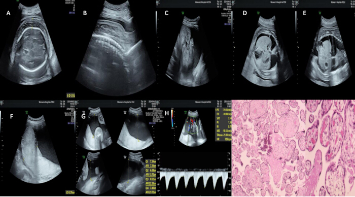
Figure 1: Representative imaging findings in mirror syndrome. (A–C) Fetal subcutaneous soft tissue edema; (D) Pleural effusion; (E) Peritoneal effusion; (F) Additional view of fetal hydrops; (G) Increased amniotic fluid index (29.8 cm); (H) Abnormal umbilical artery Doppler blood flow; (I–J) Placental histopathology showing villous edema and perivillous fibrin deposition (hematoxylin and eosin staining, original magnification ×200). Please click here to view a larger version of this figure.
Discussion
This study presents a retrospective analysis of 13 cases of mirror syndrome (MS) admitted to the Affiliated Obstetrics and Gynecology Hospital of Zhejiang University School of Medicine from January 2020 to July 2025. It systematically explores the diagnostic value of prenatal ultrasound in mirror syndrome, along with the clinical characteristics of the disease and maternal–fetal perinatal outcomes. By integrating pathological examination and laboratory parameter analysis, key findings were identified and their clinical significance elucidated. These results provide practical references for the diagnosis and management of this condition.
The clinical manifestations are distinctive, and maternal hematological indicators can support diagnosis. The primary maternal clinical features include bilateral lower-limb edema (100%), followed by hypertension (84.6%), anemia (69.2%), and proteinuria (38.4%). Laboratory findings demonstrate hemodilution, with nearly all cases showing decreased hematocrit and serum albumin levels. This contrasts with the hemoconcentration and more severe hypertension and proteinuria observed in preeclampsia, serving as an important differential diagnostic feature. Additionally, the condition is associated with notable placental morphological abnormalities: all 13 patients had placental thickness > 4 cm, 11 cases > 6 cm, and 8 cases were pathologically confirmed to have villous edema. Placental edema and villous edema represent key pathological features of the disease.
Prenatal ultrasound plays a central role in the early detection of mirror syndrome. It enables identification of characteristic findings, including fetal edema (at least two abnormal fluid accumulations or a single serous cavity effusion), placental thickening, and abnormal amniotic fluid volume. Doppler techniques allow assessment of fetal hemodynamics, including the umbilical artery and middle cerebral artery, while facilitating comprehensive fetal structural evaluation to detect associated abnormalities such as cardiac malformations and sacrococcygeal teratoma. Based on ultrasound findings of fetal edema, further investigations such as karyotype analysis and chromosomal microarray analysis (CMA) can be performed to determine etiology. In this study, among 14 fetuses (including one twin pregnancy), three cases showed chromosomal abnormalities, one case had a sacrococcygeal teratoma, and one case presented with cardiac anomalies.
Fetal prognosis is poor, whereas maternal outcomes are generally favorable. All 13 mothers recovered and were discharged without severe complications or mortality. Among the 14 fetuses, only four survived, corresponding to a neonatal mortality rate of approximately 71.4%, which remains substantially higher than that of normal pregnancies. Although this represents an improvement compared to the previously reported fetal survival rate of 7.7%21, adverse fetal outcomes remain common. Prognosis is associated with gestational age at onset and underlying etiology. Earlier onset and the presence of chromosomal or structural abnormalities are linked to poorer outcomes. In this cohort, twin pregnancies complicated by selective intrauterine growth restriction (sIUGR) resulted in stillbirth, suggesting that specific pregnancy types may represent additional risk factors.
The etiology of fetal edema is complex, with non-immune factors being the primary contributors. In this study, most cases were of unknown origin, with additional causes including chromosomal abnormalities, congenital structural malformations (cardiac and sacrococcygeal), intrauterine infection (e.g., cytomegalovirus), and complications of twin pregnancies, all classified as non-immune factors. These findings are consistent with existing literature, confirming that non-immune fetal edema is the principal trigger for mirror syndrome3.
Reports have shown that effective intrauterine treatment can prolong pregnancy in over one-third (38%) of cases22. Ultrasound-guided interventional procedures, therefore, have important clinical value. Ultrasound provides real-time guidance for procedures such as amniocentesis and umbilical cord blood sampling, enabling genetic and etiological diagnosis. It also facilitates intrauterine interventions, including thoracoabdominal drainage and selective fetal reduction. Early ultrasound screening (e.g., nuchal translucency and structural assessment) combined with timely intervention may facilitate earlier detection of fetal abnormalities and help alleviate fetal edema.
This study clarifies the diagnostic and clinical role of prenatal ultrasound. Although ultrasound demonstrates strong utility in identifying fetal edema, placental thickening, and amniotic fluid abnormalities, the findings of this study support the application of existing ultrasound-based diagnostic indicators rather than establishing new diagnostic criteria. The thresholds used for placental thickness and fetal edema were derived from widely accepted definitions, and the present results provide real-world evidence supporting their feasibility and reproducibility. However, the establishment of standardized diagnostic criteria requires further validation through larger, multicenter studies.
This study also highlights key differential diagnostic features between mirror syndrome and preeclampsia. Hemodilution (decreased hematocrit and serum albumin) represents a distinguishing laboratory characteristic. In addition, differences in placental morphology (placental thickening vs. hypoplasia), amniotic fluid volume (polyhydramnios vs. oligohydramnios), and fetal growth patterns (absence vs. presence of fetal growth restriction) contribute to improved diagnostic accuracy when integrated into a combined framework of clinical, laboratory, and ultrasonographic findings.
From a clinical management perspective, treatment strategies should focus on the underlying cause of fetal edema. Ultrasound-guided intrauterine interventions for treatable etiologies may prolong gestation and improve outcomes. In cases with unidentified etiology, early gestational age, or poor fetal prognosis, timely termination of pregnancy may be necessary to ensure maternal safety. Identified risk factors for poor outcomes include early gestational onset, chromosomal or structural abnormalities, and twin pregnancies.
Ultrasound also plays a comprehensive role in perinatal management, including diagnosis, guidance of interventions, monitoring of disease progression, assessment of therapeutic response, and determination of optimal timing of delivery. Its advantages, including lack of radiation, repeatability, and cost-effectiveness, make it an essential tool in the management of mirror syndrome.
Limitations
This study is a retrospective, single-center observational case series without a control group. The absence of a control group limits statistical power and restricts the ability to assess diagnostic performance and establish causal associations. These limitations are inherent given the rarity of mirror syndrome. Nevertheless, this study provides real-world clinical data from a relatively large case series. The findings should be interpreted with caution, and future multicenter studies with larger sample sizes are needed to validate these results and further investigate prognostic factors.
Conclusion
This study demonstrates the important role of prenatal ultrasound in the diagnosis, differential diagnosis, and management of mirror syndrome. It characterizes the clinical and laboratory features of the disease, identifies key prognostic factors, and provides clinically relevant evidence to support early diagnosis and individualized management. Further research is required to better understand the pathophysiology of mirror syndrome and to optimize diagnostic and therapeutic strategies.
Disclosures
No funding was received for this study. The authors declare no conflict of interest.
Materials
| Name | Company | Catalog Number | Comments |
|---|---|---|---|
| Samsung WS80A | Samsung Medison | WS80A | The Samsung WS80A is a 5D ultra-high-end intelligent color Doppler ultrasound diagnostic system launched by Samsung Medison. Positioned as "ultra-high-end dedicated to obstetrics and gynecology," it focuses on the diagnostic and research-teaching needs of complex cases across multiple departments including obstetrics/gynecology and neonatology. This represents one of Samsung's flagship high-end models in the ultrasound field. |
| GE-E8 | General Electric Company | Voluson™ E8 | The Voluson™ E8 is a high-end color ultrasound device launched by GE Healthcare, specializing in "4D color ultrasound" functionality. It is widely used in obstetrics, gynecology, pediatrics, and other fields, serving as a globally leading diagnostic tool for fetal imaging. |
| GE-E10 | General Electric Company | Voluson™ E10 | The Voluson E10 is a high-end intelligent device launched by GE for obstetric and gynecological ultrasound diagnosis, with "high-definition dynamic imaging" and "accurate malformation screening" as its core advantages. It supports comprehensive monitoring from pregnancy confirmation in the first trimester to fetal assessment before delivery, making it one of the preferred fetal imaging tools in clinical practice. |
References
- Li, L., Qi, H. Current research status of mirror syndrome. Chinese Journal of Maternal and Child Clinical Medicine (Electronic Edition). , (2021).
- Bianchi, D. W. Fetology: diagnosis and management of the fetal patient. , Saunders. Philadelphia. (2010).
- Seitinger, A., et al. Executable medical guidelines with Arden Syntax—applications in dermatology and obstetrics. Artif Intell Med. , (2018).
- Allarakia, S., et al. Characteristics and management of mirror syndrome: a systematic review (1956–2016). J Perinat Med. , (2017).
- Hobson, S. R., et al. Mirroring preeclampsia: the molecular basis of Ballantyne syndrome. J Matern Fetal Neonatal Med. , (2020).
- Braun, T., et al. Mirror syndrome: a systematic review of fetal-associated conditions, maternal presentation and perinatal outcomes. Fetal Diagn Ther. , (2010).
- Hirata, G., et al. Clinical characteristics of mirror syndrome: a comparative analysis of 10 cases with mirror syndrome versus non-mirror syndrome fetal hydrops cases. J Matern Fetal Neonatal Med. , (2016).
- Chen, R., et al. Clinical characteristics of mirror syndrome: a retrospective study of 16 cases. J Obstet Gynaecol. , (2021).
- Espinoza, J., et al. A role of the anti-angiogenic factor sVEGFR-1 in the “mirror syndrome” (Ballantyne’s syndrome). J Matern Fetal Neonatal Med. , (2006).
- Levine, R. J., et al. Circulating angiogenic factors and the risk of preeclampsia. N Engl J Med. , (2004).
- Katoh, Y., et al. Elevation of maternal serum sFlt-1 in pregnancy with mirror syndrome caused by fetal cardiac failure. Oxf Med Case Rep. , (2018).
- Heyborne, K. D., Chism, D. M. Reversal of Ballantyne syndrome by selective second-trimester fetal termination: a case report. J Reprod Med. , (2000).
- Gao, S., et al. Normalization of angiogenic imbalance after intrauterine transfusion for mirror syndrome caused by parvovirus B19. Fetal Diagn Ther. , (2013).
- Mathias, C. R., Rizvi, C. The diagnostic conundrum of maternal mirror syndrome progressing to preeclampsia: a case report. Case Rep Womens Health. , (2019).
- Sun, W., Sun, L. Advances in the diagnosis and treatment of mirror syndrome. China Journal of Prenatal Diagnosis (Electronic Edition). , (2024).
- Graham, N., et al. Placental expression of anti-angiogenic proteins in mirror syndrome: a case report. Placenta. , (2012).
- Bixel, K., et al. Placental origins of angiogenic dysfunction in mirror syndrome. Hypertens Pregnancy. , (2012).
- Llurba, E., Crispi, F., Verlohren, S. Update on the pathophysiological implications and clinical role of angiogenic factors in pregnancy. Fetal Diagn Ther. , (2015).
- Jain, V., et al. A case of alloimmune thrombocytopenia, hemorrhagic anemia-induced fetal hydrops, maternal mirror syndrome, and human chorionic gonadotropin-induced thyrotoxicosis. AJP Rep. , (2013).
- Gherman, R. B., et al. Ballantyne syndrome: is placental ischemia the etiology. J Matern Fetal Med. , (1998).
- Biswas, S., et al. Mirror syndrome: a systematic literature review. Am J Obstet Gynecol MFM. , (2023).
- Sichitiu, J., et al. Maternal “mirror” syndrome: evaluating the benefits of fetal therapy. Prenat Diagn. , (2024).