The ability to express viral vectors in the prepubertal brain is fundamental to studying the cellular and circuit mechanisms for brain development. Here, we describe a protocol for performing stereotaxic viral surgery in the infant mouse at postnatal days 11 or 12 (P11-P12) and provide guidelines for postoperative care.
Method Article
Stereotaxic Surgery in Infant Mice and Postoperative Care
In This Article
Summary
Abstract
Understanding the developmental, cellular, and circuit processes across the postnatal period is crucial for elucidating the complexities of brain development and its impact on behavioral and social maturation. The transition from infant to adulthood involves significant neurobiological changes, including synaptic pruning and myelination, which are fundamental for cognitive abilities, emotional regulation, and social interactions. Studying brain development in prepubertal mice enables researchers to identify critical periods that support healthy brain growth and to establish timely interventions for neurodevelopmental disorders. To precisely dissect brain function, stereotaxic surgery has been a pivotal technique in neuroscience research, offering unparalleled precision in targeting brain regions of interest and allowing the delivery of viral agents. Even though stereotaxic surgeries in adult mice are widely implemented across neurobiology labs, there are very limited guidelines for performing stereotaxic surgeries in prepubertal mice, as well as crucial postoperative care to ensure the infant can be successfully reintroduced to its dam and littermates. Here, we describe a protocol for stereotaxic surgery and viral injection in infant (P11-P12) mice, highlighting anesthesia and analgesia delivery, considerations for postoperative care, and differences from stereotaxic surgery in adults. This protocol can be implemented in any brain region of interest by updating the desired target coordinates and can be modified for brain targeting at later stages in postnatal development, such as during the juvenile period (P15-P30). Overall, this protocol provides an entry point for the study of postnatal brain development and function.
Introduction
Infancy and adolescence are crucial postnatal periods for brain development, characterized by myelination, synaptogenesis, and synaptic pruning, which facilitate circuit refinement1,2. The emergence of social behaviors, including mating, aggression, and social play, arises during the peripubertal period (P28-40)3, and is tightly regulated by circulating steroid hormones4,5. Despite significant advances in tools for dissecting the cellular underpinnings of social behaviors in adults, little is known about the neural mechanisms underlying the emergence of social behaviors across postnatal development. Reasons for the lack of understanding of developing social circuits include 1) challenges associated with performing surgeries in infant mice, including stereotaxic rigs not set up for younger mice, 2) balancing safety and effectiveness of analgesia and anesthesia during surgery6, and 3) acceptance of infants by the dam post-surgery. Furthermore, current methods utilized to deliver genetic constructs at early postnatal time points, such as in utero electroporation7, intracerebroventricular viral injection in the neonatal mouse (P0-P10) brain8, or recombinase driver and -responsive line genetic crossings9, are not ideal, as they lack precise targeting and focus on embryonic or P0 targeting.
Here, we provide a simple protocol for stereotaxic surgery in infant mice (P11-P12) and postoperative care to support successful reintegration with the dam and littermates. This protocol leverages the current stereotaxic protocol for adult mice10 and only requires minor changes to the stereotaxic setup. We also provide detailed guidance on postoperative care and on successful anesthesia and analgesia in infant mice. We selected the P11-12 timepoint for surgery as there is a 2-week incubation period for viral expression, so mice can be tested in vivo starting at P24, prior to the peripubertal period (P28-40)3. However, this same protocol can be used for stereotaxic surgery at later time points. We focus on the developing medial amygdala (MeA), an amygdala subdivision characterized by its role in a diverse set of social behaviors11,12. We specifically investigate the MeA subpopulation expressing Foxp2 (MeAFoxp2)13, a forkhead box transcription factor, as we have previously uncovered that these cells are uniquely hardwired for the in vivo processing of male sensory cues in male mice across postnatal development14. Additionally, the MeAFoxp2 subpopulation is required to bi-directionally modulate territorial aggression in males14. Overall, the current protocol allows neuroscientists to precisely deliver viral agents into targeted brain regions at key postnatal stages of brain development to study the cellular and circuit underpinnings of social and affiliative behaviors across adolescence and adulthood.
Protocol
This procedure was approved by the NIEHS Animal Care and Use Committee and was in accordance with the National Institutes of Health guidelines for the use and care of animals. Appropriate personal protective equipment, including gloves, face mask, disposable gown, hair net, and shoe covers, were used throughout the surgery and while in the presence of animals.
1. Mouse stereotaxic apparatus preparation and adjustments (Figure 1)
- Prepare the stereotaxic apparatus with stereotaxic accessories for infant mice, including a pair of non-rapture ear bars, a modified gas anesthesia head holder, and a palate bar for P12 infants.
- Sanitize all surgical tools with a glass bead sterilizer (250 °C, 30 s for smaller tools and 1 min for larger tools). Place sterile tools on a sterile drape after sterilization. Sterile tools should remain on the sterile field throughout the surgery.
- Place another sterile drape over a heating pad at a temperature of ~37 oC on the stereotaxic rig to keep the infant at a controlled temperature throughout the surgical procedure. Monitor the heating pad temperature throughout the surgical procedure.
2. Mouse surgical preparation, stereotaxic placement, leveling, and craniotomy
- Measure the body weight to provide appropriate dosage of analgesia.
- Place the mouse in a 3% isoflurane-filled induction chamber with an oxygen flow rate of 0.9 L/min. Check that the mouse's respiration has decreased to ~1 breath/s and that there is no response after testing the pedal withdrawal reflex. Switch the isoflurane/oxygen flow to the stereotaxic frame and reduce the isoflurane concentration to 2%.
NOTE: Adequate anesthesia must be confirmed by the absence of responses to the pedal withdrawal reflex every 5 min for the duration of the surgical procedure. Carefully observe the mouse's breathing pattern throughout the entire surgery. Adjust the isoflurane concentration throughout the surgery so that the lowest percentage is used to maintain deep anesthesia and avoid anesthesia overdosing. - Mount the mouse on the modified stereotaxic apparatus with stereotaxic accessories for infant mice. Place non-puncture ear bars on the side of the mouse's head and align the ear bar ruler to be equidistant for both sides with respect to the stereotaxic frame ruler. For P11 infants, set the stereotaxic ruler at 5 mm.
NOTE: Infants at P11 typically do not have their eyes open yet, so no eye lubricant is required. By P12, some infants have their eyes open. If the infant's eyes are open, ophthalmic ointment should be applied and re-applied as needed throughout the surgery. - Inject 5 mg/kg Meloxicam via the subcutaneous (s.c.) intrascapular route using a 1 mL syringe with a 27 G needle.
- Dilute 0.5% bupivacaine (5 mg/mL) with saline to 0.5 mg/mL (1:10) and inject s.c. under the skin of the head (i.e., 0.05 mL for a 5 g infant).
NOTE: Steps 2.6 to 3.15 should be performed with sterile gloves. - Perform a 1.5 cm incision with small scissors along the midline of the head.
- Use a sterile cotton swab to carefully remove the soft tissue from the surface of the skull with betadine.
- Place a pulled glass capillary (~0.8 mm in length) in a glass pipette holder. Mount the holder on the vertical stereotaxic arm.
NOTE: Steps 2.9 to 3.10 should be performed under a dissecting microscope. - Level the anterior-posterior angle of the skull surface:
- Move the capillary to the rostral confluence of the dorsal sinuses (RCS) point (Figure 2) as outlined in Chan et al.15.
- Under a microscope with 2x magnification, zero the X (Medial-Lateral), Y (Anterior-Posterior), and Z (Dorsal-Ventral) coordinates when the capillary is touching the skull in the RCS point slightly using the digital display console of the stereotaxic instrument.
- Move the capillary to lambda (at Y = -7.30 mm) and lower the capillary to slightly touch the skull (Figure 2).
- Check the value of the Z coordinates and level the anterior-posterior angle in the stereotaxic rig to change the tilt angle of the head. Repeat steps 2.9.1 to 2.9.3 until the difference in the value of the Z coordinate between RCS and lambda is <0.02 mm.
- Level the medial-lateral angle of the skull surface:
- Move the capillary to the RCS point.
- Zero the X (Medial-Lateral) and Y (Anterior-Posterior) coordinates when the capillary is slightly touching the skull in the RCS point.
- To level the head using the posterodorsal MeA (MeApd) coordinates, move the capillary to X = +2.50 mm, Y = -4.20 mm, and zero Z (Dorsal-Ventral) as the capillary slightly touches the skull.
- Move the capillary to X = -2.50 mm, Y = -4.20 mm in the other hemisphere and lower the capillary slightly to touch the skull.
- Check the value of Z and adjust the Medial-Lateral axis in the stereotaxic rig to change the tilt angle of the head. Repeat steps 2.10.1 to 2.10.4 until the difference in the Z value between the right and left hemispheres is <0.03 mm.
- Remove the capillary holder and mount a micro drill with a #75 drill bit into a drill stereotaxic holder on the vertical stereotaxic arm.
- Place the drill bit tip above the RCS point, zero the X (Medial-Lateral) and Y (Anterior-Posterior) coordinates. Move to X = ±2.03 mm, Y = -4.20 mm to target the MeApd of P11 infants.
- Drill a 0.5 mm-diameter craniotomy through the skull.
- Clean the skull surface carefully with sterile saline using delicate task wipes and cotton swabs.
NOTE: Apply sterile saline regularly to keep the skull surface hydrated.
3. Viral injection
- Fill a new capillary with mineral oil using a microfil flexible needle.
- Attach and tighten the capillary to the nanoliter injector.
- Remove the micro drill and mount the nanoliter injector on the vertical stereotaxic arm.
- Withdraw at least double the amount of virus required for surgery (here, 400 nL) at a rate of 10 nL/s.
NOTE: Any AAV virus of interest can be used for viral injection after Institutional approval. In this protocol, we utilized AAV2-DIO-mCherry (BSL-1, 2.5 E+13 vg/mL, 3 µl/tube, stored at -80 °C and then transferred to ice for surgery). - Move the capillary to the RCS point and zero the X and Y coordinates.
- Move the capillary to the target brain area of interest, here the MeApd (coordinates: X = ±2.03 mm, Y = -4.20 mm, Z= -5.08 mm from RCS).
- Lower the capillary to the surface of the skull, then zero the Z coordinate.
- Make a path by lowering the capillary to the desired depth and withdrawing the capillary back out of the brain.
- Inject ~8 nL of virus to make sure the capillary is not clogged prior to inserting back into the brain. If clogged, add sterile saline to a task wiper and gently wipe the sides and bottom of the capillary to clear the obstruction.
- Lower the capillary one more time to the desired depth and inject the desired viral amount (in this case, 150 nL of virus) at a rate of 20 nL/min.
- When done, wait 5 min before slowly withdrawing the capillary.
- If needed, repeat steps 3.4-3.11 to inject the virus in the contralateral hemisphere.
- Remove the capillary from the nanoliter injector. Discard used capillaries and viral tubes in a 50 mL conical tube with 10% bleach.
NOTE: Individual labs should follow all waste handling, spill management, and biosafety compliance guidelines from their Institutional Biological and Biosafety Branch. - Suture the skin with a non-absorbable 7-0 suture. Try lifting the hair when suturing so the sutures remain hidden.
NOTE: Typically, 3-4 sutures will suffice. In our hands, a 7-0 suture works best to avoid the dam from tearing the sutures once the infant is returned to the home cage. - Wipe the suture area with 3% hydrogen peroxide, followed by saline using sterile cotton swabs.
NOTE: The surgery typically lasts between 1 and 2 h, depending on the number of viruses injected. Viruses typically require a minimum of 2 weeks for full expression.
4. Postoperative care
- Remove the infant from the stereotaxic frame and place it in a clean cage on top of a heating pad. To prevent dehydration, injection of sterile saline solution is recommended.
- Monitor the infant until it is mobile.
- Once the infant can move around the cage, place the dam into a new clean cage for ~2 min.
- Place the infant in its original home cage alongside its littermates. Carefully wipe all infants with nest and bedding material from their home cage, then place them back inside the nest. Proceed to place the dam back into the cage.
- Monitor the dam and infants closely. Confirm that the dam returns to the nest and grooms the surgical infant but does not pull the sutures.
- If after 1 h, the dam is not accepting the surgical infant, use a foster dam. If a dam has already shown to be accepting of infants, use this dam for subsequent litters (acceptance after accepting infants in first litter has been 100%).
- Twenty-four hours post surgery, check the incision site and administer Meloxicam (5 mg/kg, s.c.).
- After successful postoperative care, leave the surgical mouse with the dam and littermates until weaned at P21. Remove any visible sutures within 10-14 days post-surgery.
NOTE: No significant differences are expected in food intake, body weight, or locomotion behavior in the surgical mice compared to littermate controls.
Infants will be monitored daily for at least 2 days post surgery and then at least 2x weekly. Humane Endpoint Criteria: If the infant is not ambulatory after 2 h post surgery, the infant will be humanely euthanized. If the infant shows decreased body weight and/or locomotion post-surgery, the infant will be humanely euthanized.
Results
Safety of the combination of isoflurane, bupivacaine, and meloxicam for infant surgeries
The safety of using anesthesia and analgesia in infant mice remains unclear6. Therefore, we tested the use of a combination of anesthesia and analgesia in infant mice to determine the survival rate. Infant mice at P11 (body weight = 6.09 ± 0.90 g, lowest body weight was 3.9 g; n=13) were placed in the induction chamber for initial anesthesia with 3% isoflurane in 0.9 L/min oxygen. Once the infants were anesthetized and non-responsive to the pedal withdrawal reflex, they were transferred to the stereotaxic apparatus, and isoflurane anesthesia was reduced to 1.5-2% isoflurane in 0.9 L/min oxygen with a warming pad (41.23 ± 2.28 °C). After placement of the infants in the stereotaxic apparatus, analgesia was administered s.c. interscapular (5 mg/kg Meloxicam) as well as topical anesthesia delivered s.c. below the skin of the head (0.05 mg/mL Bupivacaine). Thirty minutes later, we removed the infants from the stereotaxic rig and placed them in a clean cage. We observed a 100% survival rate (13/13) 30 min after analgesia delivery. Furthermore, we did not observe any complications 24 h and 72 h post surgery, nor into adulthood. These results demonstrate that combining local anesthesia with a nonsteroidal anti-inflammatory drug is safe for infant mice and reduces pain and distress.
Precise and accurate viral delivery into the MeA
To confirm the accuracy and precision of viral delivery during the infant stereotaxic surgeries, we delivered AAV2-hSyn-DIO-mCherry bilaterally into the MeApd of Foxp2cre+/- male mice at P11 (Figure 3A). Mice were then weaned at P21. At P45, mouse brains were collected for immunohistochemistry and image analysis as previously described by our group14 and others16 (Figure 3B,C). Mice were anesthetized and perfused with 30 mL of 1× PBS, followed by 30 mL of 4% PFA at 10 mL/min. Brains were post-fixed overnight at 4 °C in 4% PFA, cryoprotected in 30% sucrose, embedded in O.C.T., and sectioned at 50 µm on a cryostat. Every third section was processed for immunohistochemistry as described previously14. Free-floating sections were blocked in 10% normal donkey serum, incubated with the primary antibody overnight, washed, incubated with the secondary antibody for 4 h, washed again, and mounted with DAPI-containing medium before coverslipping. Brain sections were stained with Foxp2 antibody and Alexa Fluor 488 AffiniPure Donkey Anti-Rabbit IgG (H+L).
To confirm target efficiency, we determined the co-expression of mCherry+ and Foxp2+ cells in the MeA and observed 90.2% ± 2.1 overlap across populations, indicating that mCherry expression is restricted to Foxp2+ cells in a cre-dependent manner (Figure 3D). This result highlights the high efficiency of the viral injection using this protocol. Furthermore, we investigated the accuracy of the viral injections by analyzing the percentage of mCherry+ cells in the MeApd relative to the total percentage of mCherry across all regions. We observed that 93.7% ± 1.4 of mCherry+ cells were located within the MeApd, indicating that the virus's spatial expression is restricted to our region of interest (Figure 3E).

Figure 1: Set-up for infant stereotaxic surgery. (A) Stereotaxic rig. (B) Non-rupture ear bars. (C) Gas anesthesia head holder and palate bar for P12 infants. (D) Bead sterilizer. (E) Warming pad controller. (F) Stereotaxic arm. (G) Nanoliter injector. Please click here to view a larger version of this figure.

Figure 2: Major landmarks in the P11 mouse skull. (A) Schematic illustration of major landmarks in the infant's skull. (B) The infant's head is secured by specialized stereotaxic accessories that include non-rupture ear bars, a modified gas head holder, and a pellet bar for P12 mice. Abbreviation: RCS = Rostral confluence of the sinuses, SSS = Superior sagittal sinus, RRV = Rostral-rhinal veins, SOS = Superior olfactory sinus. Please click here to view a larger version of this figure.
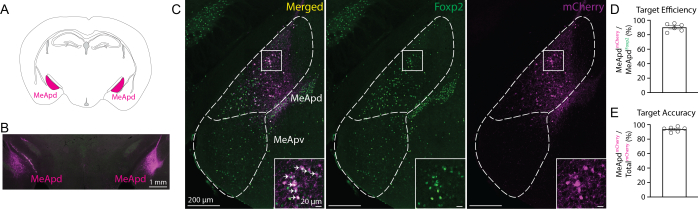
Figure 3: Targeted bilateral viral expression in the MeApd after infant stereotaxic surgery. (A) Schematic illustration of the MeApd (coronal view). (B) Representative histology image of bilateral expression of the Foxp2 antibody (green) and mCherry+ cells (magenta) in Foxp2cre+/- adult male mice. (C) Enlarged view of the MeA from both merged and individual channels. White arrows denote co-labeled Foxp2 antibody and mCherry+ cells. (D) Injection efficiency. (E) Injection accuracy. (D,E) n=3 mice, left and right hemispheres/mouse. Abbreviations: MeApd = posterodorsal medial amygdala; MeApv = posteroventral medial amygdala. Please click here to view a larger version of this figure.
| Methods | Pros | Cons | Reference |
| In utero electroporation | Larger plasmid transfection | Lack precise targeting, toxicity, need to be highly skilled | Wang, et al., Methods Mol Biol. (2013). |
| Intracerebroventricular viral injection | Fast, easy, economical | Lack precise targeting, risk of infanticide | Kim, et al., J Vis Exp. (2014). |
| Stereotactic Microinjection at P0 | Precise targeting, lower risk of maternal rejection | Cell toxicity due to virus, not ideal to study post-pubertal timepoints | Chen, et al., J Vis Exp. (2018). |
| Recombinase line genetic crossing | Non-invasive | Lack precise targeting, expensive | Robertson et al., Nat Neurosci. (2013). |
Table 1: Summary of previous methods used for studying cellular and circuit embryonic and postnatal development in rodents.
Discussion
Studying the neural mechanisms underlying social behaviors across postnatal development is essential to understanding not only their emergence but also how these mechanisms can be disrupted by environmental factors or in neurodevelopmental disorders. Our study describes a detailed protocol for stereotaxic surgery to deliver the virus precisely and efficiently into the brains of P11-12 infant mice. Therefore, this protocol allows for the manipulation, recording, and circuitry study of molecularly defined neuronal subpopulations before and after puberty using a viral strategy. We highlight pre- and postoperative modifications and care, as well as a safe combination of anesthesia and analgesia agents that are well tolerated by infant mice.
A key and challenging component of infant stereotaxic surgery is successfully reintroducing the infant to its home cage with its dam and littermates. To ensure appropriate infant care upon reintroduction to the cage, the dam should retrieve and groom the infant (without overgrooming), and any signs of potential neglect should be closely monitored. Due to the infant's incision and sutures, the dam might over-groom the head area, potentially resulting in the tearing and removal of the sutures. This would be detrimental and, hence, why monitoring the dam's behavior towards the infant post-surgery is critical to ensure survival. To increase dam acceptance of the infant post-surgery, we recommend using hydrogen peroxide and saline after suturing to clean any lingering blood residue from the skin and fur and using thinner sutures (7-0) than those used in adults (5-0). These two strategies resulted in the dam's acceptance of post-surgery infant mice.
Additionally, our study provides guidance on the safe use of anesthesia and analgesia for infant surgeries. This is critical as neonates and infant mice do not withstand analgesia and anesthesia dosages as well as adults, which can lead to detrimental respiratory and cardiac side effects6. We observed that isoflurane, an oral anesthesia, and the combination of meloxicam and bupivacaine analgesia, administered subcutaneously (intracuspular and under the skin of the head, respectively), were well tolerated by the infants and resulted in long-term survival. Furthermore, we have previously shown that the stereotaxic surgical procedure in infant mice led to similar investigation times towards conspecifics in juvenile mice 2 weeks after surgery, in comparison to age-matched controls that did not undergo surgery14. Therefore, these results indicate sufficient recovery and no overt motor deficits post-surgery in infant mice. Of note, we did not measure blood oxygenation throughout the surgery. Blood oxygen levels should be monitored with a pulse oximeter to prevent hypoxia17.
Another critical component of this protocol is the usage of the stereotaxic coordinate system based upon the lambda and the RCS at the sagittal midline15. RCS is a landmark defined by the junction of the superior sagittal sinus and the superior olfactory sinus juncture along the sagittal midline. This landmark has been shown to exhibit consistently lower positional variability than other dorsal landmarks after global alignment of the head. This is the case as the increased separation distance between RCS/lambda, compared with bregma/lambda, enhances the stability and reproducibility of the inter-animal horizontal angulation15.
Previous methods for studying neuronal development have focused on viral strategies at embryonic and P0 timepoints, or on genetic crosses for fate-mapping cells of interest (Table 1). Techniques such as in utero electroporation and intracerebroventricular viral injections lack precise targeting. Performing targeted viral injections at P018, overcomes this issue by precisely injecting a viral construct into a region of interest. However, viral constructs can become cytotoxic after several weeks, leading to cell death. Therefore, injecting a viral construct into the neonate's brain for longitudinal pubertal studies that could continue into adulthood might not be ideal. Therefore, our approach, in which the viral injection occurs a week and a half after birth, reduces the risk of cell toxicity during puberty.
Despite the advantages of our approach, several considerations remain. First, the brain coordinates used in infant mice need to be modified from those used in adults. For example, in P11 infant mice, we use the following coordinates to target the MeApd: X= +2.03 mm, Y= -4.20 mm, Z= -5.08 mm; while the coordinates used in adults are X= +2.15 mm, Y= -5.00 mm, Z= -5.10 mm. Unfortunately, there are fewer brain atlas references for prepubertal mice19 than those for adult mice (Allen Mouse Brain Atlas, mouse.brain-map.org and atlas.brain-map.org)20,21. Therefore, to successfully achieve correct viral targeting in other brain regions beyond the MeA, researchers will need to obtain an informed estimate of coordinates and perform coordinate testing. Second, the incubation time of AAVs needs to be considered. The optimal incubation time for AAV expression in vivo is at least 2-3 weeks. Considering that, in our protocol, stereotaxic surgery is performed in infant mice at P11, researchers will only be able to start testing mice post-surgery at P25, after the optimal AAV incubation period.
Overall, this protocol provides detailed guidelines for researchers to target brain regions of interest with high precision in infant mice using viral vectors. This approach is highly advantageous, as it is extremely flexible: any commercial or custom-made AAV construct can be injected. Hence, it allows studying molecularly defined cell subpopulations of interest and their anatomical circuitry, neuronal dynamics, and cellular role before and after puberty during a critical time window aligned with the emergence of social behaviors. Additionally, this approach is successful for targeting not only the amygdala, as shown here, but also other subcortical structures, such as the hypothalamus, and cortical regions.
Disclosures
The authors do not have any conflicts of interest to declare.
Acknowledgements
We thank Dr. Andrew Gorman, Dr. Donna Webb, and David Goulding from the Comparative Medicine Branch (CMB) at the National Institute of Environmental Health Sciences (NIEHS/NIH) for valuable discussions on best practices in anesthesia and analgesia for rodent surgeries. We also thank all members of the CMB at the NIEHS for their utmost care of the animal subjects and all members of the Neurobehavioral Circuits Group. This work was supported by the Intramural Research Program of the National Institute of Environmental Health Sciences, National Institutes of Health (NIEHS/NIH) (ZIA ES103403) (J.E.L.). The contributions of the NIH authors are considered Works of the United States Government. The findings and conclusions presented in this paper are those of the authors and do not necessarily reflect the views of the NIH or the U.S. Department of Health and Human Services.
Materials
| Name | Company | Catalog Number | Comments |
|---|---|---|---|
| #75 drill bit | Drill Bit City | 10WS75-850FL | |
| 27 G needle | Covetrus | 60757 | |
| 3% hydrogen peroxide | Swan | NDC 0809-0871-43 | |
| 32% paraformaldehyde | Electron Microscopy Sciences | 15714 | |
| AAV2-hSyn-DIO-mCherry | Addgene | 50459-AAV2 | |
| Alexa Fluor 488 AffiniPure Donkey (1:1000) Anti-Rabbit IgG (H+L) | Jackson Immuno Research | 711-545-152 | |
| Analytical Balance | Sartorius | 1102--15 | |
| Betadine | Avrio Pharmacy | NDC 67618-155-15 | |
| Bupivacaine | Hospira | NDC 0409-1163-18 | |
| Cryostat | Leica | CM1950 | |
| Dissecting Microscope | Zeiss | 2149-629 | |
| eye ointment | Dechra Veterinary | Puralube | |
| Fluoromount Mounting Media with DAPI | Thermo Fisher Scientific | 00-4959-52 | |
| Foxp2 Antibody (1:1000) | Atlas Antibody/Cosmo Bio | HPA000382 | |
| Foxp2cre+/- mice | The Jackson Laboratory | 30541 | |
| Gas Anesthesia Head Holder And Palate Bar For P12 Pups | David Kopf Instruments | 1934-C | |
| Glass Bead Sterilizer | Fisher Scientific | 10-000-632 | |
| Glass Capillary | World Precision Instruments | 504949 | |
| Glass Pipette Holder | David Kopf Instruments | 1975-1.2-A | |
| Heatpad | Kent Scientific Corporation | RT-0520 | |
| Isoflurane | Piramal Critical Care | NDC 66794-017-10 | |
| Meloxicam | Pivetal Veterinary Supply | NDC 46066-937-13 | |
| Micro Drill | David Kopf Instruments | model 1911 drill | |
| Microfil Flexible Needle | World Precision Instruments | MF28G67-5 | |
| Microscope Slides | Thermo Fisher Scientific | 12-550-15 | |
| Mineral Oil | Sigma-Aldrich | M5904-500ML | |
| Nanoliter Injector | World Precision Instruments | NANOLITER2020 | |
| Non-Absorbable Suture | AD Surgical | S-N718SP13 | |
| Non-Rapture Ear Bars | David Kopf Instruments | 1921-A | |
| Normal Donkey Serum | Jackson ImmunoResearch | 017-000-121 | |
| O.C.T. Compound | Sakura | 4583 | |
| Stereotaxic Rig | David Kopf Instruments | 1900 | |
| Sterile Cotton Swab | ULINE | S-21102 | |
| Sterile Drape | Busse Hospital Disposables | 696 | |
| Sterile Saline | Hospira | NDC 0409-1966-02 | |
| Sterile Water | Hospira | NDC 0409-4887-24 | |
| Sucrose | Fisher Chemical | S5-500 | |
| Syringe | Fisher Scientific | 50-252-2568 |
References
- Andersen, S. L. Trajectories of brain development: Point of vulnerability or window of opportunity. Neurosci Biobehav Rev. 27 (1-2), 3-18 (2003).
- Zeiss, C. J. Comparative milestones in rodent and human postnatal central nervous system development. Toxicol Pathol. 49 (8), 1368-1373 (2021).
- Bell, M. R. Comparing postnatal development of gonadal hormones and associated social behaviors in rats, mice, and humans. Endocrinology. 159 (7), 2596-2613 (2018).
- Auger, A. P., Olesen, K. M. Brain sex differences and the organisation of juvenile social play behaviour. J Neuroendocrinol. 21 (6), 519-525 (2009).
- Barkley, M. S., Goldman, B. D. A quantitative study of serum testosterone, sex accessory organ growth, and the development of intermale aggression in the mouse. Horm Behav. 8 (2), 208-218 (1977).
- National Research Council (US) Committee on Guidelines for the Use of Animals in Neuroscience and Behavioral Research. Guidelines for the care and use of mammals in neuroscience and behavioral research. , The National Academies Press. (2003).
- Wang, C., Mei, L. In utero electroporation in mice. Methods Mol Biol. 1018, 151-163 (2013).
- Kim, J. Y., Grunke, S. D., Levites, Y., Golde, T. E., Jankowsky, J. L. Intracerebroventricular viral injection of the neonatal mouse brain for persistent and widespread neuronal transduction. J Vis Exp. (91), e51863(2014).
- Robertson, S. D., Plummer, N. W., De Marchena, J., Jensen, P. Developmental origins of central norepinephrine neuron diversity. Nat Neurosci. 16 (8), 1016-1023 (2013).
- Geiger, B. M., Frank, L. E., Caldera-Siu, A. D., Pothos, E. N. Survivable stereotaxic surgery in rodents. J Vis Exp. (20), e880(2008).
- Prakash, N., Abu Irqeba, A., Corbin, J. G. Development and function of the medial amygdala. Trends Neurosci. 48 (1), 22-32 (2025).
- Raam, T., Hong, W. Organization of neural circuits underlying social behavior: A consideration of the medial amygdala. Curr Opin Neurobiol. 68, 124-136 (2021).
- Lischinsky, J. E., et al. Embryonic transcription factor expression in mice predicts medial amygdala neuronal identity and sex-specific responses to innate behavioral cues. Elife. 6, e21012(2017).
- Lischinsky, J. E., et al. Transcriptionally defined amygdala subpopulations play distinct roles in innate social behaviors. Nat Neurosci. 26 (12), 2131-2146 (2023).
- Chan, E., Kovacević, N., Ho, S. K., Henkelman, R. M., Henderson, J. T. Development of a high resolution three-dimensional surgical atlas of the murine head for strains 129s1/svimj and c57bl/6j using magnetic resonance imaging and micro-computed tomography. Neuroscience. 144 (2), 604-615 (2007).
- Tu, L., et al. Free-floating immunostaining of mouse brains. Methods Mol Biol. 176, 1-12 (2021).
- Thal, S. C., Plesnila, N. Non-invasive intraoperative monitoring of blood pressure and arterial pCO during surgical anesthesia in mice. J Neurosci Methods. 159 (2), 261-267 (2007).
- Chen, S. -Y., Kuo, H. -Y., Liu, F. -C. Stereotaxic surgery for genetic manipulation in striatal cells of neonatal mouse brains. J Vis Exp. (137), e57270(2018).
- Kronman, F. N., et al. Developmental mouse brain common coordinate framework. Nat Commun. 15 (1), 9072(2024).
- Dong, H. W. The Allen reference atlas: A digital color brain atlas of the C57BL/6J male mouse. , John Wiley & Sons. (2008).
- Paxinos, G., Franklin, K. B. The mouse brain in stereotaxic coordinates. , Academic Press. (2004).
Reprints and Permissions
This article has been published
Video Coming Soon