Here, we present a protocol for establishing an okadaic acid-induced Alzheimer's disease cellular model in SH-SY5Y cells and evaluating the neuroprotective effects of gastrodin through oxidative stress and AKT/GSK-3β pathway analysis.
A subscription to JoVE is required to view this content. Sign in or start your free trial.
Research Article
Gastrodin Alleviates Tau Hyperphosphorylation Associated with AKT/GSK-3β Signaling Changes in an Alzheimer's Disease Cell Model
* These authors contributed equally
In This Article
Summary
Abstract
Alzheimer's disease (AD) is a common neurodegenerative disease for which there are currently limited effective drugs. As the principal bioactive component derived from Gastrodia elata, Gastrodin (GAS) has shown clear therapeutic potential for treating AD; however, the molecular mechanism of its action remains to be elucidated. In this study, we aim to investigate the mechanism of the effect of GAS on AD model cells. Network pharmacology is employed to analyze the targets and signaling pathways affected by GAS in AD. An AD cell model is constructed by inducing human neuroblastoma SH-SY5Y cells with Okadaic acid (OA). Cell viability was assessed using the CCK-8 assay, while the levels of SOD, MDA, and T-AOC were measured. Apoptosis rate was determined through Annexin V-FITC/PI double staining, and expression of apoptosis-related factors, as well as AKT, GSK-3β, p-Tau (Ser396), and p-Tau (Thr181), was analyzed using RT-qPCR and Western blotting techniques. Network pharmacology analysis suggests that GAS has the potential to regulate cellular apoptosis and associated signaling pathways, including PI3K/AKT. Our experiments demonstrate that GAS can inhibit MDA levels, increase T-AOC and SOD in the AD model cells, and reduce cell apoptosis. Western blotting results indicate that GAS mitigates OA's inhibitory effects on p-AKT (Ser473) and p-GSK-3β (Ser9) expression. Additionally, it attenuates the overexpression of p-Tau (Ser396) and p-Tau (Thr181), suppresses Bax expression, and enhances Bcl-2 expression. GAS demonstrates the ability to ameliorate oxidative stress injury induced by OA and mitigate apoptosis. GAS may suppress Tau hyperphosphorylation, which is associated with changes in the AKT/GSK-3β signaling pathway, thereby exerting potential neuroprotective effects.
Introduction
Alzheimer's disease (AD) is a progressive neurodegenerative disorder primarily characterized by memory loss and cognitive impairment, representing the most prevalent form of dementia worldwide, accounting for approximately 60% to 80% of cases1. The underlying pathogenesis of AD remains complex and not yet fully elucidated2. Key pathological hallmarks observed in clinical AD cases include the accumulation of Senile Plaques (SP) and Neurofibrillary Tangles (NFTs) within the brain3,4. It is well recognized that the two core pathological events in AD are extracellular deposition of β-amyloid protein (Aβ) to form senile plaques and intracellular aggregation of hyperphosphorylated Tau protein to form neurofibrillary tangles5,6,7. Additionally, oxidative stress and neuroinflammation are supplementary factors that can contribute to the exacerbation of AD8,9,10,11.
As the global population aging trend intensifies, the number of dementia patients has significantly increased12,13. However, the treatment options for AD remain limited, predominantly comprising single-target drugs such as memantine, donepezil, and galantamine14. However, their therapeutic effects are primarily palliative rather than disease-modifying, and certain adverse effects such as gastrointestinal disturbances, dizziness, and bradycardia have been reported. Therefore, the development of safer and more effective therapeutic strategies remains an important research priority15,16. Recently launched monoclonal drugs, such as Lecanemab, have brought new hope for patients with mild AD. Yet their high costs and limitations fail to meet the substantial therapeutic demand. The prevention and treatment of AD continue to pose a significant challenge17,18. The findings from various studies have demonstrated that medicinal plants, particularly traditional Chinese medicines, exhibit multi-component effects that target multiple pathways involved in neurodegenerative disorders such as AD. Studies have shown that active compounds from Ganoderma lucidum can alleviate oxidative damage in the brain tissue of AD mice, inhibit apoptosis, reduce hippocampal neuron necrosis, and improve cognitive impairment19. In AD research, Bao et al. utilized network pharmacology to identify key targets of Astragaloside IV in AD-related inflammation. Their findings revealed that epidermal growth factor receptor (EGFR) and interleukin-1β (IL-1β) are crucial targets, providing potential therapeutic candidates and disease targets for the clinical treatment of neurodegenerative diseases20. Dartigues et al.21 analyzed Ginkgo biloba extract and demonstrated that it has beneficial effects on reducing the risk of dementia and mortality in elderly individuals. Patients treated with Ginkgo biloba extract exhibited a lower mortality rate22.
Gastrodia elata, belonging to the Orchidaceae family, is a traditional Chinese herb, edible and medicinal. The rhizome of Gastrodia elata is abundant in Gastrodin (GAS), gastrodia polysaccharides, and alkaloids. Recent pharmacological research has demonstrated that these active compounds possess memory-enhancing and cognitive-improving effects, as well as neuroprotective properties, and efficacy in treating insomnia and dizziness23. The main bioactive compound of Gastrodia elata is GAS, which is extracted from the tuber and possesses medicinal properties. GAS, also known as 4-hydroxybenzyl alcohol 4-O-β-d-glucoside, belongs to the class of phenolic glycosides24. The chemical structure of GAS is illustrated in Figure 1, exhibiting a molecular weight of 286.278. The relatively low molecular mass of glycosides and monoglycosides facilitates their efficient penetration through the blood-brain barrier, thus serving as a fundamental basis for GAS's neuroprotective role25. Moreover, their high absorbability further enhances their efficacy26. In recent years, numerous studies have demonstrated the potential of GAS in the treatment of various brain disorders, including AD27,28,29,30, Parkinson's disease (PD)31, and cerebral ischemia32. GAS exhibits a wide range of pharmacological properties, including antioxidative, hypolipidemic, anti-inflammatory, and anti-aging effects33,34. Its mechanism of action involves multiple signaling pathways such as Nrf2, NF-κB, and AMPK35. GAS exerts neuroprotective effects in a mouse model of Pb-induced neurological injury by modulating the Wnt/Nrf2 pathway36, and also exhibits an anti-apoptotic effect in the treatment of neurodegenerative disorders through regulation of the Nrf2 signaling pathway37. GAS also exerted a beneficial effect on AD by suppressing hippocampal BACE1 expression through the protein kinase/eukaryotic initiation factor-2α (PKR/eIF2α) signaling pathway38. Moreover, GAS exhibited inhibitory effects on TCDD-induced NF-κB signaling activation, thereby attenuating apoptosis of PC12 neurons and astrocyte activation39.
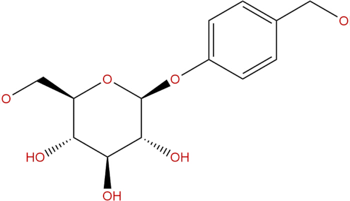
Figure 1: Chemical structural formula of GAS. Please click here to view a larger version of this figure.
OA, derived from the black sponge of the Halichondria genus, belongs to a class of long-chain fatty acids and serves as the principal component of Diarrhetic Shellfish Poison (DSP). It effectively inhibits the activities of protein phosphatases, particularly PP1A and PP2A, leading to cellular hyperphosphorylation and disruption of cellular homeostasis40,41. This characteristic renders it a valuable tool for investigating diseases associated with protein phosphorylation. Therefore, the utilization of OA enables the construction of AD cell models featuring alterations in Tau protein phosphorylation. This study aims to investigate the impact and mechanism of GAS, the primary active component of Gastrodia elata, on AD cell model induced by OA, and to clarify its neuroprotective effect on AD model cells by targeting AKT/GSK-3β signaling pathway.
Access restricted. Please log in or start a trial to view this content.
Protocol
All network pharmacology operations were conducted in accordance with the Guidelines for Network Pharmacology Evaluation Methods. All experimental procedures were performed in compliance with Zhejiang Sci-Tech University laboratory management regulations.
Network pharmacology
Prediction of potential targets of GAS: The canonical SMILES of GAS (PubChem CID: 1150) was retrieved from the PubChem database42. The SMILES structure was uploaded to the SwissTargetPrediction database (http://www.swisstargetprediction.ch/)43. The species parameter was set to Homo sapiens. Predicted targets with a probability value > 0.1 were retained to reduce false-positive predictions. All predicted protein targets were standardized to official gene symbols using the UniProt database43 (species limited to Homo sapiens), and duplicate entries were removed.
AD-related genes were collected from the GeneCards database (https://www.genecards.org/)44,45 and the OMIM database (https://www.omim.org/)46 using "Alzheimer's disease" as the search term. In GeneCards, genes with a Relevance Score > 10 were selected. Targets obtained from GeneCards and OMIM were merged, duplicates were removed, and gene names were standardized using UniProt.
The predicted GAS targets and AD-related genes were imported into the Venny 2.1 online platform to obtain overlapping targets. A total of 52 intersecting genes (See Supplementary Table 1) were identified as potential therapeutic targets of GAS in AD.
The 52 intersecting targets were uploaded to the STRING database (https://string-db.org/)47 for PPI analysis. The species was set to Homo sapiens, and the minimum required interaction score was set to 0.4 (medium confidence). Disconnected nodes were hidden. The resulting interaction data were exported and visualized using Cytoscape software (version 3.9.1).
GO and KEGG enrichment analysis: The 52 intersecting targets were uploaded to the Metascape database (https://metascape.org/)48 for Gene Ontology (GO) and Kyoto Encyclopedia of Genes and Genomes (KEGG) enrichment analyses. The species was set to Homo sapiens. The enrichment criteria were defined as p-value < 0.01, minimum overlap ≥ 3, and enrichment factor > 1.5. KEGG pathways were ranked according to p-value, and the top 20 significantly enriched pathways were selected for visualization. Enrichment results were visualized using an online bioinformatics platform.
The protein structure file of GSK-3β was obtained from the RCSB PDB database. The structure file of GAS in SDF format was downloaded from the PubChem database and subsequently converted to Mol2 format using the Open Babel GUI software. The files mentioned above were processed using AutoDock software to add hydrogen atoms, remove water molecules, and assign charges. The processed files were subsequently saved in the PDBQT format. After detecting and processing torsion bonds in the ligand molecules, the files were then stored in PDBQT format. The lowest binding energy between the two molecules was obtained by performing molecular docking using AutoDock Vina software. Subsequently, the docking results were visualized and analyzed utilizing PyMOL.
Stock solution preparation
The OA powder (25 µg) was completely dissolved in 1200 µL of 10% dimethyl sulfoxide (DMSO) prepared with sterile phosphate-buffered saline (PBS) to generate a 25 µM stock solution. The stock solution was aliquoted and stored at -80 °C. Before each experiment, OA was freshly diluted with complete Dulbecco's Modified Eagle Medium (DMEM) (see Table of Materials) to the desired working concentrations. GAS powder (20 mg) was dissolved in 1.4 mL of sterile PBS, filtered through a 0.22 µm membrane for sterilization, and prepared as a 50 mM stock solution, which was stored at -20 °C. Prior to use, the stock solution was diluted with complete DMEM to the indicated working concentrations.
Construction of the AD cell model
Human neuroblastoma SH-SY5Y cells were obtained and used in this study (see Table of Materials). SH-SY5Y cells were induced with gradient concentrations of OA (0, 10, 20, 40, 80, 160 nM) for durations of 12, 24, and 36 h, respectively, to establish the AD cell model.
Cell viability assay
To determine the optimal concentration and induction duration of OA for establishing the AD cell model, SH-SY5Y cells were treated with gradient concentrations of OA (0, 10, 20, 40, 80, and 160 nM) for different time periods (12, 24, and 36 h), and cell viability was evaluated using the CCK-8 assay49. SH-SY5Y cells were seeded into 96-well plates at a density of 7.5 × 10³ cells per well in 100 µL of complete DMEM. Plates were incubated at 37 °C in a humidified incubator with 5% CO₂ for 24 h to allow cell attachment. After 24 h attachment, 100 µL of OA working solution was added to each well to achieve final concentrations of 0, 10, 20, 40, 80, and 160 nM in a total volume of 200 µL per well. The control group (CON) received an equal volume of complete DMEM without OA, whereas the OA group received OA at the indicated concentrations. Cells were incubated for 12 h, 24 h, or 36 h under standard culture conditions. Each concentration and time point was set with six replicate wells. Three blank wells (medium only) were included for background correction.
Determination of cellular oxidative stress index
The SH-SY5Y cells in the logarithmic growth phase were seeded into a 6-well plate. Following cell treatment, the previous medium was discarded from the wells. Subsequently, the wells were washed three times with pre-chilled PBS at 4 °C. After that, cell lysis buffer was added, and the cells were lysed on ice using the appropriate method as instructed by the kit. The protein content of the samples was quantified using the BCA protein assay kit (see Table of Materials). The activities of T-AOC, SOD, and MDA were determined according to the instructions provided with the respective assay kits (see Table of Materials).
Cell apoptosis detection
Cell apoptosis was evaluated using an Annexin V-FITC/PI double staining kit (see Table of Materials) according to the manufacturer's instructions. Briefly, both floating and adherent cells were collected. Adherent cells were digested using trypsin without EDTA to avoid artificial membrane damage and combined with floating cells. Cells were washed twice with cold PBS and resuspended in 500 µL of 1× Binding Buffer at a density of 0.5–1 × 10⁶ cells.
Subsequently, 5 µL of Annexin V-FITC and 5 µL of Propidium Iodide (PI) were added to each sample, gently mixed, and incubated at room temperature in the dark for 10–20 min. Samples were placed on ice and analyzed within 1 h using a flow cytometer.
For flow cytometric analysis, unstained controls and single-stained controls (Annexin V-FITC only and PI only) were used to set compensation parameters and quadrant gates. Forward and side scatter (FSC/SSC) gating was first applied to exclude debris and cell aggregates. A minimum of 10,000 events per sample was acquired. Data were analyzed using FlowJo software.
Real-time quantitative PCR analysis (RT-qPCR)
According to the instructions provided by the AG reverse transcription kit, the extracted RNA was subjected to reverse transcription to generate complementary DNA (cDNA), which was subsequently diluted 10-fold with sterile water to serve as the template. The RT-qPCR reaction system was prepared following the protocol of the Green Master Mix (No Rox) kit, and analysis was performed using a fluorescent quantitative PCR instrument (see Table of Materials). The obtained Ct values were processed and analyzed, and the expression levels of target genes were calculated using the 2-ΔΔCt method, where ΔCt represents the difference between Ct values in the treatment group and GAPDH, and ΔΔCt represents the difference between ΔCt values in the treatment group and control group. The list of primers can be found in Supplementary Table 2.
Western blotting analysis
The cell lysis buffer, along with a protease inhibitor and a phosphatase inhibitor, was combined in a ratio of 100:1:1. The cells were lysed to extract cellular proteins. The protein concentration was determined using the BCA assay kit. Proteins (20–30 µg per lane) were separated on 10% SDS-PAGE gels at 80 V for the stacking gel and 120 V for the resolving gel. Proteins were transferred to PVDF membranes (0.45 µm) at 300 mA for 90 min using wet transfer. The membrane was blocked with 5% skim milk at room temperature for 2 h, followed by overnight incubation with primary antibody (see Table of Materials) at 4 °C. After incubating with the secondary antibody at room temperature for 1 h, the membrane was washed four times with TBST for 5 min each. The bands were visualized using enhanced chemiluminescence (ECL) detection reagents (see Table of Materials), and detection was performed with a gel imaging system. The grayscale values were analyzed using ImageJ software for data insights.
Access restricted. Please log in or start a trial to view this content.
Results
GAS-related target screening for AD treatment
The identification of GAS's targets for treating AD involved database searches and software analysis. The Swiss Target Prediction database yielded 100 potential targets, while OMIM and GeneCards (with a Relevance score > 10) were queried for AD-related targets, resulting in a total of 2376 potential targets. Subsequently, employing the Venny 2.1 online platform enabled the identification of 52 potential therapeutic targets for GAS in AD (
Access restricted. Please log in or start a trial to view this content.
Discussion
In recent years, there has been a global increase in the prevalence of AD. This condition is characterized by a chronic course, irreversible nature, disability, and elevated mortality rates, making it a significant challenge within the context of an aging global population. Despite its prominence, the etiology of AD is intricate, and its pathogenesis remains incompletely understood51.
Traditional Chinese medicine offers significant advantages in disease prevention and t...
Access restricted. Please log in or start a trial to view this content.
Disclosures
The authors have nothing to disclose.
Acknowledgements
This research was supported by the Open Fund of Zhejiang Sci-Tech University, Shaoxing Academy of Biomedicine (SXAB202007).
Access restricted. Please log in or start a trial to view this content.
Materials
| Name | Company | Catalog Number | Comments |
|---|---|---|---|
| 7500 Real-Time PCR System | APExBIO | S0080 | Used for detecting apoptotic cells |
| A450 enzyme labeling instrument | Proteintech | 50599-2-Ig | WB, dilution 1:1000 |
| Annexin V-FITC apoptosis detection kit | Proteintech | 60178-1-Ig | WB, dilution 1:1000 |
| Anti-Bax antibody | Proteintech | 10494-1-AP | WB, dilution 1:1000 |
| Anti-Bcl-2 antibody | Abcam | ab32057 | WB, dilution 1:1000 |
| Anti-GAPDH antibody | Proteintech | 28866-1-AP | WB, dilution 1:1000 |
| Anti-p-Tau (Ser396) antibody | Biofroxx | 1172 | Blocking buffer for WB |
| Anti-p-Tau (Thr181) antibody | Vazyme Biotech | A311-01/02 | Used for cell viability assay |
| Biofroxx skim milk powder | Solarbio Science & Technology | D8371 | Solvent for compounds |
| CCK-8 kit | Wisent Biotechnology | 319-105-CL | For cell culture |
| Dimethyl sulfoxide (DMSO) | APExBIO (USA) | K1231 | WB detection reagent |
| DMEM high-glucose medium | Aladdin Biochemical Technology | G111309 | Active compound |
| ECL chemiluminescent reagent | Promega | A6101 | For qPCR amplification |
| Flow cytometer | Solarbio Science & Technology | BC6410 | Lipid peroxidation assay |
| Gastrodin (GAS) powder, purity ≥98% | MedChemExpress | HY-N6785 | Inducer of tau hyperphosphorylation |
| Gel imaging system | Beyotime Biotechnology | C0221A | Wash the cells |
| GoTaq qPCR Master Mix | Proteintech | SA00001-1 | WB, dilution 1:5000 |
| MDA assay kit (malondialdehyde) | Proteintech | SA00001-2 | WB, dilution 1:5000 |
| Okadaic acid (OA) | ATCC | CRL-2266 | Cell |
| PBS | Beyotime Biotechnology | S0101M | For oxidative stress assay |
| Secondary antibody (Goat anti-mouse IgG, HRP-conjugated) | Solarbio Science & Technology | BC1315 | Antioxidant capacity detection |
| Secondary antibody (Goat anti-rabbit IgG, HRP-conjugated) | Beyotime Biotechnology | R0016 | RNA extraction |
| SH-SY5Y | Sigma-Aldrich | 9002-07-7 | Cell digestion |
| SOD assay kit (total superoxide dismutase) | Thermo Fisher Scientific | A45072 | Multifunctional microplate reader for absorbance detection |
| Total antioxidant capacity (T-AOC) kit | Becton, Dickinson and Company | 661310 | For multi-parameter single-cell analysis and cell sorting. |
| Trizol reagent | Thermo Fisher Scientific | 4351104 | gene expression analysis |
| Trypsin | Bio Rad | 1708195 | DNA/RNA/protein gel imaging |
References
- Hu, X., Wang, T., Jin, F. Alzheimer's disease and gut microbiota. Sci China Life Sci. 59 (10), 1006-1023 (2016).
- Qiu, C., Du, Y. A call for action to strengthen research on population interventions against Alzheimer's disease and senile dementia. J Shandong Univ Health Sci. 55 (10), 1-6 (2017).
- Livingston, G., et al. Dementia prevention, intervention,and care: 2020 report of the Lancet Commission. Lancet. 396 (10248), 413-446 (2020).
- Li, T. F., Guo, Z. Q., Wang, Y. Abnormal ß-amyloid metabolism and Alzheimer disease. J Neurosci Ment Health. 22 (12), 904-908 (2022).
- Zheng, J., et al. Oral administration of a fusion protein between the cholera toxin B subunit and the 42-amino acid isoform of amyloid-β peptide produced in silkworm pupae protects against Alzheimer's disease in mice. PLoS One. 9 (12), e113585(2014).
- La Spada, A. R., et al. A Becn1 mutation mediates hyperactive autophagic sequestration of amyloid oligomers and improved cognition in Alzheimer's disease. PLoS Genet. 13 (8), e1006962(2017).
- Aboelwafa, H. R., El-Kott, A. F., Abd-Ella, E. M., Yousef, H. N. The possible neuroprotective effect of silymarin against aluminum chloride-prompted Alzheimer's-like disease in rats. Brain Sci. 10 (9), 628(2020).
- Tobore, T. O. On the central role of mitochondria dysfunction and oxidative stress in Alzheimer's disease. Neurol Sci. 40 (8), 1527-1540 (2019).
- Kawamoto, E. M., et al. Oxidative state in platelets and erythrocytes in aging and Alzheimer's disease. Neurobiol Aging. 26 (6), 857-864 (2005).
- Stancu, I. C., et al. Aggregated Tau activates NLRP3–ASC inflammasome exacerbating exogenously seeded and non-exogenously seeded Tau pathology in vivo. Acta Neuropathol. 137 (4), 599-617 (2019).
- Connolly, K., et al. Potential role of chitinase-3-like protein 1 (CHI3L1/YKL-40) in neurodegeneration and Alzheimer's disease. Alzheimers Dement. 19 (1), 9-24 (2022).
- Yingquan, W., Jing-Hong, L., Ruixia, J. Alzheimer disease in China (2015-2050) estimated using the 1% population sampling survey in 2015. China J Alzheimers Dis Relat Disord. 2 (1), 289-298 (2019).
- Amat-Ur-Rasool, H., Ahmed, M., Hasnain, S., Carter, W. G. Anti-cholinesterase combination drug therapy as a potential treatment for Alzheimer's disease. Brain Sci. 11 (2), 184(2021).
- Cummings, J., Lee, G., Mortsdorf, T., Ritter, A., Zhong, K. Alzheimer's disease drug development pipeline: 2017. Alzheimers Dement (N Y). 3 (3), 367-384 (2017).
- Birks, J. S., Harvey, R. J. Donepezil for dementia due to Alzheimer's disease. Cochrane Database Syst Rev. 6 (6), CD001190(2018).
- Knopman, D. S., et al. Alzheimer's disease. Nat Rev Dis Primers. 7 (1), 33(2021).
- Silva, T., Reis, J., Teixeira, J., Borges, F. Alzheimer's disease, enzyme targets and drug discovery struggles: from natural products to drug prototypes. Ageing Res Rev. 15, 116-145 (2014).
- Liu, X., Yi, W., Xi, B., Dai, Q., Lu, L. Identification of drug-disease associations using a random walk with restart method and supervised learning. Comput Math Methods Med. 2022 (1), 7035634(2022).
- Yu, N., et al. Ganoderma lucidum triterpenoids (GLTs) reduce neuronal apoptosis via inhibition of ROCK signal pathway in APP/PS1 transgenic Alzheimer's disease mice. Oxid Med Cell Longev. 2020 (1), 1-11 (2020).
- Bao, M., et al. Astragaloside IV against Alzheimer's disease via microglia-mediated neuroinflammation using network pharmacology and experimental validation. Eur J Pharmacol. 957, 175992(2023).
- Dartigues, J. F., et al. Ginkgo biloba extract consumption and long-term occurrence of death and dementia. J Prev Alzheimers Dis. 4 (1), 16-20 (2017).
- Savaskan, E., Mueller, H., Hoerr, R., von Gunten, A., Gauthier, S. Treatment effects of Ginkgo biloba extract EGb 761® on the spectrum of behavioral and psychological symptoms of dementia: meta-analysis of randomized controlled trials. Int Psychogeriatr. 30 (3), 285-293 (2017).
- Wang, D., Liu, W., Lu, M., Xu, Q. Neuropharmacological effects of Gastrodia elata Blume and its active ingredients. Front Neurol. 16, 1574277(2025).
- El Menyiy, N., et al. Chemistry, biological activities,and pharmacological properties of gastrodin: mechanism insights. Chem Biodivers. 21 (6), e202400402(2024).
- Yu, H., et al. Research progress on classification of chemical constituents from Gastrodia elata and their pharmacological effects. Chin Tradit Herbal Drugs. 53, 5553-5562 (2022).
- Tang, C., et al. Comparative pharmacokinetics of gastrodin in rats after intragastric administration of free gastrodin, parishin and Gastrodia elata extract. J Ethnopharmacol. 176, 49-54 (2015).
- Fu, X., et al. Antioxidation effects and mechanism of gastrodin in AD rat models. Acta Anat Sin. 41 (4), 485-490 (2010).
- Shi, R., et al. Gastrodin alleviates vascular dementia in a 2-VO vascular dementia rat model by altering amyloid and Tau levels. Pharmacology. 105 (7-8), 386-396 (2020).
- Fasina, O. B., et al. Gastrodin from Gastrodia elata enhances cognitive function and neuroprotection of AD mice via the regulation of gut microbiota composition and inhibition of neuron inflammation. Front Pharmacol. 13, 814271(2022).
- Zheng, Q., et al. MicroRNA-452 promotes tumorigenesis in hepatocellular carcinoma by targeting cyclin-dependent kinase inhibitor 1B. Mol Cell Biochem. 389 (1-2), 187-195 (2014).
- Yan, J., Yang, Z., Zhao, N., Li, Z., Cao, X. Gastrodin protects dopaminergic neurons via insulin-like pathway in a Parkinson's disease model. BMC Neurosci. 20 (1), 31(2019).
- Xiao, H., et al. Gastrodin promotes hippocampal neurogenesis via PDE9-cGMP-PKG pathway in mice following cerebral ischemia. Neurochem Int. 150, 105171(2021).
- Liu, Y., et al. A review on central nervous system effects of gastrodin. Front Pharmacol. 9, 24(2018).
- Fu, J., et al. Gastrodia elata specific miRNA attenuates neuroinflammation via modulating NF-κB signaling pathway. Int J Neurosci. 134 (12), 1652-1662 (2023).
- Wang, Y., et al. Gastrodin: a comprehensive pharmacological review. Naunyn Schmiedebergs Arch Pharmacol. 397 (6), 3781-3802 (2024).
- Liu, C. M., et al. Effects of gastrodin against lead-induced brain injury in mice associated with the Wnt/Nrf2 pathway. Nutrients. 12 (6), 1805(2020).
- De Oliveira, M. R., Brasil, F. B., Fürstenau, C. R. Nrf2 mediates the anti-apoptotic and anti-inflammatory effects induced by gastrodin in hydrogen peroxide-treated SH-SY5Y cells. J Mol Neurosci. 69 (1), 115-122 (2019).
- Zhang, J. S., et al. Gastrodin suppresses BACE1 expression under oxidative stress condition via inhibition of the PKR/eIF2α pathway in Alzheimer's disease. Neuroscience. 325, 1-9 (2016).
- Jiao, M., et al. Effect of the SSeCKS–TRAF6 interaction on gastrodin-mediated protection against 2,3,7,8-tetrachlorodibenzo-p-dioxin-induced astrocyte activation and neuronal death. Chemosphere. 226, 678-686 (2019).
- Cohen, P., Holmes, C. F., Tsukitani, Y. Okadaic acid: a new probe for the study of cellular regulation. Trends Biochem Sci. 15 (3), 98-102 (1990).
- Qin, Y., et al. Okadaic acid detection through a rapid and sensitive amplified luminescent proximity homogeneous assay. Toxins (Basel). 15 (8), 501(2023).
- Kim, S., et al. PubChem 2023 update. Nucleic Acids Res. 51 (D1), D1373-D1380 (2022).
- Daina, A., Michielin, O., Zoete, V. SwissTargetPrediction: updated data and new features for efficient prediction of protein targets of small molecules. Nucleic Acids Res. 47 (W1), W357-W364 (2019).
- Stelzer, G., et al. The GeneCards suite: from gene data mining to disease genome sequence analyses. Curr Protoc Bioinformatics. 54 (1), 1.30.31-1.30.33 (2016).
- Safran, M., et al. Practical guide to life science databases. , Springer Nature. Singapore. (2021).
- Amberger, J. S., Bocchini, C. A., Schiettecatte, F., Scott, A. F., Hamosh, A. OMIM.org: Online Mendelian Inheritance in Man (OMIM®), an online catalog of human genes and genetic disorders. Nucleic Acids Res. 43 (D1), D789-D798 (2014).
- Szklarczyk, D., et al. The STRING database in 2021: customizable protein–protein networks,and functional characterization of user-uploaded gene/measurement sets. Nucleic Acids Res. 49 (D1), D605-D612 (2020).
- Zhou, Y., et al. Metascape provides a biologist-oriented resource for the analysis of systems-level datasets. Nat Commun. 10 (1), 1523(2019).
- Tominaga, H., et al. A water-soluble tetrazolium salt useful for colorimetric cell viability assay. Anal Commun. 36 (2), 47-50 (1999).
- Ton, A. M. M., et al. Oxidative stress and dementia in Alzheimer's patients: effects of synbiotic supplementation. Oxid Med Cell Longev. 2020 (1), 2638703(2020).
- Roth, K. A. Caspases, apoptosis,and Alzheimer disease: causation,correlation,and confusion. J Neuropathol Exp Neurol. 60 (9), 829-838 (2001).
- Cheng, M., et al. The effects of Porphyromonas gingivalis on the apoptosis of hippocampal cells in Sprague-Dawley rats and its underlying mechanisms. Int J Clin Exp Med. 13 (1), 300-309 (2020).
- Liu, H., et al. Development of chemical fingerprints for quality control of Xiong Ma Tang and its related preparations by high-performance liquid chromatography. J Chromatogr Sci. 54 (2), 206-215 (2016).
- Liu, S. J., et al. Gastrodin attenuates microglia activation through renin-angiotensin system and Sirtuin3 pathway. Neurochem Int. 120, 49-63 (2018).
- Hu, Y., Li, C., Shen, W. Gastrodin alleviates memory deficits and reduces neuropathology in a mouse model of Alzheimer's disease. Neuropathology. 34 (4), 370-377 (2014).
- Liu, J., et al. Systems pharmacology uncovers the multiple mechanisms of Xijiao Dihuang decoction for the treatment of viral hemorrhagic fever. Evid Based Complement Alternat Med. 2016 (1), 9025036(2016).
- Ran, B., et al. Drug-drug interactions prediction using fingerprint only. Comput Math Methods Med. 2022 (1), 7818480(2022).
- Sharma, V. K., Singh, T. G., Singh, S., Garg, N., Dhiman, S. Apoptotic pathways and Alzheimer's disease: probing therapeutic potential. Neurochem Res. 46 (12), 3103-3122 (2021).
- Zhou, W., Gao, Y., Xu, S., Yang, Z., Xu, T. Purification of a mannose-binding lectin Pinellia ternata agglutinin and its induction of apoptosis in Bel-7404 cells. Protein Expr Purif. 93, 11-17 (2014).
- Goedert, M., Spillantini, M. G., Jakes, R., Rutherford, D., Crowther, R. Multiple isoforms of human microtubule-associated protein tau: sequences and localization in neurofibrillary tangles of Alzheimer's disease. Neuron. 3 (4), 519-526 (1989).
- Hernández, F., Gómez De Barreda, E., Fuster-Matanzo, A., Lucas, J. J., Avila, J. GSK3: a possible link between beta amyloid peptide and tau protein. Exp Neurol. 223 (2), 322-325 (2010).
- Lu, Z., et al. Neuroprotection and mechanism of Gas-miR36-5p from Gastrodia elata in an Alzheimer's disease model by regulating glycogen synthase kinase-3β. Int J Mol Sci. 24 (24), 17295(2023).
- Vossel, K. A., et al. Tau reduction prevents Aβ-induced axonal transport deficits by blocking activation of GSK3β. J Cell Biol. 209 (3), 419-433 (2015).
- Singh, A. K., et al. Berberine: a plant-derived alkaloid with therapeutic potential to combat Alzheimer's disease. CNS Agents Med Chem. 19 (3), 154-170 (2019).
- Belsare, K. D., et al. Soluble TREM2 inhibits secondary nucleation of Aβ fibrillization and enhances cellular uptake of fibrillar Aβ. Proc Natl Acad Sci U S A. 119 (5), e2114486119(2022).
- Matsuda, S., et al. Roles of PI3K/AKT/GSK3 pathway involved in psychiatric illnesses. Diseases. 7 (1), 22(2019).
- Pan, S., et al. Oleanolic acid derivatives induce apoptosis in human leukemia K562 cell involved in inhibition of both Akt1 translocation and pAkt1 expression. Cytotechnology. 67 (5), 821-829 (2014).
- Zhou, F., et al. Network pharmacology-based strategy to investigate pharmacological mechanisms of Tinospora sinensis for treatment of Alzheimer's disease. J Ethnopharmacol. 259, 112940(2020).
- Liu, Z., Wang, Z. Protein tyrosine kinase 2 regulates tau phosphorylation through PI3K/AKT/Gsk3β signaling pathway. Labeled Immunoassays Clin Med. 29 (3), 464-470 (2022).
- Cope, P. J., Ourradi, K., Li, Y., Sharif, M. Models of osteoarthritis: the good, the bad and the promising. Osteoarthritis Cartilage. 27 (2), 230-239 (2019).
- Wang, X., Blanchard, J., Tung, Y. C., Grundke-Iqbal, I., Iqbal, K. Inhibition of protein phosphatase-2A (PP2A) by I1PP2A leads to hyperphosphorylation of tau, neurodegeneration,and cognitive impairment in rats. J Alzheimers Dis. 45 (2), 423-435 (2015).
- Liu, T. T., et al. Gastrodin alleviates Aβ25-35-induced glycolytic dysfunction via activating PI3K/AKT/BACH1 signaling in Alzheimer's disease models. Exp Neurol. 389, 115225(2025).
- Zuo, H. J., et al. Gastrodin regulates PI3K/AKT-Sirt3 signaling pathway and proinflammatory mediators in activated microglia. Mol Neurobiol. 61 (5), 2728-2744 (2024).
- Wang, Y. P., Mandelkow, E. Tau in physiology and pathology. Nat Rev Neurosci. 17, 22-35 (2015).
- Pardridge, W. M. Drug transport across the blood–brain barrier. J Cereb Blood Flow Metab. 32 (11), 1959-1972 (2012).
Access restricted. Please log in or start a trial to view this content.