This protocol describes the establishment of a mouse model of overload stress-induced knee osteoarthritis by combining intra-articular collagenase injections with exercise-induced overexertion, providing a minimally invasive platform for investigating degenerative pathogenesis.
Method Article
Combining Collagenase Injections with Exercise-Induced Mechanical Overload: A Mouse Model Simulating Overload Stress Injury Knee Osteoarthritis
* These authors contributed equally
In This Article
Summary
Abstract
Knee osteoarthritis (KOA) is a multifactorial degenerative joint disease driven by the complex interplay of biological tissue degradation and sustained mechanical overload. conventional surgical animal models often induce trigger post-traumatic joint destruction, which fails to accurately replicate the gradual and cumulative pathogenesis of human degenerative KOA. To bridge this translational gap, the present study establishes a novel and reproducible mouse model simulating overload stress-induced KOA by synergizing mild enzymatic cartilage degradation with sustained mechanical loading. Specifically, standardized intra-articular injections of type II collagenase were paired with daily mechanical overexertion via a strictly calibrated rotator-type fatigue apparatus over a four-week period. The efficacy of this dual-factor intervention was validated using comprehensive behavioral, histological, and molecular analyses. Automated gait analysis revealed severe locomotor deficits in the combined group. These were characterized by significant reductions in stride length, stride width, and peak stance paw area. Histomorphological evaluations using safranin O-fast green staining confirmed these functional deficits, a showing progressive cartilage structural damage, surface defects, and significantly elevated Osteoarthritis Research Society International (OARSI) scores that faithfully mirror the pathological hallmarks of early-to-mid stage human KOA. Furthermore, immunohistochemical evaluations demonstrated marked upregulation of the mechanosensitive channel Transient Receptor Potential Vanilloid 4 (TRPV4) and concurrent reduction in the crucial matrix protein Collagen Type II Alpha 1 (COL2A1) in articular chondrocytes. By integrating localized joint instability with controlled mechanical stress, this minimally invasive strategy avoids the acute trauma associated with surgical models. This protocol establishes a clinically relevant experimental platform for tracking dynamic molecular mechanisms, evaluating chronic pain phenotypes, and testing long-term therapeutic interventions for early-stage osteoarthritis.
Introduction
Osteoarthritis (OA) is a multifactorial degenerative condition characterized by pain, swelling, stiffness, and impaired function, arising from factors such as obesity, mechanical strain, and trauma1. Knee osteoarthritis (KOA) is highly prevalent and increasingly common with aging, however clinical trials are often limited in scope due to diverse clinical manifestations. Therefore, the development of effective treatments necessitates appropriate animal models2. Given the intrinsic heterogeneity of OA, no universally accepted "gold standard" model exists, requiring researchers to identify specific models that align with their scientific inquiry.
KOA is currently characterized in modern medicine as a condition that involves the interplay of mechanical and biological factors on the articular cartilage and perichondral tissue. The consequence of this intricate interplay is primarily the progressive deterioration of the articular cartilage and its surrounding matrix3. Mechanical stress serves as a crucial transduction signal governing various physiological and pathological processes within joint cells and tissues4. Maintaining articular cartilage in a healthy state necessitated appropriate mechanical loading within the physiological range. Conversely, excessive loading overload often results in the degeneration of cartilage and the subsequent onset of KOA5. Spontaneous models are hindered by substantial time and economic investments6, while surgically-induced models utilize a destructive approach that primarily represents mid- to late-stage KOA, deviating from natural degenerative progression7,8. Conversely, chemically-induced models target the collagen matrix but fail to capture the comprehensive changes of human degenerative OA when used without mechanical stress9. Combining enzymatic degradation with mechanical overexertion addresses these gaps by more faithfully recapitulating the gradual pathogenesis of overload stress injury.The goal of this study is to establish and validate a novel mouse model of KOA by combining intra-articular type II collagenase injections with overexertion using a rotating wheel fatigue apparatus.
Protocol
All animal experiments were conducted in line with protocols approved by the Animal Care and Use Committee of Shanghai University of Traditional Chinese Medicine. Experiments were performed under a project license (NO.: PZSHUTCM201016022) granted by Experimental Animal Center Of Shanghai University Of Traditional Chinese Medicine Ethics, in compliance with National Research Council (US) Committee for the Update of the Guide for the Care and Use of Laboratory Animals.
1. Animal Model Production Protocol
- Randomly assign 32 10-week-old C57BL/6 mice to four groups: control, collagenase, excessive exercise, and collagenase + excessive exercise.
- Acclimate all mice for one week under standard feeding and housing conditions before initiating the modeling procedures.
- Induce the animal models over a 4-week period according to the assigned experimental groups.
- Administer type II collagenase via intra-articular injection to the mice in the collagenase group.
- Subject the mice in the excessive exercise group to exercise training using a rotating wheel apparatus.
- Apply both the intra-articular collagenase injection and the rotating wheel exercise training to the mice in the collagenase + excessive exercise group.
- Maintain the exact same training frequency and duration across all relevant exercise groups.
2. Type II Collagenase Intra-Articular Injection Protocol
- Anesthesia
- Induce anesthesia using isoflurane delivered through a calibrated gas anesthesia machine. Perform rapid induction at a concentration of 2%–3% isoflurane.
- Maintain anesthesia at 1.5%–2% isoflurane. Ensure the proper operation of the anesthetic scavenging system to prevent occupational exposure.
- Monitor the respiratory rate and reflexes of the mouse continuously throughout the procedure.
- Skin Preparation
- Apply hair removal cream to the area surrounding the left knee joint. Remove the hair completely using a scraper.
- Clean the exposed skin thoroughly. Maintain aseptic conditions throughout the preparation.
- Type II Collagenase Preparation
- Equilibrate the type II collagenase powder to room temperature for 30 min in a sterile biosafety cabinet. Use sterile phosphate-buffered saline (PBS, pH 7.2–7.4) as the exclusive solvent and buffer for the dissolution process.
- Precisely weigh the required mass of collagenase powder using an analytical balance. Slowly add the sterile PBS to the powder to reach a final working concentration of 0.04 U/µL.
- Mix the solution gently by pipetting the mixture up and down until it is clear and free of visible particles. Avoid violent vortexing during this step to prevent the loss of enzymatic activity.
- Centrifuge the prepared 0.04 U/µL solution at 1,000 rpm for 5 min at 4 °C. This removes undissolved micro-particles and prevents needle blockage during the subsequent injection.
- Aspirate the supernatant into sterile tubes and store them in a 4 °C ice bath for immediate use. Prepare the solution freshly for each session to ensure maximum enzymatic activity.
- Intra-Articular Injection
- Grasp the dorsal skin of the mouse using the thumb and index finger of the non-dominant hand. Secure both lower limbs with the ring and little fingers to extend the knee joints naturally and fully expose the injection area.
- Hold a 10 µL microsyringe equipped with a 30 G needle. Insert the needle parallel to the skin surface into the superficial skin layer for approximately 0.2 cm at the entry point.
- Adjust the needle direction slowly to a perpendicular orientation relative to the skin when tissue resistance decreases.
- Continue advancing the needle for approximately 0.3–0.5 cm until a loss of resistance indicates entry into the joint cavity. Withdraw the syringe plunger slowly to confirm the absence of blood reflux.
- Inject 5 µL of the type II collagenase solution (concentration: 0.04 U/µL) into the joint cavity at a constant speed10.
- Maintain the needle position immediately after the injection. Gently press the skin surrounding the injection site for 10–15 s.
- Withdraw the needle slowly. Press the entry point lightly with a sterile gauze for 30 s to prevent drug leakage.
- Place the mouse into a warm and dry home cage. Allow the animal to recover naturally from anesthesia, which typically takes 5–10 min.
- Resume standard housing and feeding conditions after the mouse has fully recovered.
- Perform these intra-articular injections twice per week with a 3–4 day interval between the injections, exclusively during the 4-week modeling period (following the initial 1-week acclimatization).
- Dispose of all sharps immediately in approved biohazard containers.
NOTE: Ensure trained personnel perform all injections to minimize joint injury and experimental variability.
3. Rotating Wheel–Induced Overexertion Protocol
- Apparatus Preparation and Safety Check
- Set the rotator-type fatigue apparatus speed to 12 rpm (equivalent to approximately 0.063 m/s) and the duration to 2 h per session.
- Turn on the electrical stimulation module. Confirm the current output is within the preset safe range of 0.1–0.3 mA. Do not increase the current intensity manually.
CAUTION: Use the electrical stimulation module strictly to guide the mice back to the wheel, not as a punitive stimulus. Check the apparatus wiring for damage and ensure good electrode contact before operation to prevent short circuits and electrical shock injuries.
- Acclimatization Training
- Ensure the rotating wheel chambers of the fatigue apparatus are clean, dry, and free of debris. Deactivate the electrical stimulation electrodes at the wheel entrance to eliminate any potential stimulation-related stress.
- Handle each mouse gently by the base of the tail or scoop them using sterile soft tissue. Place mice individually into the central region of the rotating wheel’s running surface.
- Position the mouse in a natural quadrupedal standing posture on the horizontal running surface. Orient the body parallel to the wheel’s central rotation axis with the front paws forward and hind paws slightly back.
- Use a temporary, non-restrictive soft barrier to prevent the mouse from entering the narrow electrode zone during the first 2 min. Remove the barrier after this period to allow the mouse to explore the entire chamber freely.
- Leave the mice in the stationary apparatus for 10 min daily for the first 3 days. Do not start the wheel rotation or initiate any electrical stimulation during this period.
- Monitor the mouse for signs of severe stress such as persistent freezing or excessive rearing. Reposition the mouse gently to the central wheel area if it exhibits abnormal behavior to restore a natural posture.
- Start the wheel at a speed of 5 rpm for a 30 min duration on day 4. This step acclimates the mice to the movement rhythm of the apparatus without the use of electrical cues.
- Guide the mice manually back to the center of the wheel if they fall or stop during the rhythmic adaptation phase. Ensure the electrical stimulation remains deactivated throughout the entire 4-day acclimatization period.
CAUTION: Avoid using electrical stimulation during the acclimatization phase to prevent fear-induced stress responses and ensure future training compliance.
- Formal Exercise Execution
- Conduct the formal training at a fixed time each day (e.g., 9:00 AM–11:00 AM). Wear insulated gloves to place the mice into the rotator-type fatigue apparatus.
- Close the apparatus door. Start the preset parameters of 12 rpm (~0.063 m/s) for a maximum duration of 2 h per session (See Figure 1).
CAUTION: Monitor the mice continuously during the training. Pause the training for 5 min if a mouse falls continuously (more than 3 times within 1 min) to prevent severe exhaustion and stress-induced injury.
- Fatigue Endpoint Determination and Stimulation
- Monitor the mice continuously to evaluate their fatigue state. Terminate the training session early, without forcing the full 2 h duration, if a mouse meets at least two of the four specific fatigue criteria to avoid over-stress.
- Define Criterion 1 as the inability to remain on the running wheel for more than 5 s before falling. Define Criterion 2 as a significantly increased falling frequency of 3 or more times within a 1 min period.
- Define Criterion 3 as a lack of voluntary intention to return to the wheel, where the mouse remains immobile or moves only briefly despite electrical stimulation.
- Define Criterion 4 as the presentation of abnormal locomotor postures. Look for obvious signs such as dragging limbs or a staggering gait.
- Allow the apparatus to automatically trigger a mild electrical stimulation when a mouse falls to the spinner entrance. Ensure the electrical stimulation stops immediately once the mouse returns to the wheel11.
CAUTION: Terminate the training immediately if the mouse exhibits abnormal conditions such as convulsions or dyspnea. Remove the mouse from the apparatus and provide symptomatic treatment if necessary.
- Post-Training Processing and Frequency
- Turn off the apparatus power after the training ends. Wear insulated gloves to remove the mice and return them to their home cages.
- Provide the mice with adequate drinking water. Observe the animals for any abnormal activity, mental status, or limb issues (e.g., claudication or muscle tremors).
- Record any abnormalities and provide timely treatment.
- Maintain this training frequency of once daily, 2 h per session, for 4 consecutive weeks. Keep the training parameters and safety monitoring consistent throughout the period.
NOTE: Verify that the fatigue apparatus and electrical stimulation module are functioning properly before each daily training session to ensure consistent stimulation intensity.
4. Gait Analysis
- System Overview
- Perform the gait acquisition and analysis using an automated gait imaging system.
- Conduct all gait procedures on 32 10-week-old C57BL/6 mice following the 4-week modeling period.
- Position the high-speed camera beneath the transparent treadmill belt to capture ventral images during locomotion.
- Acclimatization and Setup
- Acclimate all mice to the treadmill apparatus prior to formal data collection to minimize stress.
- Set the treadmill speed to 20 cm/s to match the physiological walking pace of the mice.
- Record three independent valid trials per mouse with a 2-min rest interval between trials.
- Trial Recording and Stride Selection
- Allow the mouse to walk continuously for a 60-second acquisition period during each trial.
- Perform a replacement trial if a recording is invalid due to "freezing," jumping, or turning.
- Initiate data collection only after the mouse achieves stable, consistent locomotion for at least 10 s.
- Select a minimum of 4 consecutive full strides from each 60-second trial for analysis.
- Ensure two independent blinded observers perform the stride selection to eliminate subjective bias.
- Confirm the mouse maintains a natural quadrupedal posture without skipping or sudden speed fluctuations.
- Verify the stride includes both a weight-bearing stance phase and a forward-propulsion swing phase.
- Check for clear paw-belt contact images without tracking loss, blurring, or contamination.
- Image Processing and Parameter Extraction
- Use the system software's built-in algorithms to digitize captured images and detect paw-belt contact points.
- Calculate the Swing/stance ratio as the ratio of swing phase duration to stance phase duration.
- Measure Stride length (cm) as the linear distance traveled during a single full stride.
- Measure Stride width (cm) as the horizontal distance between the medial edges of contralateral hind paws at peak stance.
- Determine the Paw area at peak stance (cm2) representing the total contact area at maximum weight-bearing.
- Calculate Paw area variability (cm2) as the coefficient of variation of the contact area across consecutive strides.
- Calculate the average value of the 3 valid trials for each individual mouse and export the final datasets.
NOTE: Exclude trials with irregular stopping, jumping, or turning behaviors from the analysis.
5. Histology
- Tissue Collection and Fixation
- Euthanize all mice humanely by cervical dislocation following the 4-week induction period. Dissect the left knee joint carefully and remove the surrounding excess soft tissue.
- Immerse the specimen immediately in pre-cooled 4% paraformaldehyde (PFA) fixative (w/v). Ensure the fixative volume is 10–15 times the specimen volume to submerge the tissue completely.
- Fix the tissue at room temperature for 48 h. Agitate the fixation container gently every 8 h to ensure adequate fixative penetration.
- Tissue Decalcification
- Rinse the fixed specimen with slowly running water for 10 min. Repeat this rinsing process three times to remove the residual PFA.
- Transfer the specimen into a 0.5 mol/L ethylenediaminetetraacetic acid (EDTA) decalcification solution (pH 7.2–7.4). Maintain the decalcification solution volume at 20 times the specimen volume and store it at 4 °C.
- Decalcify the tissue for 14–21 days. Replace the old fluid with fresh EDTA decalcification solution every 3 days.
- Test the decalcification endpoint by gently piercing the subchondral bone with a sterile needle. Conclude the decalcification process when the needle penetrates smoothly without resistance.
- Wash the completely decalcified specimen under running water for 2 h to remove the residual EDTA. Wash the specimen subsequently with phosphate-buffered saline (PBS) three times for 5 min each.
- Embedding and Sectioning
- Dehydrate the decalcified specimen sequentially using a graded ethanol series (70%, 80%, and 90% for 2 h each; 95% for 1 h; absolute ethanol twice for 1 h each).
- Clear the dehydrated tissue in xylene twice for 10 min each. Embed the cleared tissue in paraffin.
- Cut the paraffin-embedded tissue into 5 µm thick sagittal continuous sections using a microtome. Mount the sections onto poly-L-lysine-coated glass slides and bake them in a slide drying hotplate at 60 °C for 2 h.
- Histomorphological Evaluation
- Stain the sections with safranin O-fast green to evaluate the articular cartilage histomorphology.
- Assess the cartilage degeneration using the Osteoarthritis Research Society International (OARSI) scoring system under double-blind conditions12.
- Immunohistochemistry (IHC) Core Workflow
- Deparaffinize the sections in xylene twice for 10 min each. Rehydrate the sections sequentially through a graded ethanol series to deionized water.
- Immerse the sections in 0.01 mol/L citrate buffer (pH 6.0) for microwave-induced heat epitope retrieval. Heat the buffer to a boil on high power, then maintain a gentle simmer on medium power for 15 min.
- Cool the sections naturally to room temperature. Wash the slides with PBS three times for 5 min each
- Incubate the sections in a 3% hydrogen peroxide (H2O2) methanol solution for 20 min at room temperature to block endogenous peroxidase activity. Wash the slides with PBS three times for 5 min each.
- Apply a 5% goat serum blocking solution to the sections. Incubate for 60 min at room temperature to block non-specific binding sites.
- Discard the blocking solution without washing. Apply the specific primary antibody working solution (Transient Receptor Potential Vanilloid 4 (TRPV4) at 1:100, Caspase-3 at 1:200, or Collagen Type II Alpha 1 (COL2A1) at 1:150 dilution).
- Incubate the sections with the primary antibody in a humidified chamber at 4 °C overnight for 12–16 h.
- Equilibrate the sections to room temperature for 30 min the following day, then wash with PBS three times for 5 min each. Apply a horseradish peroxidase (HRP)-conjugated goat anti-rabbit or anti-mouse IgG secondary antibody (1:500 dilution).
- Incubate the sections with the secondary antibody for 60 min at room temperature. Wash the slides with PBS three times for 5 min each.
- Apply a 3,3'-diaminobenzidine (DAB) chromogen solution and incubate in the dark for 3–5 min at room temperature. Stop the reaction immediately with deionized water when a clear brown signal appears under microscopic observation.
- Counterstain the sections with a hematoxylin solution for 5 min. Rinse under running water for 10 min to allow the stain to turn blue.
- Differentiate the sections using a 1% acid alcohol solution for 30 s. Rinse the slides under running water for 5 min.
- Dehydrate the sections rapidly through a graded ethanol series and clear the tissue in xylene twice for 10 min each.
- Mount the slides using neutral balsam and cover them with optical resin coverslips. Allow the mounted slides to dry at room temperature.
- IHC Controls and Image Analysis
- Establish a negative control by replacing the primary antibody with PBS during the staining procedure. Include a known positive cartilage tissue section as a positive control to verify antibody efficacy.
- Acquire the stained section images using a whole slide scanner at 40× and 100× magnifications.
NOTE: Perform all histological evaluations independently by at least two blinded observers to reduce subjective bias.
6. Statistical Analysis
- Data Handling and Overall Testing
- Conduct all data collection and analysis under blinded conditions to minimize experimental bias.
- Analyze the continuous variables among the four experimental groups using a one-way analysis of variance (ANOVA) to test for overall differences.
- Apply the Kruskal-Wallis H test to any variables that do not meet the necessary parametric assumptions.
- Post Hoc Testing and Significance Thresholds
- Perform post hoc pairwise comparisons using the Tukey test to identify specific group differences if the initial ANOVA indicates statistical significance.
- Rely solely on the Tukey test to control for the family-wise error rate. Do not combine this with any additional multiple-comparison correction methods.
- Define statistical significance uniformly as p < 0.05
Results
Gait Analysis
Gait cycle parameters: The swing/stance ratio—defined as the ratio of the duration of the non-weight-bearing swing phase to the weight-bearing stance phase of a single stride—was evaluated to reflect the balance of weight-bearing and propulsion. Compared to the control group (2.10 ± 0.06), mice that received an intra-articular injection of collagenase (1.45 ± 0.05) or underwent excessive exercise (1.68 ± 0.05) showed significantly lower swing/stance ratios. It is worth noting that this reduction was most pronounced in the collagenase + excessive exercise group (0.93 ± 0.05) compared to either the collagenase group or the excessive exercise group alone (p < 0.05) (See Figure 2).
Distance parameters: Distance parameters included stride length (the linear forward propulsion distance traveled during a single stride) and stride width (the horizontal distance between the medial edges of the contralateral hind paws at peak stance). Compared to the control group (stride length: 2.01 ± 0.23 cm; stride width: 1.97 ± 0.26 cm), mice that received an intra-articular injection of collagenase alone (stride length: 1.70 ± 0.18 cm; stride width: 1.69 ± 0.12 cm) or in combination with excessive exercise exhibited significantly reduced stride lengths and stride widths. Furthermore, these reductions were most pronounced in the collagenase + excessive exercise group (stride length: 1.34 ± 0.21 cm; stride width: 1.36 ± 0.15 cm) compared to the other experimental groups (p < 0.05) (See Figure 2).
Kinetic parameters: Kinetic parameters assessed included the paw area at peak stance (total contact area reflecting weight-bearing distribution) and paw area variability (coefficient of variation of the contact area). Compared to the control group (paw area: 0.20 ± 0.03 cm2; variability: 0.07 ± 0.01 cm2), mice that received a collagenase joint cavity injection or underwent overtraining had a significantly reduced paw area (0.17 ± 0.01 cm2 for both single-intervention groups) and variability (0.06 ± 0.01 cm2 for both) at peak stance. Notably, these reductions were more pronounced in the collagenase + overexertion group (paw area: 0.12 ± 0.02 cm2; variability: 0.04 ± 0.01 cm2) than in either the collagenase injection group alone or the overexertion group (p < 0.05) (See Figure 2).
Joint histology
Safranin O-fast green: The continuity of the articular cartilage surface was still intact in the control group of mice, and no obvious cartilage defects were found after 4 weeks. Meanwhile, the cartilage surface was slightly damaged, and the cartilage layer was thinned in the collagenase group. In addition, the cartilage surface of the mice in the overexertion group was slightly roughened, but still relatively intact. In contrast, the mice in the collagenase + overexertion group showed cartilage damage and defects, combined with unsmooth cartilage surfaces (See Figure 3A).
OARSI score: Following a 4-week period of intervention, the OARSI scores for the mouse knee joints exhibited notable increases compared to the control group (0.21 ± 0.27). Scores were significantly elevated in the collagenase (1.75 ± 0.46) and overexertion (1.25 ± 0.46) groups. This increase was particularly pronounced when collagenase was combined with excessive exercise (2.86 ± 0.69), demonstrating early pathologic alterations that resemble those observed in KOA (p < 0.05) (See Figure 3B).
Immunohistochemistry
Immunohistochemical staining revealed a marked upregulation of TRPV4 expression, accompanied by a decrease in COL2A1 expression, in the cartilage of mouse knee joints following interventions with collagenase and/or overexercise, compared with the control group. Notably, the collagenase + overexercise group exhibited pronounced alterations in the expression levels of the relevant proteins, distinguishing it from both the collagenase and overexercise groups (See Figure 4).
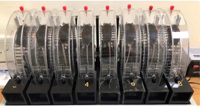
Figure 1. Overexertion in mice using a rotator-type fatigue apparatus. Overexertion induction in mice using a rotator-type fatigue apparatus. Representative photograph of the multi-lane motorized treadmill system used to implement the forced-running protocol. The apparatus consists of eight independent enclosed lanes (labeled 1–8) designed to ensure consistent mechanical loading and treadmill-induced overexertion across all experimental subjects. Please click here to view a larger version of this figure.
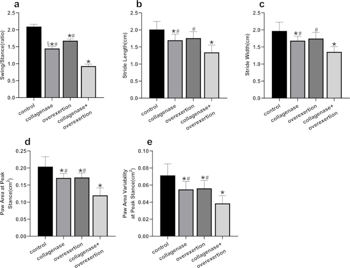
Figure 2. Gait Analysis. Gait cycle, distance, and kinetic parameters are presented as individual data points with mean ± standard deviation (n = 7–8 mice per group). Comparisons among multiple groups were made using an ANOVA followed by a Tukey's post hoc test. p-values less than 0.05 represent statistical significance. *p < 0.05, compared with the control group. #p < 0.05, compared with the collagenase + overexertion group. §p < 0.05, compared with the overexertion group. Please click here to view a larger version of this figure.
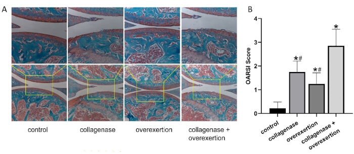
Figure 3. Joint histology. A: Representative images of safranin O-fast green staining in the mouse knee joints (Top panel: scale bar = 200 µm, magnification 40×; Bottom panel: scale bar = 100 µm, magnification 100×). B: OARSI scores are presented as individual data points with mean ± standard deviation (n = 7–8 mice per group). Comparisons among multiple groups were performed using an ANOVA followed by a Tukey post hoc test. Statistical significance was defined as p < 0.05. *p < 0.05, compared with the control group. #p < 0.05, compared with the collagenase + overexertion group. Please click here to view a larger version of this figure.
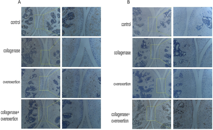
Figure 4. Immunohistochemistry. Representative immunohistochemical staining images of (A) COL2A1 and (B) TRPV4 expression in the articular cartilage (Left panel: scale bar = 200 µm, magnification 40×; Right panel: scale bar = 100 µm, magnification 100×). Please click here to view a larger version of this figure.
Discussion
In this study, we developed a novel mouse model of KOA that mimics the effects of excessive stress on the joint by combining intra-articular injection of collagenase with wheel-based fatigue-inducing exercise training. These findings once again substantiate the notion that KOA arises from the interplay of biological and mechanical factors. Understanding and identifying these factors hold promise for devising individualized treatment plans and preventive strategies to alleviate symptoms and improve the quality of life for individuals suffering from knee osteoarthritis13. Moreover, collagenase could also degrade the collagen matrix within tendons and ligaments, resulting in joint instability14. Excessive exercise could induce mechanical overload and ultimately trigger KOA15. Compared to previous models of KOA, this model exhibited a closer resemblance to human mechanically induced KOA in terms of its pathogenic mechanisms. Furthermore, intra-articular collagenase injection combined with excessive exercise in juvenile animals (10 weeks old) led to joint changes within a limited time frame (several weeks) that resemble those observed in spontaneously occurring osteoarthritis in aged mice. In summary, the use of juvenile mice and a time-effective experimental setup enabled convenient and cost-effective investigation of experimental osteoarthritis.
The initial description of OA could be traced back to 1886 when John K. Spender in England characterized it as a condition involving degenerative alterations in the knee joint, leading to joint pain and restricted range of motion16. Exploring the pathogenesis of OA has remained a topic of investigation for centuries. The Clinical Practice Guidelines for the Non-Surgical Treatment of Osteoarthritis of the Knee, Hip, and Multiple Joints, published by the OARSI in 2019, recognized osteoarthritis as an ailment characterized by irreversible degenerative changes, often associated with injuries, among which mechanical overload represents a specific subtype17. Established factors contributing to KOA encompassed heightened loading (e.g., obesity or physical labor) and ligamentous injury-induced instability18. Biomechanical aspects, widely acknowledged in the academic realm, were considered crucial causative elements in the development of KOA19,20. Furthermore, biomechanical factors were deemed the foremost contributors amidst various causes, resulting in an imbalance within the knee joint structure and leading to concentrated stress distribution21,22. As abnormal joint kinematics and/or loading were associated with OA, the prevalence of OA generally rose along with diminished joint stability and consistency23.
The stability of the knee joint emanated from three distinct sources: structural stability, conferred by the configuration of the articular surface and meniscus; static stability, provided by ligaments and the joint capsule; and dynamic stability, maintained by the surrounding muscles24,25,26. While bone structure did contribute to stabilizing the knee joint, ligaments and muscles surrounding the joint played the predominant role in maintaining stability27. These muscles not only participated in knee movement but also counteracted external loads exerted on the knee joint, contributing to its stabilization28. Lax ligaments and joint instability exerted unequal and elevated forces on the bones. The presence of soft tissue lesions surrounding the joint altered the magnitude, direction, or point of force application, subsequently impacting the collective force and influencing stress distribution within the joint29. Repetitive loading, referred to as strain, could induce joint degeneration30. This biomechanical factor played a pivotal role in the progression of KOA. Although chemical, enzymatic, and metabolic factors could diminish the resilience of articular cartilage, the occurrence of cartilage wear necessitated mechanical forces31. An imbalanced distribution of forces on the joint intensified the load exerted on the joint surface, expediting cartilage degeneration and establishing a self-perpetuating cycle32.
Furthermore, this study introduced gait analysis to evaluate behavioral aspects in mice. Gait analysis is an essential method for assessing walking ability and gait patterns. The swing/stance ratio reflects the balance between standing and swinging during the process of walking in the context of KOA. Additionally, stride width and stride length are important parameters for evaluating gait stability and symmetry, respectively. In patients with KOA, knee pain and functional impairments can restrict the stance phase, stride width, and stride length, while reducing the swing phase. These changes ultimately result in slowed walking speed, incoherent walking rhythm, diminished walking stability, and compensatory movements in other body regions.
Compared to traditional surgical models such as destabilization of the medial meniscus (DMM) or partial meniscectomy (PMX) that induce rapid post-traumatic osteoarthritis33, the combined collagenase and exercise model offers distinct translational advantages. By synergizing mild enzymatic cartilage degradation with cumulative mechanical loading, this approach faithfully mimics the gradual pathogenesis of human degenerative KOA rather than acute joint trauma. Crucially, the progressive nature of this model preserves early-to-mid stage KOA characteristics over a four-week period, providing a superior and extended window for tracking dynamic molecular mechanisms, evaluating chronic pain phenotypes, and testing long-term therapeutic interventions34 Furthermore, utilizing minimally invasive intra-articular injections alongside controlled exercise significantly reduces procedural complications and enhances animal welfare compared to highly invasive surgical techniques.
The success and stability of this model depend on the standardized implementation of several critical procedures and proactive troubleshooting. Accurate intra-articular injection requires precise anatomical positioning approximately 0.5 cm above the patellar ligament attachment. To prevent injection deviation and drug leakage, the knee should be kept slightly flexed to reduce joint cavity pressure, and a pre-rinsed 30 G needle must be used with thoroughly dissolved and centrifuged collagenase. Administering the 5 µL dose at 1 µL/s, followed by immediate post-injection compression, prevents joint pressure spikes and minimizes drug loss. For consistent mechanical loading, the fatigue apparatus must be calibrated to a stable 12 rpm. Enforcing a 3-day gradient acclimatization training mitigates low animal compliance and fear-induced refusal to run. During exercise, electrical stimulation must be strictly capped at 0.1–0.3 mA; if movement is not guided successfully, electrode contact should be checked rather than blindly increasing the current. Reliable gait analysis requires an extended treadmill acclimatization period and an undisturbed, temperature-controlled environment to reduce stress-induced abnormal movements. The treadmill belt and mouse paws must be thoroughly cleaned of debris prior to testing at a stable 20 cm/s speed to ensure the accurate image recognition of at least four uninterrupted strides. Finally, optimal tissue processing necessitates immediate fixation in 4% paraformaldehyde followed by carefully monitored decalcification in 0.5 mol/L EDTA. A needle puncture test should be frequently utilized during the 14–21-day decalcification period to prevent over-decalcification, which can severely compromise sectioning quality and tissue antigenicity.
While the outcomes are positive, several limitations and reproducibility considerations must be acknowledged. This model primarily simulates early-to-mid stage KOA and cannot fully replicate the extensive cartilage loss and osteophyte formation typical of late-stage disease. Furthermore, the inherent anatomical differences between mice and humans, combined with the exclusive use of male C57BL/6 mice and fixed concentration-intensity settings, omit common clinical risk factors such as aging, obesity, and gender differences. The absence of a saline-injected vehicle control cohort also prevents the absolute exclusion of localized micro-inflammatory responses caused by the needle insertion. In future studies, the experimental design will be enriched by exploring collagenase/exercise gradients and incorporating multi-dimensional observables—such as advanced imaging, biochemical assays, and multi-omics approaches—to further elucidate the dynamic molecular pathology of KOA from a holistic perspective. To enhance external reproducibility, standardized operator training and blinded behavioral assessments remain essential for minimizing technical variability across different laboratory settings.
In summary, combining intra-articular collagenase injections with overexertion successfully establishes a highly relevant overload stress injury KOA model in mice.
Disclosures
The authors have no conflicts of interest to declare.
Acknowledgements
The authors would like to thank the participants for their time and effort, and all the animals in the study. This work was supported by the Incentive Funding Application for hanghai Yueyang Hospital Ruian Branch Major Discipline Construction Incentive Fund - Tuina Department [QY71.42.06] and The National Administration of Traditional Chinese Medicine High-Level Key Discipline-Science of Tuina [zyyzdxk -2023061].
Materials
| Name | Company | Catalog Number | Comments |
|---|---|---|---|
| 4% Paraformaldehyde fixative | Beyotime | P0099 | |
| Acetic acid | Sinopharm | 631-61-8 | |
| DigiGait imaging system for mice | RWD Life Science | MSI-DIG-MS | |
| EDTA solution | Procell | PB180320 | |
| Embedding cassette | Leica | IP C | |
| Ethanol | Sinopharm | 64-17-5 | |
| Fast green stain | Leyan | 1085995 | |
| Hair removal cream | Veet | G20161330 | |
| Hematoxylin | Adamas | 61753A | |
| High-speed refrigerated centrifuge | Hermle | Z366K | |
| Incubator | TAITEC | M-210FN | |
| Inhalation anesthesia machine | Ranger | RZ-Viking | |
| Isoflurane | Webio | w100 | |
| Microtome | Ted Pella | 10180-220 | |
| Mouse rotator-type fatigue apparatus | Beijing Zhishu Duobao Biological Technology | YLS-10B | |
| Neutral balsam | Sinopharm | 96949-21-2 | |
| Safranin O stain | Bioss | S0062 | |
| Semi-automatic tissue embedding center | Thermo Fisher | HistoStar | |
| Slide drying hotplate | Electron Microscopy Sciences | XH-2002 | |
| Tissue flotation water bath | BIOBASE | BT-I | |
| Type II collagenase | Sigma-Aldrich | C4-22-1G | |
| Whole slide scanner | Leica | Aperio VERSA | |
| Xylene | Sinopharm | 1330-20-7 |
References
- Friedman, B., et al. Adenosine A2A receptor signaling promotes FoxO associated autophagy in chondrocytes. Scientific Reports. 11, 968-968 (2021).
- O'Brien, P., et al. What are the patient factors that impact on decisions to progress to total knee replacement? BMJ Open. 9, e031310-e031310 (2019).
- Amirkhizi, F., et al. Higher dietary phytochemical index is associated with lower odds of knee osteoarthritis. Scientific Reports. 12, 9059-9059 (2022).
- Liu, Q., et al. Effects of mechanical stress on chondrocyte phenotype and chondrocyte extracellular matrix expression. Scientific Reports. 6, 37268-37268 (2016).
- Halonen, K. S., et al. Workflow assessing the effect of gait alterations on stresses in the medial tibial cartilage. Scientific Reports. 7, 17396-17396 (2017).
- Xu, L., Kazezian, Z., Pitsillides, A. A., Bull, A. M. J. A synoptic literature review of animal models for investigating the biomechanics of knee osteoarthritis. Frontiers in Bioengineering and Biotechnology. 12, 1408015-1408015 (2024).
- Kim, J. E., et al. Development and characterization of various osteoarthritis models for tissue engineering. PLoS One. 13, e0194288-e0194288 (2018).
- Lampropoulou-Adamidou, K., et al. Useful animal models for the research of osteoarthritis. European Journal of Orthopaedic Surgery & Traumatology. 24, 263-271 (2014).
- Park, J., et al. A pathophysiological validation of collagenase II-induced biochemical osteoarthritis animal model in rabbit. Tissue Engineering and Regenerative Medicine. 15, 437-444 (2018).
- van der Kraan, P. M., et al. Degenerative knee joint lesions in mice after a single intra-articular collagenase injection. Journal of Experimental Pathology. 71, 19-31 (1990).
- Li, J. A mouse model of osteoarthritis originating from lateral instability of the ankle joint. , Suzhou University. (2017).
- Glasson, S. S., et al. The OARSI histopathology initiative - recommendations for histological assessments of osteoarthritis in the mouse. Osteoarthritis and Cartilage. 18, Suppl 3. S17-S23 (2010).
- Perera, J. R., Gikas, P. D., Bentley, G. The present state of treatments for articular cartilage defects in the knee. Annals of the Royal College of Surgeons of England. 94, 381-387 (2012).
- Zhang, J., et al. Tetrahydrohyperforin prevents articular cartilage degeneration and affects autophagy in rats with osteoarthritis. Experimental and Therapeutic Medicine. 15, 5261-5268 (2018).
- Sun, Y., Wang, C., Gong, C. Repairing effects of glucosamine sulfate in combination with etoricoxib on articular cartilages of knee osteoarthritis. Journal of Orthopaedic Surgery and Research. 15, 150-150 (2020).
- Spender, J. K. On some hitherto undescribed symptoms in the early history of osteoarthritis: the so-called rheumatoid arthritis. British Medical Journal. 1, 781-783 (1888).
- Bannuru, R. R., et al. OARSI guidelines for the non-surgical management of knee, hip, and polyarticular osteoarthritis. Osteoarthritis and Cartilage. 27, 1578-1589 (2019).
- Felson, D. T. Osteoarthritis as a disease of mechanics. Osteoarthritis and Cartilage. 21, 10-15 (2013).
- van Tunen, J. A. C., et al. Association of malalignment, muscular dysfunction, and abnormal joint loading with tibiofemoral knee osteoarthritis. BMC Musculoskeletal Disorders. 19, 273-273 (2018).
- Zhang, L., et al. Knee joint biomechanics in physiological conditions and how pathologies can affect it: a systematic review. Applied Bionics and Biomechanics. 2020, 7451683-7451683 (2020).
- Amin, S., et al. Knee adduction moment and development of chronic knee pain in elders. Arthritis and Rheumatism. 51, 371-376 (2004).
- Foroughi, N., Smith, R., Vanwanseele, B. The association of external knee adduction moment with biomechanical variables in osteoarthritis. The Knee. 16, 303-309 (2009).
- Henak, C. R., Anderson, A. E., Weiss, J. A. Subject-specific analysis of joint contact mechanics: application to the study of osteoarthritis. Journal of Biomechanical Engineering. 135, 021003-021003 (2013).
- Menge, T. J., et al. Anterior horn meniscal repair using an outside-in suture technique. Arthroscopy Techniques. 5, e1111-e1116 (2016).
- Zhang, J., et al. Feasibility study of early prediction of postoperative MRI findings for knee stability after ACL reconstruction. BMC Musculoskeletal Disorders. 22, 649-649 (2021).
- Kawada, M., et al. Contribution of hip and knee muscles to lateral knee stability during gait. Journal of Physical Therapy Science. 32, 729-734 (2020).
- Shim, J. K., Choi, H. S., Shin, J. H. Effects of neuromuscular training on knee joint stability after anterior cruciate ligament reconstruction. Journal of Physical Therapy Science. 27, 3613-3617 (2015).
- Hirschmüller, A., et al. Rehabilitation before regenerative cartilage knee surgery: a new prehabilitation guideline. Archives of Orthopaedic and Trauma Surgery. 139, 217-230 (2019).
- Hartmann, H., Wirth, K., Klusemann, M. Analysis of the load on the knee joint and vertebral column with changes in squatting depth. Sports Medicine. 43, 993-1008 (2013).
- Fong, I. C. D., et al. Effect of foot progression angle adjustment on the knee adduction moment in runners with knee osteoarthritis. Gait & Posture. 61, 34-39 (2018).
- Du, J., et al. Effect of high fat diet and excessive compressive mechanical force on pathologic changes of temporomandibular joint. Scientific Reports. 10, 17457-17457 (2020).
- Hunt, M. A., et al. Varus thrust in medial knee osteoarthritis: quantification and effects of different gait-related interventions. Arthritis Care & Research. 63, 293-297 (2011).
- Christiansen, B. A., et al. Non-invasive mouse models of post-traumatic osteoarthritis. Osteoarthritis and Cartilage. 23, 1627-1638 (2015).
- Adães, S., et al. Intra-articular injection of collagenase in the knee of rats as an alternative model to study nociception. Arthritis Research & Therapy. 16, R10-R10 (2014).