This review examines brain organoids as platforms for modeling selected neurotropic viral infections and highlights their applications in viral pathogenesis, antiviral discovery, innate immune responses, and central nervous system (CNS)-targeted drug delivery.
A subscription to JoVE is required to view this content. Sign in or start your free trial.
Review Article
Brain Organoids as Emerging Platforms for Modeling CNS Infections: Neuropathogenesis, Therapeutic Discovery, and Drug Delivery
* These authors contributed equally
In This Article
Summary
Abstract
Neurotropic viruses remain a persistent global health challenge, and the mechanisms by which they damage the human brain are not yet fully understood. Animal models are often limited by human-specific aspects of CNS biology, whereas two-dimensional (2D) cell cultures cannot recapitulate the complex three-dimensional (3D) cellular interactions that occur during viral infection of the brain. Over the past decade, brain organoids derived from human stem cells have emerged as physiologically relevant models that address these limitations. These 3D cultures self-assemble into structures containing neurons, astrocytes, and progenitor cells arranged in patterns that resemble early brain development. This review examines the application of brain organoids to the study of infections caused by Zika virus (ZIKV), severe acute respiratory syndrome coronavirus 2 (SARS-CoV-2), herpes simplex virus (HSV), and human immunodeficiency virus type 1 (HIV-1). Studies were screened from PubMed and shortlisted based on their relevance to organoid-based CNS infection modeling, antiviral drug screening, CNS-targeted drug delivery, and neuroinflammation. Organoid-based antiviral screening has identified promising compounds from libraries containing more than 1,000 candidates. Drug delivery strategies are also discussed, with particular emphasis on nanoparticles, polymer-based carriers, and extracellular vesicles (EVs) evaluated in organoid and spheroid models for their ability to cross the blood-brain barrier (BBB) and deliver therapeutic cargo to neural cells. The roles of damage-associated and pathogen-associated molecular patterns (DAMPs and PAMPs) in neuroinflammation, complement evasion, and chronic post-infection damage, including long COVID, are also examined. Current limitations, including the lack of functional vasculature, incomplete BBB components, and reproducibility challenges, are discussed. Despite these limitations, CNS organoids bridge the gap between basic research and clinical application, advancing the development of effective therapies for viral infections of the brain.
Introduction
The central nervous system (CNS) is a complex system comprising distinct functional regions of the brain and spinal cord. CNS organoids, often referred to as "mini-brains," are three-dimensional (3D) multicellular structures that mimic the developing human brain. They are primarily generated from human pluripotent stem cells (hPSCs), which can self-assemble into tissues that represent diverse brain regions. These mini-brains recapitulate the epigenetic patterns, structure, and transcriptional markers of the developing brain1,2,3.
The generation of cerebral organoids using patient-derived induced pluripotent stem cells (iPSCs) was first developed in 20134. Prior to this discovery, access to human brain tissue was limited because of the ethical issues associated with conducting experimental studies, particularly those involving fetal or in utero brain tissue2. The development of cerebral organoids demonstrated the ability to mimic human neurodevelopment, including the formation of distinct brain regions such as the cerebral cortex. Lancaster et al. provided the first experimental evidence that stem cells can form complex neural structures in vitro, offering a model that more accurately reflects human physiology than conventional two-dimensional (2D) cell cultures5. Importantly, certain human neurodevelopmental conditions, such as microcephaly, have proven difficult to replicate in rodent models, further emphasizing the need for human-derived systems6.
CNS organoids contain multiple cell types that differentiate and form multilayered structures resembling the developing brain7, making them well-suited for high-throughput studies of neurogenesis, neuronal migration, and developmental abnormalities8. The generation of CNS organoids begins with pluripotent stem cells (PSCs), which include iPSCs and embryonic stem cells. Embryonic stem cells are exposed to growth factors and signaling inhibitors to promote differentiation into neural cells and mimic early embryonic development2. Once the cells begin to aggregate, they undergo self-organization within a 3D environment and are often embedded in an extracellular matrix to provide structural stability. Organoids are then typically maintained under dynamic culture conditions, such as spinning bioreactors or orbital shaking, to enhance nutrient and oxygen delivery, and support continued growth9. Region-specific organoids of the forebrain, midbrain, and hindbrain can also be generated using guided protocols in miniaturized spinning bioreactors, with distinct brain-like regions typically emerging over several weeks to months9,10,11. CNS organoids capture many key structural features of the developing human brain, including ventricular regions containing radial glia that scaffold migrating neurons and intermediate progenitors that amplify neuronal populations12. Early neurons form cortical-like layers with emerging synaptic networks. Although organoids do not fully replicate the size or organization of the mature brain, they are more physiologically relevant than traditional monolayer cultures.
A major advantage of CNS organoids is their ability to model developmental timing. Cultures progress from weeks to months and parallel human fetal brain development. This allows researchers to observe neurological processes such as neurogenesis, axon extension, and early neural circuit formation in real time. Organoids can also be programmed to include microglia, blood vessel-like cells, or even blood-brain barrier (BBB) components, such as astrocytes, endothelial cells, and pericytes. These organoids can support studies of immune responses and substance transport13. Additionally, organoids can be combined to create assembloids, which are self-organizing systems that model communication between different brain regions14. With these capabilities, CNS organoids offer a direct way to investigate how neurotropic viruses invade, replicate within, and damage the brain, questions that have long been difficult to address using conventional models.
There are a vast number and diversity of neurotropic viruses that can affect the human brain, including flaviviruses, severe acute respiratory syndrome coronavirus 2 (SARS-CoV-2), herpes simplex virus (HSV), and human immunodeficiency virus type 1 (HIV-1). The development of effective antiviral therapies is crucial yet remains challenging due to the complexity of virus-brain interactions. The brain is considered an immune-privileged organ, supported by the BBB, which prevents large immune cells and molecules from entering the brain from the bloodstream.
Neurotropic viruses have evolved diverse strategies to breach this barrier, including transcellular migration, disruption of tight junctions, and the "Trojan horse" mechanism, whereby infected immune cells carry the virus across the endothelium4. As a result, immune surveillance in the brain is restricted, making viral clearance more difficult. Furthermore, the cellular components of the BBB, including endothelial cells, astrocytes, and pericytes, express pattern recognition receptors that detect viral pathogen-associated molecular patterns (PAMPs) and mount innate immune responses, although pro-inflammatory signaling can paradoxically worsen BBB disruption15. Additionally, limited access to human brain tissue hampers studies of viral interactions with neural cells and the delivery of potent antiviral agents to the CNS. These combined challenges hinder the discovery of new antiviral agents to treat viral infections at all stages of development and across the lifespan, making it essential to better understand how viruses interact with the brain and to develop new therapeutic strategies to combat these infections16. Overall, this review emphasizes the utility of CNS organoids for studying neurotropic viral infections, mechanisms of neuroinflammation, and antiviral drug discovery. Figure 1 provides an overview of the major applications of brain organoids in modeling neurotropic viral infections, investigating host-pathogen interactions, and advancing antiviral therapeutic development.
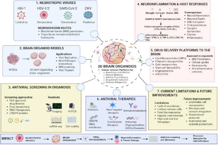
Figure 1: Brain organoids as human-relevant platforms for modeling neurotropic CNS viral infections. The figure provides an overview of the key topics addressed, with a focus on three-dimensional (3D) brain organoids as experimental models. Neurotropic viruses discussed in this review include HIV-1, HSV-1/2, SARS-CoV-2, and ZIKV. Different brain organoid models enable studies of viral pathogenesis, host-pathogen interactions, blood-brain barrier (BBB) dysfunction, and neuroinflammatory responses involving neurons, astrocytes, microglia, and brain endothelial cells. Brain organoids provide physiologically relevant platforms for antiviral drug screening and evaluation of antiviral therapies. Created in BioRender. Kashanchi, F. (2026) https://BioRender.com/y7eenju Please click here to view a larger version of this figure.
Access restricted. Please log in or start a trial to view this content.
Review and Perspective
Therapeutic discovery in virus-infected brain organoids
Antiviral screening
There is a lack of approved therapies for many neurotropic viral infections, limiting treatment options beyond supportive care. Brain organoids are a valuable tool for discovering novel antiviral compounds by enabling drug screening in a physiologically relevant, 3D environment that resembles human brain tissue. In an initial study, a high-content chemical screening of 1,120 FDA-approved drugs and drug candidates was performed against ZIKV in fetal-like brain organoids. Antiviral activity was assessed based on cell proliferation and suppression of ZIKV infection. Of all the compounds tested, nine showed potential, with two compounds—hippeastrine hydrobromide (HH) and amodiaquine dihydrochloride dihydrate (AQ)—standing out because of their strong antiviral effects, significantly reducing infection by suppressing viral RNA production17.
Several subsequent studies explored compounds targeting different stages of the viral life cycle. In a study by Watanabe et al., 25-hydroxycholesterol (25HC), a compound that blocks viral entry by inhibiting membrane fusion, reduced ZIKV mRNA levels in cortical organoids by ~74% and decreased ZIKV-positive cells by ~72%. Furthermore, the same study evaluated antiviral compounds targeting the AXL receptor expressed on neural progenitor cells. A small-molecule inhibitor of the AXL tyrosine kinase domain, R428, produced a modest reduction in ZIKV mRNA levels (~41%). However, treatment with an anti-AXL antibody did not significantly reduce ZIKV mRNA levels18. Finally, three anti-flaviviral drugs, duramycin, ivermectin, and azithromycin, were tested in organoids. Duramycin and ivermectin significantly reduced ZIKV mRNA levels, whereas azithromycin showed no protective effect against ZIKV, as determined by RT-qPCR and immunostaining18.
In a separate study, Xu et al. screened 173 compounds as suppressors of ZIKV-induced caspase-3 activity and identified emricasan as the most potent neuroprotective candidate without affecting cell proliferation in brain organoids. Two additional compounds, niclosamide and PHA-690509, demonstrated significant anti-ZIKV activity by inhibiting viral replication at a post-entry stage in a dose-dependent manner. Furthermore, co-treatment with emricasan and PHA-690509 produced an additive effect and preserved astrocyte viability following ZIKV infection19. In another study, enoxacin, an RNA interference (RNAi) enhancer, demonstrated direct antiviral activity in RNAi-competent cells and completely prevented ZIKV-induced microcephalic phenotypes in brain organoids, suggesting that enhancing intrinsic antiviral pathways in neural progenitor cells may represent a viable therapeutic strategy20.
Pettke et al. subsequently screened 110 structural analogues of TH3289 at multiple doses in ZIKV-infected organoids21. Two compounds, TH3289 and TH6744, showed the strongest antiviral activity, and the most active hits shared a benzimidazolone core. This study was significant because the organoid system enabled not only rapid compound screening but also mechanistic follow-up. For example, TH6744 was found to interfere with late stages of the ZIKV life cycle21.
Brain organoids have also been used to identify therapeutic candidates for SARS-CoV-2 infection. An FDA-approved anti-hepatitis C drug, sofosbuvir, improved neuronal survival and rescued impaired synaptogenesis in SARS-CoV-2-infected brain organoids22. Brain organoids have also contributed to understanding HSV-1-mediated CNS infection; however, antiviral drug screening for HSV-1 in organoid models remains limited compared with studies of ZIKV and SARS-CoV-2. A landmark study using iPSC-derived neurons demonstrated that TLR3 deficiency renders human neurons susceptible to HSV-1 infection, revealing a human-specific phenomenon that rodent models had failed to capture23. More recently, Bellizzi et al. demonstrated that clustered regularly interspaced short palindromic repeats (CRISPR)-Cas9 targeting of HSV-1 genomic sequences suppressed both primary infection and viral reactivation in 3D organoid cultures, establishing gene editing as a feasible antiviral strategy for herpesvirus CNS infection24. Given that acyclovir resistance is an emerging clinical problem and no curative therapy exists for HSV encephalitis25, the absence of large-scale antiviral compound screening against HSV-1 in brain organoids represents a significant knowledge gap.
In addition to small molecules, antisense oligonucleotides (ASOs), which target RNA and modify gene expression, are emerging therapeutic tools for treating various diseases, and cerebral organoids can be employed to evaluate novel ASO therapies26. Given the utility of organoids for ASO studies, important characteristics such as biodistribution, toxicity, and therapeutic efficacy could be investigated in virus-infected brain organoids.
One of the most important applications of brain organoids is assessing the neurotoxicity of existing antiviral drugs. This is particularly important because commonly used HIV treatments have been shown to be toxic to brain cells both in vitro and in vivo27. Akay et al. demonstrated the neurotoxicity of antiviral drugs in primary cortical cultures containing microglia, astrocytes, and neurons. MAP2-positive neurons showed a dose-dependent decrease following exposure to ritonavir and saquinavir. Treatment with combinations of antiretroviral drugs (cART) resulted in acute neurotoxicity, and cultures treated with zidovudine (AZT) exhibited increased production of reactive oxygen species (ROS)28. Although these findings were obtained using 2D cultures, brain organoids could expand this research by providing a more physiologically relevant platform for high-throughput neurotoxicity screening of individual or combination antiretroviral therapies. To investigate the mechanisms underlying antiviral-induced neurotoxicity in these 3D models, techniques such as morphological analysis, functional assays, and transcriptomic profiling can be employed29.
In addition to neurotoxicity screening, brain organoids have been developed to study HIV-associated neurocognitive disorder (HAND), which continues to affect 30%–50% of people living with HIV despite effective antiretroviral therapy (ART). Microglial cells are the primary reservoir of HIV-1 in the CNS, and organoid models have been instrumental in elucidating how microglial infection drives neuronal damage. Kong et al. used iPSC derived cerebral organoids containing microglia and observed a marked increase in CCL2 and CXCL10 chemokine gene expression, together with activation of multiple type I interferon-stimulated genes (MX1, ISG15, ISG20, IFI27, IFITM3, and others) following HIV-1 infection30. The inflammatory gene expression profile was propagated to bystander cells, including uninfected neurons. These neurons exhibited a marked decline in neurotransmitter transporter expression together with increased expression of genes associated with cellular senescence and cell death. The use of brain organoids for HIV research was pioneered by Dos Reis et al., who incorporated HIV-infected microglia into human brain organoids31. This model reproduced the low-level viral replication typically observed in the human brain. Incorporation of HIV-infected microglia triggered a neuroinflammatory response associated with astrogliosis, neurodegenerative pathology, and degeneration and loss of neuronal synaptic processes. Donadoni et al. subsequently used human induced pluripotent stem cell (hiPSC)-derived cerebral organoids containing microglia and astrocytes to model NeuroHIV and evaluate the effects of combination antiretroviral therapy (cART) in a 3D organoid system32. Collectively, these organoid-based studies provide important mechanistic insights into HAND and establish a valuable model system for investigating neuroprotective therapies.
In conclusion, brain organoids provide a unified model system for screening antiviral compounds while simultaneously assessing neurotoxic effects early in the drug development process. Identification of effective antiviral compounds is only the first step toward clinical translation (Table 1). Equally important is the development of drug-delivery strategies capable of delivering therapeutic agents to infected cells within the CNS.
| Virus | Organoid Model | Cell Types | Key Findings | Limitations | Reference | |
| ZIKV | hPSC-derived fetal-like organoids | Neural progenitors, neurons | HH and AQ reduced viral RNA from 1120-compound screen | No immune cells; fetal stage | 17 | |
| Cortical organoids | Neural progenitors | 25HC reduced infection by ~74%; R428 reduced ZIKV mRNA by ~41% | No vasculature | 18 | ||
| Brain organoids | Neurons, astrocytes | Emricasan neuroprotective; niclosamide and PHA-690509 inhibit replication | No microglia | 19 | ||
| Brain organoids | Neural progenitors | Enoxacin prevented ZIKV-induced microcephaly via RNAi | No immune cells | 20 | ||
| Brain organoids | Neurons | TH3289 and TH6744 (benzimidazolone core) interfere with late ZIKV life cycle | No immune cells | 21 | ||
| SARS-CoV-2 | Cerebral organoids | Neurons | Sofosbuvir rescued synaptogenesis and neuronal survival | No BBB | 22 | |
| HSV-1 | iPSC-derived neurons | Neurons | TLR3 deficiency leaves human neurons susceptible; human-specific phenomenon | 2D neurons only | 23* | |
| 3D organoid cultures | Neurons | CRISPR-Cas9 suppressed primary infection and reactivation | Limited maturation | 24 | ||
| HIV-1 | iPSC cerebral organoids | Neurons, microglia | CCL2, CXCL10 upregulation; type I interferon gene activation; neuronal senescence | No vasculature | 30 | |
| Human Brain ORGanoids with microglia | Neurons, microglia | Neuroinflammation, astrogliosis, synaptic degeneration | No vasculature | 31 | ||
| hiPSC cerebral organoids | Neurons, microglia, astrocytes | cART effects evaluated in 3D NeuroHIV model | Early stage model | 32 | ||
Table 1: Brain organoid models used to study neurotropic viral infections. This table summarizes the virus studied, organoid model, cell types included, key findings, limitations, and reference for representative brain organoid-based studies of ZIKV, SARS-CoV-2, HSV-1, and HIV-1 infections.
Drug delivery platforms
Finding the most effective antiviral compounds for treating CNS infections is essential, but without efficient delivery to the brain, these compounds may fail to achieve therapeutic benefit. Brain organoids can be used to evaluate drug delivery systems for their ability to penetrate neural tissue and deliver antiviral payloads.
Lipid-based nanoparticles (LNPs) are widely used as drug carriers. These drug carriers were shown to increase drug bioavailability and enable targeted drug delivery to the disease site, especially across the BBB33. LNP-based mRNA formulations were successfully utilized in COVID-19 vaccination34,35. Brain organoids can be used to evaluate the effectiveness and safety of new LNP-based formulations. LNPs can also be vectorized to the CNS by surface modification with targeting ligands. For example, transferrin or lactoferrin were conjugated to LNPs to facilitate receptor-mediated transcytosis and enhance transport across the BBB36,37. Collectively, the potential drug delivery efficacy, neurotoxicity, and changes in nanoformulation stability after BBB penetration can be investigated in brain organoids38.
In addition to LNPs, dual-targeted polypeptide nanocarriers were also studied as a drug delivery system in human brain organoids. Targeted poly(L-glutamic acid) nanoparticles (NPs) showed excellent cytocompatibility and the ability to enter midbrain-like organoids. It was demonstrated that alanine and glutathione could efficiently direct NPs to cerebral endothelial cells and penetrate cerebral organoids, resulting in drug delivery to the brain39. The permeability of polymer-based nanoparticles was investigated in human 3D microvasculature organoids, focusing on the effects of their size and surface chemistry. The generated organoids consisted of human iPSC-derived endothelial cells, astrocytes, and primary brain pericytes. Confocal images revealed that conjugation of the NPs with the brain-associated ligand holo-transferrin (Tf) significantly increased their permeability40.
Next, the absorption, penetration, and distribution of gold nanoparticles (AuNPs) were investigated in BBB brain spheroids. These spheroids are composed of six brain cell types: neurons, astrocytes, pericytes, endothelial cells, microglia, and oligodendrocytes. The authors reported that transient hypoxia led to BBB opening and increased transport of AuNPs into the spheroid interior. The nanoparticles were able to enter cells and nuclei easily41. In a similar study using the same spheroid model system, concurrent tracking of nanocarrier penetration kinetics and their therapeutic payload release was evaluated by confocal laser scanning microscopy42. In another study, Park et al. demonstrated that brain-derived neurotrophic factor (BDNF)-conjugated AuNPs successfully entered cerebral organoids without causing significant toxicity43.
Beyond targeting moieties, the shape and size of nanoparticles can also play a significant role in a nanoformulation's ability to deliver its therapeutic payload to the brain44. Polystyrene 200 nm spheres vectorized with antibodies against VCAM-1 accumulated 1.2- to 1.5-fold greater than the control non-vectorized NPs. Interestingly, rod-shaped particles exhibited even greater accumulation of 2.1- to 2.5-fold over corresponding controls, suggesting that rod-shaped particles offer an advantage over spheres in terms of cell binding. Cerebral organoids can provide detailed insights into how particle geometry can affect brain penetration. Additionally, the effects of hypoxia and other biologically active compounds on BBB permeability could be further investigated in neurovascular brain organoids45,46.
Stem cell-derived extracellular vesicles (EVs) represent a biological alternative to synthetic drug carriers such as LNPs. Branscome et al. explored the reparative potential of EVs against HIV-1 infection in human neurospheres47. In this study, it was demonstrated that EVs produced from mesenchymal stem cells (MSCs) and iPSCs can naturally penetrate neurospheres, rescue cellular viability, and reduce the expression of inflammatory cytokines in HIV-1-infected neurospheres, highlighting the neuroprotective and anti-inflammatory properties of stem cell EVs47. In another study by Kim et al., small extracellular vesicles (sEVs) were engineered for antiviral delivery to treat SARS-CoV-2 infection48. The nanocarriers were loaded with soluble ACE2 (sACE2) via genetic modification of the donor cells with a plasmid encoding eGFP-CD9ΔTM4-sACE2(WT), resulting in the expression of sACE2 on the sEV surface. It was demonstrated that sACE2-vectorized sEVs protected against WT and mutant S protein pseudotyped virus48. The protection mechanism could be tested in SARS-CoV-2-infected brain organoids. Additionally, another study showed that EVs released from HSV-1-infected cells carry innate immune components such as STING, which can activate innate immune responses in recipient cells, resulting in a potent inhibition of HSV-1 replication. A crucial separation of EVs from infectious viral particles was achieved by density gradient centrifugation49.
Cerebral organoids can also be used to test the potential neurotoxicity of new drug nanoformulations. For instance, when pegylated AuNPs were injected into brain organoids, significant inflammation, mediated by ROS production, was observed. In contrast, polymeric poly-lactic acid NPs exhibited efficient drug delivery and low cytotoxicity due to their slow uptake and degradability, enabling penetration into the brain organoid without eliciting an immune response50. The neurotoxicity of AuNPs, graphene, and carbon dots was also assessed in 3D neurovascular organoids. These organoids consisted of neural cells, microglia, and an endothelial cell surface. These studies revealed that microglia could play a pivotal role in the permeability and toxicity of these formulations51. Furthermore, high concentrations of ZnO NPs have also been shown to cause significant neuronal death. The mechanism of toxicity was linked to intracellular accumulation of Zn ions, which decreased LC3B protein levels, suggesting defective autophagy52. Overall, these studies show that 3D brain organoids can capture how nanoformulations interact with neural tissue, from uptake and penetration to immune activation and toxicity. The success of CNS-targeted drug delivery strategies ultimately depends not only on reaching target cells but also on modulating the inflammatory pathways activated during infection, many of which are initiated by DAMPs and PAMPs signaling.
DAMPs and PAMPs in viral infections
Brain organoids can be used to study DAMPs and PAMPs that are released by stressed, injured, or dying cells in the CNS. DAMPs include heat shock proteins, ATP, and uric acid, whereas PAMPs include bacterial, viral, fungal, and parasitic macromolecules. Hence, these molecular patterns alert the innate immune system and activate several signal transduction pathways in response to foreign antigens or any sign commonly associated with infection. The mechanisms underlying their protective effects in response to viral infections are not fully understood, and detailed investigations of these processes can be carried out in brain organoids53. Released DAMPs and PAMPs bind to a large group of receptors, including Toll-like receptors (TLRs), NOD-like receptors (NLRs), AIM2-like receptors, RIG-I-like receptors (RLRs), and C-type lectin receptors (CLRs), which are expressed on the plasma membrane or intracellularly54. These receptors are crucial for cytokine and interferon production in response to neurotropic viral infections, including EV-71, HIV-1, HSV-1, flaviviruses, West Nile virus (WNV), and others54.
Slowikowski et al. investigated how DAMPs, chemokines, and adhesion molecules released by damaged neural cells coordinate leukocyte entry into the CNS during flavivirus encephalitis55. In particular, it was revealed that WNV increases the expression of highly inflammatory necroptosis and pyroptosis cell death markers that promote the release of DAMPs in the infected brain. When such molecules, for example, high mobility group box 1 (HMGB1) protein, are released, they induce multiple signaling pathways, leading to CXCR4-dependent migration of activated monocytes, macrophages, and dendritic cells to infected tissues56,57. Furthermore, it was shown that DAMPs can be released in response to infection, followed by upregulation of TLRs and exacerbation of CNS inflammation and tissue damage58. Overall, these studies indicate that viral infections appear to trigger overlapping DAMP-PAMP signaling pathways, leading to neuroinflammation and tissue damage. Collectively, brain organoids containing microglia and astrocytes could serve as a model for tracing immune cascade responses in human neural tissue, especially the interplay between DAMP release and TLR-mediated neuroinflammation.
The complement system is a major element of innate immunity that responds rapidly to infection. However, many viruses have developed mechanisms to overcome this defense system59. Certain viruses evade the complement system by producing proteins that bind to complement components and either inhibit or sequester them. This has been demonstrated in a variety of viruses, including human T-lymphotropic virus-1 (HTLV-1), human cytomegalovirus (HCMV), HIV-1, simian virus 5 (SV5), and mumps virus (MuV), which incorporate host complement proteins into their virions (i.e., CD55/DAF, CD59, CD64, and MCP). These proteins inhibit the innate immune response, promoting viral replication and persistence60. Thus, brain organoids can be used to identify the specific mechanisms by which viruses evade the complement system. Growing evidence suggests that DAMPs and PAMPs not only shape the initial antiviral response but also serve as key mediators of the persistent inflammation that underlies chronic post-viral conditions.
Role of DAMPs/PAMPs in persistent inflammation caused by viral infection
Beyond the acute inflammatory phase, viral infections can cause chronic exposure to DAMPs/PAMPs, resulting in long-term inflammatory consequences, particularly in the brain. Cerebral organoids can become a powerful tool to study the underlying mechanisms of syndromes related to the post-acute phase of infection.
Most currently known human non-persistent viruses activate immune responses during the acute phase of infection, followed by pathogen elimination. However, in some cases, patients experience persistent symptoms that may differ from those observed during the acute phase and can linger for months or even years. Specifically, patients with long COVID-19 report chronic fatigue, "brain fog," fever, pain, and other neurological complications61. Moreover, chronic inflammation may increase mortality and morbidity, as well as the risk of atherothrombosis, cancer, and cognitive impairment62,63.
Additionally, the persistence of viral proteins and RNA can lead to elevated levels of pro-inflammatory cytokines, such as TNF-α, IL-1α, IL-1β, IFN-γ, and IL-6, thereby causing tissue damage, premature aging, and immune system exhaustion64. Slow replication or traces of viral components could trigger the release of DAMPs/PAMPs that interact with their specific receptors, causing persistent low-grade inflammation65. In some cases, HSV-1 infection of the brain can lead to neuronal damage followed by encephalitis and may eventually contribute to the development of neurodegenerative disorders such as Alzheimer's disease66. Disrupted autophagy mechanisms can be a major trigger of chronic inflammation in patients with HSV-2, in which defective protective mechanisms lead to persistent viral replication, the continuous presence of viral components in the brain, and neurodegeneration67. These viral components activate innate immune sensors through PAMPs and can manipulate signaling pathways to bypass the antiviral effects of innate immune activation. Importantly, HCMV and Epstein-Barr virus (EBV) have been shown to establish chronic viral infections that promote immune dysfunction68,69. Sustained infection-induced senescence may also contribute to the long-term inflammation associated with various viral infections. 3D brain organoids with integrated immune components provide relevant platforms for understanding the complex mechanisms underlying chronic exposure to DAMPs/PAMPs and their role in the long-term complications of viral infections in the CNS.
Limitations and perspective:
Despite their advantages over traditional in vitro models, brain organoids have significant limitations that must be addressed to support their broader application. Specifically, most organoids lack resident immune cells, which are major contributors to viral disease modeling and to understanding innate and adaptive immune responses during viral infection70. In particular, standard organoid protocols lack incorporated microglia, the primary CNS immune cells and reservoir of HIV-1, which limits the modeling of neuroinflammation and viral persistence71. Another shortcoming is incomplete tissue complexity, including the absence of vasculature, which plays an important role in nutrient and oxygen delivery, as well as viral dissemination72. Thus, organoid growth is limited because cells in the center cannot receive adequate nutrients. Additionally, excretion of metabolic waste is impaired, often leading to hypoxic or necrotic cores in large organoids. Moreover, organoids do not fully reproduce the interactions among neurons, endothelial cells, and stromal cells that can influence viral infection and pathogenesis. Furthermore, at this stage of development, organoids cannot recapitulate systemic infection, interorgan spread, and whole-body immune responses73. Another limitation is that many epithelial organoids have limited accessibility for viral inoculation due to their inward-facing apical surface74. As a result, viral inoculation requires microinjection, leading to variability and low throughput. Thus, inoculation of organoids with different diameters makes the multiplicity of infection difficult to control. Additionally, many organoids resemble fetal tissue rather than adult organs, which can affect viral tropism, replication kinetics, and host responses73. This is particularly evident in the case of HSV-1, as neuronal maturation impacts latency establishment and reactivation processes that fetal-stage organoids may not fully recapitulate23. Next, organoids may vary considerably in composition and function, leading to varied outcomes, low reproducibility, and the need for numerous experimental replicates. For example, organoids may differ in size, cellular composition, architecture, and maturation state. This makes standardization difficult, if not impossible, across laboratories7,75. Finally, the high cost and limited scalability of organoid generation and maintenance remain significant barriers. As a result, a major challenge is the development of high-throughput platforms compatible with organoid technology74.
Despite these shortcomings and limitations, organoids hold significant potential for modeling viral infections, toxicology, and drug discovery. Overcoming these limitations could significantly reduce reliance on animal models and facilitate fundamental mechanistic studies76. One of the notable shortcomings of existing organoid models is their limited physiological complexity and lack of diverse cellular composition. Future developments will need to focus on incorporating immune, neuronal, vascular, and stromal components into organoid models. This could be achieved using co-culture models that integrate organoids with the aforementioned cell types77,78. Specifically, incorporating immune cells will enable modeling of systemic immune responses and inflammation during viral infections. Furthermore, vascularized organoids can be further developed to improve physiological complexity and better recapitulate in vivo conditions79,80. Thus, cerebral organoids incorporating endothelial tissues will support investigations of blood-brain barrier function and drug delivery during viral infections13,81. Additionally, 3D brain organoids can replicate several key aspects of viral infection in the brain. In fact, several organoid models with intricate structures that closely represent various brain regions are currently under investigation82. Finally, inclusion of neuronal and stromal components would further increase the fidelity of organoids to native organ systems. Reproducibility remains another serious barrier to the broader application of organoid technologies. Therefore, generating robust, highly reproducible organoids is crucial for the further translation of organoid applications. Various methods for cell isolation and seeding, as well as the selection of matrices and soluble factors, should be standardized across the field83. Of note, the composition of the culture medium, cell density, and culture time may affect the outcomes of these models. Therefore, strategies to reduce matrix variability, including synthetic hydrogels composed of fully defined polymer networks with precisely controlled chemical and mechanical characteristics, have been introduced as a promising alternative84. These organoids exhibit minimal batch-to-batch variability, enabling controlled studies. Furthermore, the creation of organoid biobanks representing diverse patient populations would become a valuable resource for drug discovery and biomarker identification. Emerging artificial intelligence (AI) technologies are becoming important tools in organoid research85. Further application of these technologies may accelerate analyses and reduce bias in data interpretation. Nevertheless, AI applications in this field remain in their early stages. Incorporation of AI approaches into organoid development, such as AI-driven image analysis86 or predictive optimization of culture conditions87, would significantly facilitate organoid research.
Access restricted. Please log in or start a trial to view this content.
Conclusions
This review has examined how brain organoids, 3D neural cultures that closely mimic the human neurovascular unit, are being used to study the invasion, replication, and CNS damage caused by neurotropic viruses. Although each virus has unique genetic and structural features, many of the pathological processes triggered by viruses in the brain are shared, and organoid models have proven well-suited for dissecting these common and different mechanisms. Excessive inflammation can worsen nerve cell damage; however, when the i...
Access restricted. Please log in or start a trial to view this content.
Disclosures
ChatGPT (OpenAI; GPT-5.5) was used in a limited capacity to assist with literature screening and language refinement. All literature selection, fact-checking, reference verification, and scientific interpretation were performed independently by the authors.
Acknowledgements
The authors gratefully acknowledge all members of the Kashanchi Laboratory, with special appreciation to Gwen Cox for her invaluable support. This study was also supported by National Institutes of Health (NIH) Grants R01MH134389, AI078859, and AI074410 R21DA057887 and Department of Defense HT9425-24-1-0876 to F.K.
Access restricted. Please log in or start a trial to view this content.
References
- Lancaster MA, Knoblich JA. Generation of cerebral organoids from human pluripotent stem cells. Nature Protocols. 2014;9(10):2329-40.
- Chhibber T et al. CNS organoids: an innovative tool for neurological disease modeling and drug neurotoxicity screening. Drug Discovery Today. 2020;25(2):456-65.
- Eichmüller OL, Knoblich JA. Human cerebral organoids — a new tool for clinical neurology research. Nature Reviews Neurology. 2022;18(11):661-80.
- Al-Obaidi MMJ et al. Disruption of the blood brain barrier is vital property of neurotropic viral infection of the central nervous system. Acta Virologica. 2018;62(1):16-27.
- Lancaster MA et al. Cerebral organoids model human brain development and microcephaly. Nature. 2013;501(7467):373-9.
- Kelava I, Lancaster MA. Dishing out mini-brains: Current progress and future prospects in brain organoid research. Developmental Biology. 2016;420(2):199-209.
- Velasco S et al. Individual brain organoids reproducibly form cell diversity of the human cerebral cortex. Nature. 2019;570(7762):523-7.
- Giandomenico SL et al. Cerebral organoids at the air–liquid interface generate diverse nerve tracts with functional output. Nature Neuroscience. 2019;22(4):669-79.
- Lancaster MA, Knoblich JA. Organogenesis in a dish: Modeling development and disease using organoid technologies. Science. 2014;345(6194):1247125.
- Elvira R, Tan EK, Zhou ZD. Three-dimensional midbrain organoids: a next-generation tool for Parkinson’s disease modelling and drug discovery. Stem Cell Research & Therapy. 2025;16(1):502.
- Kim S, Chang MY. Application of Human Brain Organoids—Opportunities and Challenges in Modeling Human Brain Development and Neurodevelopmental Diseases. International Journal of Molecular Sciences. 2023;24(15):12528.
- Noctor SC et al. Dividing Precursor Cells of the Embryonic Cortical Ventricular Zone Have Morphological and Molecular Characteristics of Radial Glia. The Journal of Neuroscience. 2002;22(8):3161-73.
- Sun XY et al. Generation of vascularized brain organoids to study neurovascular interactions. eLife. 2022;11:e76707.
- Onesto MM, Kim J, Pasca SP. Assembloid models of cell-cell interaction to study tissue and disease biology. Cell Stem Cell. 2024;31(11):1563-73.
- Liu D et al. Innate Immune Effectors Play Essential Roles in Acute Respiratory Infection Caused by Klebsiella pneumoniae. Journal of Immunology Research. 2020;2020:5291714.
- Louveau A, Harris TH, Kipnis J. Revisiting the Mechanisms of CNS Immune Privilege. Trends in Immunology. 2015;36(10):569-77.
- Zhou T et al. High-Content Screening in hPSC-Neural Progenitors Identifies Drug Candidates that Inhibit Zika Virus Infection in Fetal-like Organoids and Adult Brain. Cell Stem Cell. 2017;21(2):274-283.e5.
- Watanabe M et al. Self-Organized Cerebral Organoids with Human-Specific Features Predict Effective Drugs to Combat Zika Virus Infection. Cell Reports. 2017;21(2):517-32.
- Xu M et al. Identification of small-molecule inhibitors of Zika virus infection and induced neural cell death via a drug repurposing screen. Nature Medicine. 2016;22(10):1101-7.
- Xu YP et al. Zika virus infection induces RNAi-mediated antiviral immunity in human neural progenitors and brain organoids. Cell Research. 2019;29(4):265-73.
- Pettke A et al. Broadly Active Antiviral Compounds Disturb Zika Virus Progeny Release Rescuing Virus-Induced Toxicity in Brain Organoids. Viruses. 2020;13(1):37.
- Mesci P et al. SARS-CoV-2 infects human brain organoids causing cell death and loss of synapses that can be rescued by treatment with Sofosbuvir. PLOS Biology. 2022;20(11):e3001845.
- Lafaille FG et al. Impaired intrinsic immunity to HSV-1 in human iPSC-derived TLR3-deficient CNS cells. Nature. 2012;491(7426):769-73.
- Bellizzi A et al. Suppression of HSV-1 infection and viral reactivation by CRISPR-Cas9 gene editing in 2D and 3D culture models. Molecular Therapy. Nucleic Acids. 2024;35(3):102282.
- Karrasch M et al. Rapid acquisition of acyclovir resistance in an immunodeficient patient with herpes simplex encephalitis. Journal of the Neurological Sciences. 2017;384:89-90.
- Collotta D, Bertocchi I, Chiapello E, Collino M. Antisense oligonucleotides: a novel Frontier in pharmacological strategy. Frontiers in Pharmacology. 2023;14:1304342.
- Robertson K, Liner J, Meeker RB. Antiretroviral neurotoxicity. Journal of NeuroVirology. 2012;18(5):388-99.
- Akay C et al. Antiretroviral drugs induce oxidative stress and neuronal damage in the central nervous system. Journal of NeuroVirology. 2014;20(1):39-53.
- Fan P et al. The Application of Brain Organoids in Assessing Neural Toxicity. Frontiers in Molecular Neuroscience. 2022;15:799397.
- Kong W et al. Neuroinflammation generated by HIV-infected microglia promotes dysfunction and death of neurons in human brain organoids. PNAS Nexus. 2024;3(5):pgae179.
- dos Reis RS et al. Modeling HIV-1 neuropathogenesis using three-dimensional human brain organoids (hBORGs) with HIV-1 infected microglia. Scientific Reports. 2020;10(1):15209.
- Donadoni M et al. Modeling HIV-1 infection and NeuroHIV in hiPSCs-derived cerebral organoid cultures. Journal of Neurovirology. 2024;30(4):362-79.
- Mehta M et al. Lipid-Based Nanoparticles for Drug/Gene Delivery: An Overview of the Production Techniques and Difficulties Encountered in Their Industrial Development. ACS Materials Au. 2023;3(6):600-19.
- Liu J et al. BNT162b2-elicited neutralization of Delta plus, Lambda, Mu, B.1.1.519, and Theta SARS-CoV-2 variants. npj Vaccines. 2022;7(1):41.
- Wilson B, Geetha KM. Lipid nanoparticles in the development of mRNA vaccines for COVID-19. Journal of Drug Delivery Science and Technology. 2022;74:103553.
- Khare P et al. Lipid nanoparticle-mediated drug delivery to the brain. Advanced Drug Delivery Reviews. 2023;197:114861.
- Zhao Z et al. Red Blood Cell Anchoring Enables Targeted Transduction and Re-Administration of AAV-Mediated Gene Therapy. Advanced Science. 2022;9(24):2201293.
- Bergmann S et al. Blood–brain-barrier organoids for investigating the permeability of CNS therapeutics. Nature Protocols. 2018;13(12):2827-43.
- Mészáros M et al. Targeting Human Endothelial Cells with Glutathione and Alanine Increases the Crossing of a Polypeptide Nanocarrier through a Blood–Brain Barrier Model and Entry to Human Brain Organoids. Cells. 2023;12(3):503.
- Lee SWL et al. Modeling Nanocarrier Transport across a 3D In Vitro Human Blood-Brain-Barrier Microvasculature. Advanced Healthcare Materials. 2020;9(7):e1901486.
- Sokolova V et al. Transport of ultrasmall gold nanoparticles (2 nm) across the blood–brain barrier in a six-cell brain spheroid model. Scientific Reports. 2020;10(1):18033.
- Kostka K et al. The Application of Ultrasmall Gold Nanoparticles (2 nm) Functionalized with Doxorubicin in Three-Dimensional Normal and Glioblastoma Organoid Models of the Blood–Brain Barrier. Molecules. 2024;29(11):2469.
- Park SB et al. Gold nanoparticle-assisted delivery of brain-derived neurotrophic factor to cerebral organoids. Nano Research. 2022;15(4):3099-105.
- Da Silva-Candal A et al. Shape effect in active targeting of nanoparticles to inflamed cerebral endothelium under static and flow conditions. Journal of Controlled Release. 2019;309:94-105.
- Park TE et al. Hypoxia-enhanced Blood-Brain Barrier Chip recapitulates human barrier function and shuttling of drugs and antibodies. Nature Communications. 2019;10(1):2621.
- Mantle JL, Lee KH. Immunoglobulin G transport increases in an in vitro blood–brain barrier model with amyloid-β and with neuroinflammatory cytokines. Biotechnology and Bioengineering. 2019;116(7):1752-61.
- Branscome H et al. Retroviral infection of human neurospheres and use of stem Cell EVs to repair cellular damage. Scientific Reports. 2022;12(1):2019.
- Kim HK et al. Engineered small extracellular vesicles displaying ACE2 variants on the surface protect against SARS-CoV-2 infection. Journal of Extracellular Vesicles. 2022;11(1):e12179.
- Deschamps T, Kalamvoki M. Extracellular Vesicles Released by Herpes Simplex Virus 1-Infected Cells Block Virus Replication in Recipient Cells in a STING-Dependent Manner. Journal of Virology. 2018;92(18):e01102-18.
- Leite PEC et al. Suitability of 3D human brain spheroid models to distinguish toxic effects of gold and poly-lactic acid nanoparticles to assess biocompatibility for brain drug delivery. Particle and Fiber Toxicology. 2019;16(1):22.
- Kumarasamy M, Sosnik A. Heterocellular spheroids of the neurovascular blood-brain barrier as a platform for personalized nanoneuromedicine. iScience. 2021;24(3):102183.
- Liu L et al. The cytotoxicity of zinc oxide nanoparticles to 3D brain organoids results from excessive intracellular zinc ions and defective autophagy. Cell Biology and Toxicology. 2023;39(1):259-75.
- Venegas C, Heneka MT. Danger-associated molecular patterns in Alzheimer’s disease. Journal of Leukocyte Biology. 2017;101(1):87-98.
- Lester SN, Li K. Toll-Like Receptors in Antiviral Innate Immunity. Journal of Molecular Biology. 2014;426(6):1246-64.
- Slowikowski E, Willems C, Marques PE. Leukocyte recruitment in flavivirus-induced encephalitis. Frontiers in Immunology. 2025;16:1650903.
- Rendon-Mitchell B et al. IFN-γ Induces High Mobility Group Box 1 Protein Release Partly Through a TNF-Dependent Mechanism. The Journal of Immunology. 2003;170(7):3890-7.
- Schiraldi M et al. HMGB1 promotes recruitment of inflammatory cells to damaged tissues by forming a complex with CXCL12 and signaling via CXCR4. Journal of Experimental Medicine. 2012;209(3):551-63.
- Soong L et al. Type 1-skewed neuroinflammation and vascular damage associated with Orientia tsutsugamushi infection in mice. PLOS Neglected Tropical Diseases. 2017;11(7):e0005765.
- Shastri A, Bonifati DM, Kishore U. Innate Immunity and Neuroinflammation. Mediators of Inflammation. 2013;2013:1-19.
- Saifuddin M et al. Role of virion-associated glycosylphosphatidylinositol-linked proteins CD55 and CD59 in complement resistance of cell line-derived and primary isolates of HIV-1. The Journal of Experimental Medicine. 1995;182(2):501-9.
- Bennett JM, Reeves G, Billman GE, Sturmberg JP. Inflammation–Nature’s Way to Efficiently Respond to All Types of Challenges: Implications for Understanding and Managing “the Epidemic” of Chronic Diseases. Frontiers in Medicine. 2018;5:316.
- Deeks SG, Tracy R, Douek DC. Systemic Effects of Inflammation on Health during Chronic HIV Infection. Immunity. 2013;39(4):633-45.
- Zapata HJ, Shaw AC. Aging of the human innate immune system in HIV infection. Current Opinion in Immunology. 2014;29:127-36.
- Peluso MJ et al. Markers of Immune Activation and Inflammation in Individuals With Postacute Sequelae of Severe Acute Respiratory Syndrome Coronavirus 2 Infection. The Journal of Infectious Diseases. 224(11):1839-48.
- Brodin P. Immune determinants of COVID-19 disease presentation and severity. Nature Medicine. 2021;27(1):28-33.
- Marcocci ME et al. Herpes Simplex Virus-1 in the Brain: The Dark Side of a Sneaky Infection. Trends in Microbiology. 2020;28(10):808-20.
- Hait AS et al. Defects in LC3B2 and ATG4A underlie HSV2 meningitis and reveal a critical role for autophagy in antiviral defense in humans. Science Immunology. 2020;5(54):eabc2691.
- Collins-McMillen D, Buehler J, Peppenelli M, Goodrum F. Molecular Determinants and the Regulation of Human Cytomegalovirus Latency and Reactivation. Viruses. 2018;10(8):444.
- Pizzigallo E, Racciatti D, Gorgoretti V. EBV CHRONIC INFECTIONS. Mediterranean Journal of Hematology and Infectious Diseases. 2010;2(1):e2010022.
- Piazzidan K et al. Immune and Immune-Integrated Organoids as NextGeneration Platforms for Disease Modeling. MedComm. 2025;6(12):e70531.
- Ormel PR et al. Microglia innately develop within cerebral organoids. Nature Communications. 2018;9(1):4167.
- Huang Y et al. Research Progress, Challenges, and Breakthroughs of Organoids as Disease Models. Frontiers in Cell and Developmental Biology. 2021;9:740574.
- Chu JTS, Lamers MM. Organoids in virology. npj Viruses. 2024;2(1):5.
- Aguilar C et al. Organoids as host models for infection biology – a review of methods. Experimental & Molecular Medicine. 2021;53(10):1471-82.
- Volpato V et al. Reproducibility of Molecular Phenotypes after Long-Term Differentiation to Human iPSC-Derived Neurons: A Multi-Site Omics Study. Stem Cell Reports. 2018;11(4):897-911.
- Kim J, Koo BK, Knoblich JA. Human organoids: model systems for human biology and medicine. Nature Reviews Molecular Cell Biology. 2020;21(10):571-84.
- Sabate-Soler S, Kurniawan H, Schwamborn JC. Advanced brain organoids for neuroinflammation disease modeling. Neural Regeneration Research. 2023;19(1):154-5.
- Buonfiglioli A et al. A microglia-containing cerebral organoid model to study early life immune challenges. Brain, Behavior, and Immunity. 2025;123:1127-46.
- Zhu Z et al. Advances in the Development and Application of Human Organoids: Techniques, Applications, and Future Perspectives. Cell Transplantation. 2025;34:09636897241303271.
- Kistemaker L, Bodegraven EJ, Vries HE, Hol EM. Vascularized human brain organoids: current possibilities and prospects. Trends in Biotechnology. 2025;43(6):1275-85.
- Zhang S, Wan Z, Kamm RD. Vascularized organoids on a chip: strategies for engineering organoids with functional vasculature. Lab on a Chip. 2021;21(3):473-88.
- Kshirsagar et al. Multi-Region Brain Organoids Integrating Cerebral, Mid-Hindbrain, and Endothelial Systems. Advanced Science. 2025. Available from: https://advanced.onlinelibrary.wiley.com/doi/10.1002/advs.202503768.
- Zhao HH, Haddad G. Brain organoid protocols and limitations. Frontiers in Cellular Neuroscience. 2024;18.
- Gan Z et al. Recent advances in defined hydrogels in organoid research. Bioactive Materials. 2023;28:386-401.
- Shi H et al. Organoid intelligence: Integration of organoid technology and artificial intelligence in the new era of in vitro models. Medicine in Novel Technology and Devices. 2024;21:100276.
- Okamoto T et al. Integration of human inspection and artificial intelligence-based morphological typing of patient-derived organoids reveals interpatient heterogeneity of colorectal cancer. Cancer Science. 2022;113(8):2693-703.
- Kowalczewski A et al. Design optimization of geometrically confined cardiac organoids enabled by machine learning techniques. Cell Reports Methods. 2024;4(6):100798.
Access restricted. Please log in or start a trial to view this content.