This protocol outlines an established approach for isolating olfactory progenitor cells from adult human superior turbinate tissue to generate olfactory epithelial organoids. It also details a chemical induction strategy to drive the differentiation of these organoids into mature, olfactory sensory neurons, thereby enabling the modeling of human olfactory neurogenesis.
Method Article
Generation and Differentiation of Human Olfactory Epithelial Organoids from Adult Stem Cells
* These authors contributed equally
In This Article
Summary
Abstract
The human olfactory epithelium possesses remarkable neurogenic capacity, sustained by a resident population of basal stem cells; however, establishing a dynamic in vitro model that faithfully recapitulates this regenerative process has been proven challenging. Here, we present an established protocol for generating olfactory organoids from progenitor cells isolated from adult human superior turbinate tissue. These organoids maintain stemness, as evidenced by SOX2 expression, and display a spatially organized differentiation pattern, characterized by uniform expression of the immature neuronal marker GAP43 and peripheral localization of the mature olfactory sensory neuron marker OMP. Subsequent targeted differentiation with the Notch inhibitor LY411575, the Wnt activator CHIR-99021, and retinoic acid generated marker-positive olfactory sensory neuron–like cells that co-expressed OMP and the neuronal marker Tuj1 (class III β-tubulin). This human-derived model circumvents the limitations of murine systems and provides a robust in vitro platform for studying olfactory neurogenesis, COVID-19–associated anosmia, and other olfactory-related neurodegenerative disorders.
Introduction
The human olfactory system supports continuous neurogenesis,1 a process primarily driven by basal stem cells in the olfactory epithelium, specifically, horizontal basal cells (HBCs) and globose basal cells (GBCs).2,3 GBCs constitutively differentiate into olfactory sensory neurons (OSNs), whereas HBCs remain quiescent until activated by injury.4,5 Olfactory perception begins when odorant molecules bind to receptors on the cilia of OSNs, triggering a signal transduction cascade that relays information to the olfactory bulb and cortex.6 The development and maintenance of this sensory function are governed by the olfactory epithelium.
Olfactory dysfunction caused by physical, chemical, or biological insults often resolves spontaneously.7 Nevertheless, it affects 1.5% to 25% of the global population8 and can result from diverse etiologies, including trauma,9 infections,10 and neurodegenerative disorders such as Alzheimer’s and Parkinson’s disease11; in these cases, spontaneous recovery is frequently incomplete or absent. Current therapeutic approaches, such as olfactory training and corticosteroid administration, show limited efficacy.12,13 This therapeutic gap stems from an incomplete understanding of the mechanisms governing olfactory neuroepithelium survival and regeneration following injury, a knowledge gap further exacerbated by the inadequacy of existing in vitro models for studying human olfactory biology.
In recent years, the development of diverse human organoid models has created new technological platforms for studying human diseases.14 Organoids derived from adult stem cells (AdSCs), also referred to as tissue-resident stem cells, have been successfully generated for a range of endoderm-derived tissues, including the stomach, liver, pancreas, lungs, and bladder.15,16,17,18,19 In contrast, establishing human olfactory epithelial organoids from the ectoderm-derived olfactory placode remains technically challenging. A major practical challenge is the difficulty of consistently obtaining viable olfactory epithelial tissue from the human superior turbinate. The location and extent of the olfactory region vary considerably among individuals and undergo pronounced age-related degeneration. Inadvertent collection of respiratory rather than olfactory epithelium compromises culture outcomes. To address this, we refined the 2021 protocol by Ren et al., who successfully cultured cell clusters from human olfactory mucosa,20 to reliably generate human olfactory epithelial organoids. Our approach generates human olfactory epithelial organoids with a three-dimensional, hierarchically organized architecture and supports their differentiation into olfactory sensory neurons. This model provides a valuable experimental platform for investigating the mechanisms that determine the fate of olfactory epithelial stem cells and for screening potential therapeutic agents for olfactory disorders.
To summarize explicitly, the primary objective of this method is to establish a standardized workflow for generating and differentiating human olfactory epithelial organoids from adult progenitor cells. The rationale for this approach is to address the critical gap created by the absence of physiologically relevant in vitro models of the human olfactory system. Compared with traditional two-dimensional (2D) monolayer cultures or murine models, this three-dimensional (3D), human-derived system offers the distinct advantage of faithfully recapitulating the complex spatial organization and human-specific genetic context of the olfactory mucosa. For researchers to consider this protocol, it is essential to recognize that precise isolation of viable olfactory epithelium from the human superior turbinate, while avoiding contamination with respiratory epithelium, is exceptionally challenging and highly influenced by patient age and individual anatomical variation. Consequently, this protocol is particularly well-suited for investigators who have consistent access to human clinical specimens and seek to model human olfactory neurogenesis, virus-induced anosmia, or olfactory deficits associated with neurodegenerative disorders.
Protocol
This study was conducted with approval from the Institutional Review Board (IRB) of West China Hospital of Sichuan University (Approval No. 2024 (31)). Written informed consent was obtained from all participating patients prior to tissue collection and data publication. Superior turbinate tissue specimens were obtained from patients undergoing endoscopic skull base surgery or ethmoid sinus surgery for cerebrospinal fluid rhinorrhea.
1. Tissue sample processing and transport
- Immediately after collection, place the tissue samples in a prechilled, sterile transport medium, composed of DMEM/F12/HEPES basal medium supplemented with 10% fetal bovine serum and 5% penicillin–streptomycin solution, and transport them to the laboratory at 4 °C.
NOTE: The serum helps maintain cell viability, while the antibiotic mixture prevents potential microbial contamination during transit. Keep transportation time under 2 h to preserve cell viability.
2. Tissue preprocessing (performed on ice)
- Transfer the sample to a prechilled, sterile culture dish, remove the transport medium, and wash the tissue with a modified PBS buffer containing 5% penicillin-–streptomycin solution. Perform the wash as follows: add 5 mL of prechilled wash solution to the dish, gently tap the tissue 5–7x, then completely aspirate the liquid. Repeat this wash cycle 3x to remove blood cells and tissue debris.
- Using sterile microscissors, mince the tissue into fragments of approximately 1–2 mm3.
NOTE: During this step, maintain tissue hydration, secure it with blunt-tipped forceps, and minimize trimming time to preserve cell viability.
3. Enzymatic digestion
- Transfer the mechanically dissociated tissue into a 15 mL centrifuge tube and add 3 mL of prewarmed (37 °C) compound digestion buffer (containing 0.125% trypsin and 10 U/mL DNase I), maintaining a 3:1 enzyme-to-tissue volume ratio. Incubate the tube in a 37 °C water bath with gentle agitation for 20 min. Inspect the sample every 5 min to monitor digestion progress and prevent tissue clumping.
- Stop the digestion by adding an equal volume of termination solution, DMEM/F12/HEPES basal medium supplemented with 10% fetal bovine serum. Gently pipette the mixture up and down 10x to ensure complete mixing and effective contact between the enzymes and the inhibitory components.
NOTE: The α1-antitrypsin present in the serum efficiently neutralizes trypsin activity.
4. Preparation of single-cell suspension
- Place a 40 µm cell strainer on top of a 50 mL centrifuge tube. Using a trimmed 1 mL pipette tip, transfer the digested tissue and liquid from the 15 mL tube onto the strainer. Gently swirl the material on the filter with the pipette tip to facilitate passage, then rinse the strainer with an additional 1–2 mL of termination solution. Once all liquid has passed through the strainer, transfer the filtrate from the 50 mL tube into a new 15 mL centrifuge tube.
- Centrifuge the suspension at 300 × g for 5 min at 4 °C. Observe the distinct cell pellet at the bottom of the tube and carefully aspirate the supernatant, leaving approximately 100 µL to avoid disturbing or losing the cell pellet.
5. Establishment of three-dimensional culture system
- Without resuspending the cell pellet in culture medium, directly combine it with Matrigel using a prechilled, low-binding pipette tip to achieve a precise density of 10,000 cells/µL. Dispense 40 µL of this cell–Matrigel mixture into each well of a 24-well plate and 20 µL into each glass-bottom confocal dish. Incubate the plates at 37 °C for 30 min to allow the Matrigel to polymerize and form a three-dimensional scaffold.
- After Matrigel solidification, gently add 500 µL of serum-free olfactory neural stem cell culture medium along the well wall to avoid disrupting the gel. Maintain cultures in a humidified incubator at 37 °C with 5% CO₂, replacing the medium every 3 days with fresh prewarmed medium.
NOTE: The olfactory neural stem cell culture medium consists of DMEM/F12/HEPES supplemented with the following components: R-spondin 1 (200 ng/mL), Noggin (100 ng/mL), Wnt3a (50 ng/mL), EGF (50 ng/mL), Y-27632 (10 µM), a primary antimicrobial agent (100 µg/mL), N2 supplement (1%), B27 supplement (2%), and L-glutamine (1%).
6. Passaging and differentiation
- Prechill the organoid recovery solution on ice for 10 min before use; aspirate the culture medium from the wells, then add 1 mL of ice-cold recovery solution to each well. Tilt the plate to a 45° angle and gently aspirate and dispense the solution 5–7x to dislodge organoids from the basement membrane matrix and transfer the resulting suspension to a 15 mL centrifuge tube. Incubate the tube on ice for 3–5 min, then centrifuge at 300 × g for 5 min at 4 °C, and carefully remove the supernatant, leaving behind approximately 100 µL of liquid to prevent loss of the pellet.
- Add 1 mL of organoid dissociation reagent directly to the cell pellet and incubate the tube at 37 °C with gentle shaking at 220 rpm for 5 min. Immediately terminate digestion by adding an equal volume of Stop Solution and gently pipette the mixture up and down 5x to ensure homogeneity. Centrifuge at 300 × g for 5 min at 4 °C to collect the dissociated cells.
- Resuspend the cell pellet in Matrigel at a volume of 40 µL per well for 24-well plates or 20 µL per well for glass-bottom confocal culture dishes; mix thoroughly using a prechilled, low-binding pipette tip to ensure a uniform suspension. After seeding into the appropriate plates or dishes, incubate the constructs at 37 °C for 30 min to allow Matrigel polymerization and three-dimensional scaffold formation. Then, gently add 500 µL of specialized serum-free spheroid culture medium for olfactory neural stem cells along the well walls to avoid disturbing the gel. Maintain cultures in an incubator at 37 °C with 5% CO₂ and 95% relative humidity.
- Approximately 7 days after passaging, once the new generation of olfactory epithelial organoids has reformed into spheroids, aspirate the expansion medium from the glass-bottom confocal dishes and replace it with differentiation medium. Replace the differentiation medium with fresh, pre warmed medium every 3 days. Morphologically distinct olfactory neurons typically become apparent under microscopy after approximately 7 days of differentiation.
NOTE: The differentiation medium consists of DMEM/F12/HEPES supplemented with R-Spondin 1 (200 ng/mL), Noggin (100 ng/mL), Wnt3a (50 ng/mL), EGF (50 ng/mL), Y-27632 (10 µM), LY411575 (5 µM), CHIR99021 (3 µM), retinoic acid (RA, 1 µM), a primary antimicrobial agent (100 µg/mL), N2 supplement (1%), B27 supplement (2%), and L-glutamine (1%).
7. Immunofluorescence
- Aspirate the culture medium and gently wash the samples once with 500 µL of PBS. Remove the PBS, then add 500 µL of 4% paraformaldehyde and fix the samples at room temperature for 15 min. Aspirate and discard the fixative; then wash the fixed samples with PBS (3 x 500 µL) and store them at 4 °C for up to 48 h.
- Aspirate the PBS and add 500 µL of permeabilization solution (0.2% Triton X-100 in PBS), Incubate for 15 min at room temperature, then aspirate and discard the solution. Wash the samples with 0.02% PBST (0.02% Triton X-100 in PBS; 3 x 500 µL).
- Add 500 µL of blocking solution (3% BSA in PBS) and incubate for 1 h at room temperature.
- Aspirate the blocking solution and replace it with 500 µL of primary antibody solution, prepared by diluting the appropriate primary antibody (e.g., anti-GAP43 at 1:160, anti-OMP at 1:50, or anti-SOX2 at 1:50) in blocking solution, and incubate overnight at 4 °C. The following day, aspirate the primary antibody solution and wash the samples with 0.02% PBST (3 x 500 µL).
- Add 500 µL of secondary antibody solution, containing an Alexa Fluor 488– or Alexa Fluor 555–conjugated secondary antibody diluted 1:500 in blocking solution, and incubate at room temperature in the dark for 1 h. Aspirate the secondary antibody solution and perform additional washes with 0.02% PBST (3 x 500 µL).
- Add 500 µL of DAPI working solution (DAPI diluted in PBS) and incubate in the dark for 10 min to stain nuclei. Aspirate the DAPI solution and wash the samples with 0.02% PBST (3 x 500 µL). Store the stained samples at 4 °C for up to 48 h prior to imaging.
8. Confocal microscopy imaging
- Perform confocal microscopy using a Z-axis size of 1 µm for organoids with a diameter of ≤100 µm, and 0.5 µm for organoids larger than 100 µm. Acquire images in multi-channel sequential mode, scanning in the following order: DAPI, FITC, TRITC, and Cy5. Import the acquired Z-stack image sequence files into the 3D rendering software (e.g., Imaris). Utilize the 3D volume rendering function to generate a complete three-dimensional model of the organoid. Observe this reconstructed model to visualize the spatial distribution of specific cellular markers (e.g., SOX2, GAP43, and OMP) and to qualitatively confirm the hierarchical architecture and neuronal differentiation status of the olfactory epithelial organoids as the final endpoint.
Results
Isolation of olfactory epithelial tissue and culture of primary olfactory epithelial organoids
To establish a culture system for olfactory epithelial organoids, we isolated olfactory epithelium from human superior turbinate tissue and embedded it in a three-dimensional Matrigel matrix (Figure 1). Building on the defined culture conditions reported by Ren et al., we validated and fine-tuned the concentrations of key growth factors, EGF, Noggin, Wnt3a, and R-pondin 1, together with the ROCK inhibitor Y-27632. This combination robustly supported clonal expansion and proliferation of olfactory epithelial organoids. Under these conditions, single olfactory epithelial cells survived in Matrigel and developed into a classic spherical organoid within 7–12 days of primary culture (Figure 2). Organoids cultured for 12 days were fixed and subjected to immunofluorescence staining for SOX2, a transcription factor critical for maintaining neural stem cell regenerative capacity, as well as GAP43 and OMP, markers of immature and mature olfactory sensory neurons, respectively. The results showed that organoids generated using our protocol co-expressed SOX2 with GAP43, and SOX2 with OMP. GAP43 expression was relatively uniform throughout the organoid, whereas OMP was predominantly localized to cells at the periphery. These findings indicate that the cultured organoids retain stemness and display a spatially organized, intermediate differentiation state (Figure 3). The three-dimensional spatial architectures and hierarchical marker distributions of these organoids can be dynamically viewed in Supplemental Video S1 (GAP43 and SOX2) and Supplemental Video S2 (OMP and SOX2). Supplemental Figure S1, an additional immunofluorescence image of mouse olfactory epithelium, confirms the specificity and reliability of the antibodies used in this study. SOX2 (red) and OMP (green) display the expected, on-target layered staining pattern in native in vivo tissue. This result strongly supports the idea that the cellular features observed in our organoid cultures reflect genuine in vitro phenotypes rather than artifacts caused by nonspecific or failed antibody staining. Nevertheless, we acknowledge that further validation is required to fully interpret the biological significance and the precise molecular dynamics of this marker overlap within the in vitro organoid system. Notably, prolonged passaging leads to morphological changes and a progressive loss of both stemness maintenance and differentiation capacity. Therefore, passaging is recommended only for early-stage expansion (e.g., passages 1–2) to obtain sufficient cells for differentiation.
Differentiation and identification of primary olfactory epithelial organoids
To induce neuronal differentiation, we formulated a specialized medium containing small molecules known to drive neurogenesis in organoid systems: the Notch signaling inhibitor LY411575, the Wnt pathway activator CHIR-99021, and retinoic acid (RA), a well-established regulator of neurodifferentiation-associated genes. By day 7 of differentiation, cells exhibiting characteristic neuronal morphology were observed adhering to the bottom of the culture dish. In the later stages of the protocol, the compact organoid structures gradually dissociated, and differentiating cells migrated outward from the three-dimensional spheres, adopting a two-dimensional, adherent growth pattern on the surface of the glass-bottom confocal dishes. Immunofluorescence staining of fixed cultures revealed that these bipolar cells, derived from primary olfactory epithelial organoids and grown as a monolayer, co-expressed OMP and the pan-neuronal marker Tuj1 (class III β-tubulin). This co-expression confirms the successful generation of mature olfactory sensory neurons from the organoid system (Figure 4). A three-dimensional visualization of these differentiated 2D-adherent neurons is provided in Supplemental Video S3.
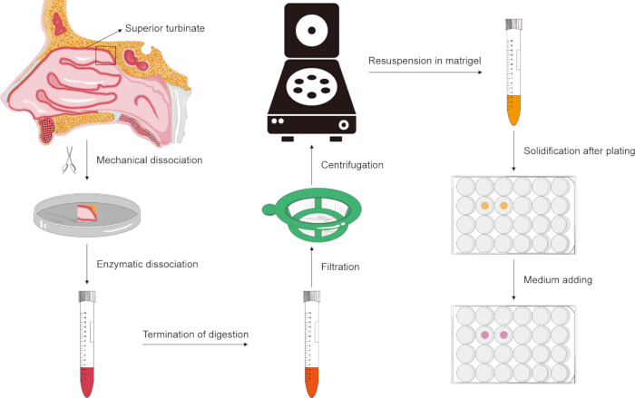
Figure 1: Schematic diagram of the experimental workflow for establishing human olfactory epithelial organoids. Please click here to view a larger version of this figure.
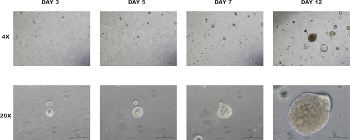
Figure 2: Establishment of human olfactory epithelial organoids. Bright-field images illustrating organoid development in a specialized culture medium from day 0 to day 12. Representative images were acquired on days 3, 5, 7, and 12 using live-cell imaging. Scale bar: 100 µm. Please click here to view a larger version of this figure.
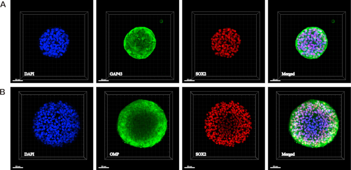
Figure 3: Immunofluorescence characterization of human olfactory epithelial organoids. Confocal microscopy images showing optical sections of three-dimensional reconstructed models of organoids immunostained for (A) the immature olfactory sensory neuron marker GAP43 (green) and the neural stem cell marker SOX2 (red), and (B) the mature olfactory sensory neuron marker OMP (green) and SOX2 (red). Scale bar: 50 µm. Please click here to view a larger version of this figure.
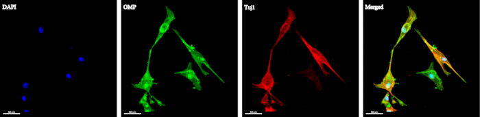
Figure 4: Immunofluorescence identification of human olfactory sensory neurons. Confocal microscopy image depicting differentiated cells derived from organoids exhibiting 2D adherent growth, co-stained for the mature olfactory sensory neuron marker OMP (green) and the pan-neuronal marker Tuj1 (red). Scale bar: 50 µm. Please click here to view a larger version of this figure.
Supplemental Figure S1. Validation of antibody specificity in mouse olfactory epithelium. Immunofluorescence staining of a mouse olfactory epithelium tissue section demonstrating the highly specific, on-target expression patterns of the antibodies used in this study. In the natural in vivo tissue, the neural stem cell/progenitor marker SOX2 (red) is strictly localized to the nuclei of the basal cell layer, while the mature olfactory sensory neuron marker OMP (green) is robustly expressed in the mature sensory neurons. Nuclei were counterstained with DAPI (blue).Please click here to download this file.
Supplemental Video S1. Three-dimensional reconstruction of an olfactory epithelial organoid (GAP43 and SOX2). Confocal Z-stack 3D reconstruction video (acquired at 40X magnification) demonstrating the spatial distribution of the immature olfactory sensory neuron marker GAP43 (green) and the neural stem cell marker SOX2 (red) within the spherical structure of the organoid.Please click here to download this file.
Supplemental Video S2. Three-dimensional reconstruction of an olfactory epithelial organoid (OMP and SOX2). Confocal Z-stack 3D reconstruction video (acquired at 40X magnification) demonstrating the spatial distribution of the mature olfactory sensory neuron marker OMP (green) and the neural stem cell marker SOX2 (red) within the spherical structure of the organoid.Please click here to download this file.
Supplemental Video S3. Three-dimensional visualization of differentiated olfactory sensory neurons (OMP and Tuj1). Confocal Z-stack 3D reconstruction video (acquired at 20X magnification) showing the morphological features of differentiated cells derived from the organoids. These adherent cells co-express the mature olfactory sensory neuron marker OMP (green) and the pan-neuronal marker Tuj1 (red).Please click here to download this file.
Discussion
Organoids represent a unique in vitro model because their self-organizing, three-dimensional architecture closely recapitulates that of native human tissues and, in some cases, can be histologically indistinguishable from actual organs.14 Establishing human organoid models is therefore critical for studying human diseases. Here, we present a protocol for generating an organoid culture system from primary human olfactory epithelial cells that reliably differentiate into mature olfactory sensory neurons. This approach overcomes the limitations of conventional methods, which have relied exclusively on mouse olfactory epithelium for organoid generation,21,22,23,24,25,26 and provides a robust platform for investigating the spectrum of human olfactory disorders and their underlying pathological mechanisms.
Previous studies have reported that organoids embedded in Matrigel require extremely high concentrations of primary antibody for effective protein detection during fixation, thereby increasing background fluorescence. Furthermore, directly mounting coverslips onto slides compresses and disrupts organoid structures, compromising analytical accuracy. These factors make immunofluorescence staining of organoids particularly challenging.27,28 To preserve three-dimensional organoid architecture while enabling high-quality imaging, we seeded organoids directly into glass-bottom confocal culture dishes for immunofluorescence. Subsequent three-dimensional reconstruction using Imaris software substantially improved image quality and accurately resolved the spatial morphology of the organoids.
An important consideration is that differentiating human olfactory epithelial organoids poses a significantly greater challenge compared to those derived from mice. In this protocol, we introduced a combination of specific small molecules to promote differentiation: the Notch signaling pathway inhibitor LY411575,29 the Wnt signaling pathway activator CHIR-99021,30 and RA, a regulator of neural differentiation31. The coordinated action of these molecules successfully induced the differentiation of cells resembling olfactory sensory neurons.
A limitation of our current approach is that the human olfactory epithelial organoids we established do not yet support long-term expansion. This underscores the need for further optimization to establish a stable, long-term culture system for human olfactory epithelial organoids. Another critical limitation is that the successful construction and neuronal differentiation of the organoids have been assessed solely by morphological analysis and immunofluorescence staining. While this protocol establishes a standardized workflow for morphological and phenotypic characterization suitable for publication in a methodology journal, it falls short of fully confirming the physiological functionality of the differentiated olfactory sensory neurons.
Future work should incorporate functional assays, such as calcium imaging and patch-clamp recordings, to rigorously validate the electrophysiological activity and maturation of the differentiated neurons. Precise quantitative analyses, such as determining the proportion of marker-positive cells or comparing OMP fluorescence intensity, were not performed in this study due to technical limitations inherent to whole-mount 3D imaging. Conducting accurate single-cell-level quantification within dense 3D organoid spheres is highly prone to error. Without tissue-clearing treatments, cells within organoids are densely packed and overlapping, making precise 3D single-cell segmentation computationally challenging. Additionally, limitations in laser penetration depth lead to inevitable attenuation of the fluorescence signal in deeper regions along the Z-axis. Consequently, our current protocol primarily focuses on qualitative and morphological demonstrations.
Future efforts should aim to refine the culture and differentiation induction conditions to establish a human olfactory epithelial organoid model capable of sustained, long-term propagation. Furthermore, integrating rigorous functional validations will be essential to confirm the physiological functionality of the differentiated neurons. This comprehensive approach will ensure the development of a reliable and versatile model for studying human olfactory disorders and their underlying mechanisms.
Disclosures
The authors have no conflicts of interest to declare.
Acknowledgements
The authors sincerely thank all patients who generously donated superior turbinate tissue specimens, this study would not have been possible without their contribution. Financial support for the research, authorship, and/or publication of this article was received. This work was supported by the National Natural Science Foundation of China (Ba L: 82160209 and 82460217), the Sichuan University–Zigong Municipal Government Science and Technology Cooperation Special Fund (Yang H: 2023CDZG-18), and the Natural Science Foundation of the Tibet Autonomous Region (Cong TC: XZ2023ZR-ZY10(Z)).
Materials
| Name | Company | Catalog Number | Comments |
|---|---|---|---|
| ALEXA FLUOR 488 DONKEY | Thermo | A11055 | 1:500 |
| ALEXA FLUOR 555 DONKEY | Thermo | A31572 | 1:500 |
| Anti-GAP43 antibody | Abcam | ab75810 | 1:160 |
| Anti-OMP antibody | SANTA CRUZ | sc-365818 | 1:50 |
| Anti-Sox-2 antibody | SANTA CRUZ | sc-365823 | 1:50 |
| B-27 Supplement | Gibco | 17504044 | 2% |
| CHIR99021 | Novoprotein | HY-10182 | 3 μM |
| DMEM/F12/HEPES | Procell | PM150310 | / |
| Glutamax (L-glutamine supplement) | Gibco | 35050061 | 1% |
| LY411575 | Novoprotein | HY-50752 | 5 μM |
| Matrigel (basement membrane matrix) | Corning | 356231 | 4% (Vol/Vol) |
| N-2 Supplement | Gibco | 17502048 | 1% |
| Organoid Dissociation Reagent | Precedo | PRS-ODR | / |
| Organoid Recovery Solution | Thewellbio | MS04-100 | / |
| Primocin (primary antimicrobial agent) | Invivogen | ant-pm-05 | 100 μg/mL |
| Recombinant Human EGF | Novoprotein | C029 | 50 ng/mL |
| Recombinant Human Noggin | Novoprotein | CB89 | 100 ng/mL |
| Recombinant Human RSPO1 | Novoprotein | CX83 | 200 ng/mL |
| Recombinant Human Wnt3a | Novoprotein | C18K | 50 ng/mL |
| Retinoic acid | MCE | HY-14649 | 1 μM |
| Y27632 | MCE | HY-10071 | 10 μM |
References
- Hummel, T., et al. Olfactory dysfunction: etiology, diagnosis, and treatment. Dtsch Arztebl Int. 120 (9), 146-154 (2023).
- Firestein, S. How the olfactory system makes sense of scents. Nature. 413 (6852), 211-218 (2001).
- Peterson, J., et al. Activating a reserve neural stem cell population in vitro enables engraftment and multipotency after transplantation. Stem Cell Rep. 12 (4), 680-695 (2019).
- Schnittke, N., et al. Transcription factor p63 controls the reserve status but not the stemness of horizontal basal cells in the olfactory epithelium. Proc Natl Acad Sci U S A. 112 (36), E5068-E5077 (2015).
- Schwob, J. E., et al. Stem and progenitor cells of the mammalian olfactory epithelium: taking poietic license. J Comp Neurol. 525 (4), 1034-1054 (2017).
- Takeuchi, H. Olfactory cilia, regulation and control of olfaction. Physiol Rep. 12 (19), e70057 (2024).
- Lin, B., et al. Injury induces endogenous reprogramming and dedifferentiation of neuronal progenitors to multipotency. Cell Stem Cell. 21 (6), 761-774.e5 (2017).
- Huang, T., Wei, Y., Wu, D. Effects of olfactory training on posttraumatic olfactory dysfunction: a systematic review and meta-analysis. Int Forum Allergy Rhinol. 11 (7), 1102-1112 (2021).
- Hura, N., et al. Treatment of post-viral olfactory dysfunction: an evidence-based review with recommendations. Int Forum Allergy Rhinol. 10 (9), 1065-1086 (2020).
- Doty, R. L. Olfactory dysfunction in neurodegenerative diseases: is there a common pathological substrate?. Lancet Neurol. 16 (6), 478-488 (2017).
- Patel, Z. M., et al. International consensus statement on allergy and rhinology: olfaction. Int Forum Allergy Rhinol. 12 (4), 327-680 (2022).
- Liu, J., et al. A novel surgical treatment for long lasting unilateral peripheral parosmia: olfactory cleft blocking technique. Auris Nasus Larynx. 48 (6), 1209-1213 (2021).
- Seo, B. S., et al. Treatment of postviral olfactory loss with glucocorticoids, ginkgo biloba, and mometasone nasal spray. Arch Otolaryngol Head Neck Surg. 135 (10), 1000 (2009).
- Kim, J., Koo, B. K., Knoblich, J. A. Human organoids: model systems for human biology and medicine. Nat Rev Mol Cell Biol. 21 (10), 571-584 (2020).
- Lee, S. H., et al. Tumor evolution and drug response in patient-derived organoid models of bladder cancer. Cell. 173 (2), 515-528.e17 (2018).
- Loomans, C. J. M., et al. Expansion of adult human pancreatic tissue yields organoids harboring progenitor cells with endocrine differentiation potential. Stem Cell Rep. 10 (3), 712-724 (2018).
- Huch, M., et al. Long-term culture of genome-stable bipotent stem cells from adult human liver. Cell. 160 (1-2), 299-312 (2015).
- Schlaermann, P., et al. A novel human gastric primary cell culture system for modelling Helicobacter pylori infection in vitro. Gut. 65 (2), 202-213 (2016).
- Bartfeld, S., et al. In vitro expansion of human gastric epithelial stem cells and their responses to bacterial infection. Gastroenterology. 148 (1), 126-136.e6 (2015).
- Ren, W., et al. Expansion of murine and human olfactory epithelium/mucosa colonies and generation of mature olfactory sensory neurons under chemically defined conditions. Theranostics. 11 (2), 684-699 (2021).
- Li, X., et al. Age-dependent activation and neuronal differentiation of Lgr5+ basal cells in injured olfactory epithelium via notch signaling pathway. Front Aging Neurosci. 12, 602688 (2020).
- Dai, Q., et al. Notch signaling regulates Lgr5+ olfactory epithelium progenitor/stem cell turnover and mediates recovery of lesioned olfactory epithelium in mouse model. Stem Cells. 36 (8), 1259-1272 (2018).
- Wang, L., et al. Chitinase-like protein Ym2 (Chil4) regulates regeneration of the olfactory epithelium via interaction with inflammation. J Neurosci. 41 (26), 5620-5637 (2021).
- Ma, Z., et al. TMEM59 ablation leads to loss of olfactory sensory neurons and impairs olfactory functions via interaction with inflammation. Brain Behav Immun. 111, 151-168 (2023).
- Li, W., et al. A single-cell transcriptomic census of mammalian olfactory epithelium aging. Dev Cell. 59 (22), 3043-3058.e8 (2024).
- Chen, Z. H., et al. Matrix metalloprotease-mediated cleavage of neural glial-related cell adhesion molecules activates quiescent olfactory stem cells via EGFR. Mol Cell Neurosci. 108, 103552 (2020).
- Shechtman, L. A., et al. Generation and culture of lingual organoids derived from adult mouse taste stem cells. J Vis Exp. (170), e62300 (2021).
- Liu, M., et al. Multimodal spatiotemporal monitoring of basal stem cell-derived organoids reveals progression of olfactory dysfunction in Alzheimer’s disease. Biosens Bioelectron. 246, 115832 (2024).
- Abranches, E., et al. Neural differentiation of embryonic stem cells in vitro: a road map to neurogenesis in the embryo. PLoS One. 4 (7), e6286 (2009).
- McLean, W. J., et al. Clonal expansion of Lgr5-positive cells from mammalian cochlea and high-purity generation of sensory hair cells. Cell Rep. 18 (8), 1917-1929 (2017).
- Freemantle, S. J., et al. Developmentally-related candidate retinoic acid target genes regulated early during neuronal differentiation of human embryonal carcinoma. Oncogene. 21 (18), 2880-2889 (2002).