This study demonstrates that Jiangtangle-containing Tibetan medicine serum protects RIN-m5F pancreatic β-cells from high-glucose-induced mitochondrial dysfunction. High-resolution LC-MS identified 90 compounds, including six absorbed into circulation. The serum improved cell viability and restored mitochondrial and glycolytic function, ATP production, oxygen consumption, and respiration, supporting its therapeutic potential for type 2 diabetes mellitus.
Review Article
Enhancing Recovery in Stroke Survivors through Sleep Interventions: A Comprehensive Review
In This Article
Summary
Abstract
Sleep disorders are common complications following stroke, with a global meta-analysis indicating a prevalence as high as 53%. These disorders primarily include insomnia, obstructive sleep apnea (OSA), and circadian rhythm disruptions. By impairing neuroplasticity, exacerbating inflammatory responses, and interfering with the effectiveness of rehabilitation training, such disorders significantly delay neurological recovery, increase the risk of secondary stroke, and elevate the incidence of depression and anxiety. Consequently, they have become a critical factor limiting stroke rehabilitation. This article systematically reviews the application and progress of sleep interventions in stroke rehabilitation. A comprehensive literature search was conducted in PubMed, Web of Science, and CNKI databases from inception to December 2025, using keywords including “stroke,” “sleep disorders,” “sleep interventions,” and “neurorehabilitation.” Studies were included if they reported original data on sleep interventions in stroke patients, with no restrictions on study design. Data extraction and quality assessment were performed independently by two reviewers. Core strategies reviewed include cognitive behavioral therapy for insomnia (CBT-I), continuous positive airway pressure (CPAP), pharmacotherapy, and Traditional Chinese Medicine interventions such as acupuncture. Among these, CBT-I, as a first-line approach, has been shown to significantly improve sleep quality and emotional state when administered for over 8 weeks. CPAP can reduce the risk of recurrent vascular events by 55% in patients with OSA while also promoting neurocognitive recovery. Studies confirm that standardized sleep interventions, by optimizing sleep architecture and enhancing neural repair capacity, synergistically improve limb motor function and activities of daily living. Current research still faces challenges such as variable intervention adherence and insufficient mechanistic studies on non-OSA subtypes. Future efforts should rely on multidisciplinary collaboration to develop personalized intervention plans, incorporate novel technologies to optimize treatment strategies, further explore the clinical value of sleep interventions in stroke rehabilitation, and provide an evidence-based foundation for improving long-term patient prognosis.
Introduction
Stroke, as one of the leading global causes of disability and mortality, imposes a substantial burden on patients, families, and society due to its high incidence, mortality, and disability rates1. Stroke survivors often experience varying degrees of neurological deficits, such as motor impairments, cognitive decline, speech difficulties, and dysphagia. These long-term sequelae significantly affect patients’ abilities to perform daily activities and their overall quality of life2. Among the numerous complications following stroke, sleep disturbances represent a highly prevalent yet frequently overlooked issue. It has been reported that the incidence of post-stroke sleep disorders ranges from 20% to 78%, manifesting in various forms, including insomnia, sleep apnea, hypersomnia, and circadian rhythm disruptions3. Sleep disturbances are not only common consequences of stroke but are also closely associated with anxiety, depressive symptoms, and cognitive decline. Moreover, they can further impede neurological recovery, increase the risk of stroke recurrence, and create a vicious cycle4. Notably, sleep-related breathing disorders, particularly obstructive sleep apnea, have been identified as independent risk factors for de novo ischemic stroke, with evidence indicating that untreated sleep apnea significantly increases the risk of recurrent vascular events and is associated with unfavorable functional outcomes following ischemic stroke5. A growing body of evidence suggests that the intermittent hypoxia and sleep fragmentation characteristic of sleep-related breathing disorders contribute to endothelial dysfunction, systemic inflammation, and hemodynamic instability, thereby promoting both the initiation and progression of cerebrovascular pathology6. A follow-up study indicated that stroke patients with chronic insomnia face a 66% higher mortality risk7. Therefore, a deeper understanding and effective intervention for post-stroke sleep disorders have become crucial for improving patient prognosis.
Contemporary neuroscience research has demonstrated that sleep, particularly slow-wave sleep and rapid eye movement (REM) sleep, plays an essential role in the transformation of memory traces, integrating and consolidating short-term memory fragments into long-term memories8. During sleep, the brain actively reactivates neural activity patterns associated with learning, a process especially important for skill acquisition and motor recovery. Simultaneously, sleep facilitates synaptic optimization and pruning, clears metabolic toxins accumulated during wakefulness (such as β-amyloid), and creates favorable conditions for plasticity within neural circuits. In the context of stroke, these sleep-dependent processes acquire heightened significance. Preclinical studies using rodent models of ischemic stroke have demonstrated that post-stroke sleep fragmentation impairs motor skill consolidation, while sleep enhancement through optogenetic manipulation promotes functional recovery9. Clinical studies have further shown that sleep architecture following stroke directly correlates with motor outcome measures: patients with preserved slow-wave activity during the subacute phase exhibit greater gains in upper extremity function at three months post-stroke10. Neuroplasticity serves as the cornerstone of functional recovery after stroke, and sleep supports the functional remodeling and compensation of damaged brain regions by modulating the expression of molecules such as brain-derived neurotrophic factor (BDNF) and maintaining the balance of neurotransmitters (e.g., acetylcholine, norepinephrine, and serotonin)11,12. This evidence suggests that high-quality sleep fosters a favorable neurochemical environment for stroke rehabilitation.
Given the high prevalence of sleep disturbances in the stroke population and their profound impact on neuroplasticity, actively exploring sleep-targeted intervention strategies to accelerate stroke recovery holds significant theoretical and clinical value. The methodological framework followed the Preferred Reporting Items for Systematic Reviews and Meta-Analyses (PRISMA) guidelines. A systematic search was performed in PubMed, Web of Science, and CNKI databases for articles published up to December 2025, using the following search strategy: (“stroke” OR “cerebrovascular accident” OR “cerebral infarction” OR “intracranial hemorrhage”) AND (“sleep disorders” OR “insomnia” OR “sleep apnea” OR “circadian rhythm” OR “sleep disturbance”) AND (“rehabilitation” OR “recovery” OR “neuroplasticity” OR “functional outcome”) AND (“sleep intervention” OR “CBT-I” OR “CPAP” OR “light therapy” OR “pharmacotherapy”). Inclusion criteria were: (1) original research articles including randomized controlled trials, cohort studies, case-control studies, and cross-sectional studies; (2) adult stroke patients (≥18 years); (3) reported data on sleep disorders or sleep interventions; (4) reported outcomes related to neurological recovery, functional status, or quality of life. Exclusion criteria were: (1) non-English or non-Chinese publications; (2) conference abstracts, case reports, editorials, or commentaries; (3) animal studies or in vitro studies; (4) studies without original data. Two independent reviewers screened titles, abstracts, and full texts against the eligibility criteria. Disagreements were resolved by consensus or consultation with a third reviewer. Data extraction was performed using standardized forms, capturing study characteristics, population demographics, intervention details, outcome measures, and key findings. Quality assessment was conducted using the Cochrane Risk of Bias Tool for randomized controlled trials and the Newcastle-Ottawa Scale for observational studies.
Review and Perspective
Post-stroke sleep disorders: Types, impact, and pathophysiological mechanisms
Primary types and epidemiology of post-stroke sleep disorders
Post-stroke sleep disorders (PSSD) represent a highly prevalent yet frequently overlooked complication following stroke, characterized by diverse and complex manifestations, with an overall incidence ranging from 20% to 78%13. PSSD not only occurs significantly more frequently than in the general population but also encompasses a wide spectrum of clinical subtypes, which can be broadly categorized into respiratory-related and non-respiratory-related sleep disorders. Among respiratory-related disorders, obstructive sleep apnea (OSA) is the most common14. Studies have indicated that over 50% of patients in the acute phase of stroke present with OSA, typically characterized by recurrent apnea or hypopnea during sleep, leading to intermittent hypoxia and fragmentation of sleep architecture15. Central sleep apnea (CSA) is relatively less common and is closely associated with damage to respiratory centers such as the medulla oblongata.
Non-respiratory-related sleep disorders encompass a broader range of conditions. Insomnia is one of the core types, with an incidence as high as 30% to 68% in the acute stroke phase, primarily manifesting as difficulty initiating sleep, maintaining sleep, early morning awakening, and poor sleep quality. Daytime hypersomnia is another common issue, affecting approximately 36.96% of stroke patients, potentially linked to damage in the ascending reticular activating system or the hypothalamic orexin pathway16. Circadian rhythm sleep-wake disorders are also prevalent among stroke patients, who often exhibit symptoms such as day-night reversal, nocturnal insomnia, and daytime sleepiness. Furthermore, stroke can induce or exacerbate sleep-related movement disorders, such as restless legs syndrome (RLS) and periodic limb movement disorder (PLMD), with an incidence of approximately 12.4%, as well as parasomnias, such as rapid eye movement sleep behavior disorder (RBD), which is particularly common in patients with brainstem infarction17,18. The timing of PSSD onset follows specific patterns. Patients without impaired consciousness frequently develop sleep disturbances within 3–5 days post-stroke, while those with impaired consciousness often experience them within 3 days of regaining consciousness. The peak incidence typically occurs within 3–4 months following the stroke19.
Multidimensional negative impacts of sleep disorders on stroke rehabilitation outcomes
Motor function
Sleep deprivation or disrupted sleep architecture diminishes patients' physical strength and endurance, leading to a lack of motivation and concentration for participation in rehabilitation training. Studies have demonstrated that insomnia, RLS, and sleep fragmentation are frequently associated with poor balance, slower recovery of walking ability, and suboptimal overall recovery outcomes20. A prospective cohort study of 89 ischemic stroke patients found that those with sleep efficiency below 70% during the first week post-stroke had significantly lower Fugl-Meyer Assessment scores at six months (mean difference 15.3 points, 95% CI 8.7–21.9), independent of initial stroke severity and age21. The underlying mechanism may involve the interference of sleep with the offline consolidation process of motor skills, whereby sleep insufficiency hinders the brain's ability to convert motor skills learned during the day into long-term memory22.
Cognitive function
Sleep, particularly slow-wave sleep and REM sleep, is crucial for memory consolidation, information integration, and synaptic plasticity. PSSD can lead to impaired concentration, decreased alertness, forgetfulness, and slowed thinking in patients. These cognitive deficits severely hinder patients' ability to learn new compensatory strategies and adapt to life with disability, directly delaying the recovery of neurocognitive function23,24. A longitudinal study of 142 stroke patients demonstrated that those with moderate-to-severe sleep apnea at baseline had a 3.2-fold increased risk of developing post-stroke cognitive impairment at 12 months (adjusted OR 3.22, 95% CI 1.48–7.01), with the strongest effects observed in executive function and processing speed domains25.
Emotional disorders
Post-stroke insomnia and poor sleep quality are significant risk factors for the development of depression and anxiety. Prolonged poor sleep exacerbates patients' feelings of pessimism, anxiety, and fear. These negative emotions, in turn, further worsen sleep, creating a vicious cycle wherein sleep disturbances promote emotional problems, which then impede neurological recovery, significantly reducing patients' life satisfaction and confidence in rehabilitation26.
By hindering the recovery of motor and cognitive functions and inducing emotional problems, sleep disorders lead to a marked decline in patients' activities of daily living (ADL), social participation, and overall well-being. More critically, PSSD also significantly increases the risk of stroke recurrence. Research indicates that the mortality risk in stroke patients with chronic insomnia can be increased by 66%. Sleep-disordered breathing, such as OSA, elevates the risk of secondary stroke through pathways including triggering nocturnal blood pressure surges, inducing cardiac arrhythmias, and exacerbating atherosclerosis27.
Exploration of the bidirectional pathophysiological mechanisms
A complex bidirectional pathophysiological link exists between stroke and sleep disorders, forming the basis for a vicious cycle between the two. The pathophysiology of stroke-related insomnia can be visually represented in a schematic diagram (Figure 1). This figure illustrates the core pathophysiological mechanisms of stroke-related insomnia, highlighting the dysregulation of circadian rhythms governed by photic and non-photic zeitgebers following stroke. This dysregulation involves stroke-induced brain injury, inflammatory responses, medication effects, dysfunction of peripheral oscillators, and alterations in cellular oscillator function, with concurrent sleep-disordered breathing further exacerbating the imbalance in sleep homeostasis.
On one hand, the stroke lesion can directly damage key brain centers that regulate the sleep-wake cycle. Regions such as the thalamus, hypothalamus, brainstem reticular formation, and basal forebrain are primarily responsible for maintaining normal sleep rhythms28. When a stroke affects these areas, it can directly lead to the disintegration of sleep architecture. For instance, infarction of the thalamus or brainstem often causes insomnia or hypersomnia; damage to the pontine tegmentum can induce RBD; and medullary infarction is prone to causing central sleep apnea. The topography of stroke lesions is a critical determinant of post-stroke sleep-disordered breathing. Clinical evidence indicates that patients with acute stroke localized to the capsular or pontine regions exhibit a significantly higher prevalence of sleep-related breathing disorders, particularly in those with a history of smoking29. This finding underscores the importance of lesion location in modulating respiratory control during sleep and suggests that patients with subcortical and brainstem strokes may warrant more intensive screening for sleep apnea. Such structural damage constitutes the most direct pathological basis for PSSD30.
On the other hand, sleep disorders are not merely passive consequences but actively impede neural remodeling and functional recovery through multiple mechanisms. First, sleep disorders (particularly the intermittent hypoxia caused by OSA) can exacerbate central nervous system inflammation, promoting the release of pro-inflammatory cytokines such as tumor necrosis factor-alpha (TNF-α) and interleukin-6 (IL-6). These inflammatory factors have neurotoxic effects, hindering the repair of surviving brain tissue31. Second, sleep is the critical period for the efficient clearance of metabolic waste (e.g., beta-amyloid) by the brain's glymphatic system. Sleep disorders severely impair this cleansing function, leading to the accumulation of neurotoxic substances and disrupting the stability of the neural microenvironment32. Direct evidence for this mechanism in stroke comes from animal studies demonstrating that glymphatic function is markedly impaired in the peri-infarct region during the acute phase, and that sleep deprivation exacerbates this impairment, resulting in increased infarct volume and worsened neurological deficits33. In human stroke patients, cerebrospinal fluid biomarker studies have shown that poor sleep quality is associated with elevated levels of neurotoxic metabolites, providing translational support for this pathway34,35.
Furthermore, an imbalance in neurotransmitter systems is also a core mechanism. Following stroke, the synthesis, release, and balance of neurotransmitters associated with wakefulness (e.g., dopamine, norepinephrine, orexin) and those promoting sleep (e.g., serotonin 5-HT, melatonin, gamma-aminobutyric acid GABA) are disrupted36. Serum melatonin levels are often significantly reduced in patients with post-stroke insomnia. Such disturbances in the neurochemical environment collectively contribute to the dysregulation of the sleep-wake cycle.
Thus, stroke leads to sleep disorders through direct damage to sleep centers and induced neurotransmitter imbalances. Conversely, sleep disorders, through mechanisms such as exacerbating neuroinflammation, impeding brain waste clearance, interfering with neurotrophic factor secretion, and disrupting skill consolidation, in turn, hinder post-stroke neural remodeling and functional recovery. This bidirectional vicious cycle underscores the extreme importance and urgency of intervening in sleep disorders within comprehensive stroke management.
Beyond the structural damage to sleep centers and the bidirectional vicious cycle described above, an emerging body of literature has highlighted the interplay between sleep-disordered breathing, cerebral white matter alterations, and underlying cerebrovascular risk factors. White matter hyperintensities, commonly observed on neuroimaging in aging populations, are associated with cumulative vascular burden and have been linked to both the presence and severity of sleep apnea37. Notably, a cluster of silent vascular risk factors, including hypertension, diabetes, and subclinical atherosclerosis, may interact with sleep-related breathing disturbances to promote progressive white matter damage, thereby contributing to cognitive decline and increased stroke susceptibility over the course of aging38. This interrelationship suggests that sleep-disordered breathing may serve not only as a consequence of stroke but also as a modifiable contributor to vascular brain injury through its effects on cerebral small vessel pathology.
Sleep intervention strategies for promoting stroke rehabilitation: Evidence and evaluation
Effective management of post-stroke sleep disorders is a crucial component for optimizing rehabilitation outcomes. Current sleep intervention strategies for stroke survivors are characterized by diversification, encompassing non-pharmacological interventions, pharmacological and innovative physical interventions, as well as integrated strategies, all aimed at promoting neurofunctional remodeling through the improvement of sleep quality. The specifics are outlined in Table 1. Table 2 summarizes key findings from representative studies on sleep interventions in stroke patients.
Continuous Positive Airway Pressure (CPAP)
Given the high prevalence of obstructive sleep apnea (OSA) in stroke populations, affecting over 50% of patients in the acute phase, and its established role as a risk factor for both initial and recurrent stroke, CPAP therapy represents a critical intervention in post-stroke sleep management39. CPAP functions by delivering pressurized air through a mask to maintain upper airway patency during sleep, thereby preventing apneic events, reducing intermittent hypoxia, and stabilizing sleep architecture40.
Clinical evidence supporting CPAP in stroke patients has accumulated substantially. A landmark randomized controlled trial (the Sleep Apnea Cardiovascular Endpoints study) demonstrated that CPAP treatment in patients with cardiovascular disease and OSA reduced the risk of recurrent stroke by 55% among adherent users (hazard ratio 0.45, 95% CI 0.22–0.92)41. In the context of stroke rehabilitation, observational studies have shown that CPAP-adherent patients exhibit greater improvements in cognitive function, particularly in attention and executive domains, compared to non-adherent patients42. Furthermore, CPAP has been associated with reduced depressive symptoms and improved quality of life in stroke survivors with OSA43.
Despite these benefits, CPAP adherence remains a significant challenge in stroke populations. Reported adherence rates range from 40% to 60% in clinical trials, with barriers including mask discomfort, claustrophobia, cognitive impairment limiting device management, and caregiver burden44. Strategies to improve adherence include mask optimization, humidification, telemonitoring with remote feedback, and gradual desensitization protocols. Early initiation of CPAP during the subacute phase, before hospital discharge, has been associated with higher long-term adherence rates.
Non-pharmacological interventions
Non-pharmacological interventions, owing to their high safety profile and minimal side effects, constitute foundational treatment options for post-stroke sleep disorders. Their core principle lies in optimizing sleep architecture and quality through behavioral, cognitive, and environmental adjustments. Cognitive Behavioral Therapy for Insomnia (CBT-I) is internationally recommended as the first-line non-pharmacological treatment. CBT-I corrects patients' irrational beliefs and fears about sleep through cognitive restructuring, while concurrently employing behavioral interventions such as sleep restriction therapy (reducing time spent in bed awake) and stimulus control therapy (strengthening the association between bed and sleep)45. A study on post-ischemic stroke patients with insomnia demonstrated that adding CBT-I to eszopiclone treatment resulted in a significantly lower Pittsburgh Sleep Quality Index (PSQI) score (6.31 ± 0.76) in the combined therapy group compared to the medication-only group (9.36 ± 0.91) after 2 weeks. Moreover, more significant improvements were observed in Hamilton Anxiety (HAMA) and Depression (HAMD) scale scores in the combined group46. The advent of digital CBT-I (e.g., the Sleepio program) has enhanced treatment accessibility, with its standardized courses facilitating remote patient guidance. CBT-I not only improves subjective sleep perception but may also provide a more optimized process for the consolidation of motor skills acquired during daytime learning by regulating the sleep-wake cycle, thereby indirectly promoting motor function recovery47. However, the application of CBT-I in stroke populations presents unique feasibility challenges. Post-stroke aphasia, present in up to 30% of stroke survivors, may limit participation in cognitively demanding components such as cognitive restructuring and sleep diary completion48. Cognitive deficits, including impaired attention, executive dysfunction, and memory impairment, may hinder patients' ability to adhere to complex behavioral protocols such as sleep restriction and stimulus control.
Circadian rhythm modulation therapy primarily targets the common post-stroke disruption of the sleep-wake cycle. Scheduled morning (6:00–9:00) exposure to natural or simulated bright light (15–60 min) can effectively suppress melatonin secretion and reset the biological clock in the suprachiasmatic nucleus of the hypothalamus, leading to a phase advance of sleep50. Studies indicate this method can reduce sleep onset latency by 20–30 min and improve sleep quality by approximately 15%. For patients with adjustment difficulties or shift workers, supplementation with exogenous melatonin (0.5–3 mg) 1–2 h before bedtime can serve as a short-term adjuvant, particularly useful for regulating rhythm disorders caused by direct damage to sleep centers from stroke lesions51. These therapies help stabilize the internal environment, creating favorable conditions for neuroplasticity.
Other non-pharmacological therapies include sleep hygiene education, relaxation training, and physical therapies. Sleep hygiene education focuses on establishing regular sleep-wake schedules, optimizing the sleep environment (reducing noise, maintaining suitable temperature and humidity), and avoiding caffeine and alcohol intake before bedtime52. Relaxation training techniques, such as progressive muscle relaxation and meditation, can help alleviate post-stroke anxiety and reduce hyperarousal levels. Repetitive Transcranial Magnetic Stimulation (rTMS), a non-invasive neuromodulation technique, has been shown to improve sleep quality and cognitive function when applying low-frequency (1 Hz) stimulation to the right dorsolateral prefrontal cortex53. Cranial Electrotherapy Stimulation (CES), which applies microcurrents via earclip electrodes to influence neurotransmitter activity, has also been proven to increase alpha waves and enhance sleep quality54.
Pharmacological and innovative physical interventions
Pharmacological therapy holds value for rapidly stabilizing sleep. Benzodiazepines (e.g., diazepam) are no longer recommended as first-line agents due to their addictive potential, muscle-relaxant effects, and risk of exacerbating cognitive impairment. Non-benzodiazepine receptor agonists (e.g., eszopiclone) and melatonin receptor agonists (e.g., ramelteon) offer a relatively higher safety profile. Certain antidepressants with sedating properties (e.g., mianserin, trazodone) may be considered for stroke patients with comorbid depression or anxiety and insomnia55. Paroxetine, a selective serotonin reuptake inhibitor (SSRI), is sometimes used for post-stroke depression and may have indirect effects on sleep; however, its use requires caution due to potential activating effects in some patients and the risk of adverse events in stroke populations56. The choice of pharmacotherapy should be guided by the predominant clinical presentation, potential drug interactions, and the patient's overall medical status.
Hyperbaric oxygen therapy (HBOT), by increasing arterial oxygen partial pressure and scavenging free radicals, improves cerebral metabolism while simultaneously prolonging total sleep time and shortening sleep latency57. Emerging interventions, such as synchronized brain-mimetic electrical stimulation within the hyperbaric chamber, combine neuromodulation with environmental intervention. Preliminary research suggests potential in improving stroke-related sleep disorders and mood, although further evidence is required for validation58.
Integrated and combined Chinese-Western medicine intervention strategies
Single interventions are often insufficient to address the multifactorial nature of post-stroke sleep disorders. Multimodal interventions integrating CBT-I, circadian rhythm modulation, and moderate exercise can more comprehensively target diverse symptoms. The concept of sequential intervention emphasizes initiating intensive, task-oriented motor rehabilitation training promptly after effectively improving sleep quality through means such as dCBT59. This strategy aims to utilize the optimized sleep window to maximize the consolidation of motor memory, thereby establishing a virtuous cycle of improved sleep, facilitating optimized learning, which in turn promotes functional recovery.
Combined Chinese-Western medicine approaches demonstrate unique advantages in clinical practice. Western medicine excels at rapidly controlling symptoms, while Traditional Chinese Medicine (TCM) emphasizes holistic regulation and treatment based on pattern differentiation. Chinese patent medicines (e.g., sedative pills, granules) show clear efficacy in improving post-stroke insomnia with a relatively low incidence of adverse reactions. A network meta-analysis indicated that combined treatment with Chinese patent medicines and Western drugs was superior to either Western drugs or Chinese medicines alone in terms of increasing clinical effectiveness rate (surface under the cumulative ranking curve SUCRA 0.999) and improving PSQI scores (SUCRA 0.982)60. Acupuncture therapy, targeting acupoints such as Baihui (GV20), Shenmen (HT7), and Sishencong (EX-HN1), or employing methods like umbilical herb moxibustion and Governor Vessel moxibustion, can regulate meridians and visceral functions, promoting tranquility and sleep61. TCM techniques such as acupoint massage and auricular stimulation have also been proven effective in enhancing sleep quality. This combined model achieves synergy between rapid symptom control and long-term regulation, helping to alleviate patient anxiety and improve the adherence and sustainability of rehabilitation training.
Post-stroke sleep interventions have evolved into a diversified strategic system, progressing from single to integrated approaches and from symptomatic to causative management. Clinical selection should be based on the specific type of sleep disorder, stroke phase, comorbidities, and patient preferences. Non-pharmacological interventions should be prioritized, with pharmacological or integrated Chinese-Western medicine approaches incorporated judiciously when necessary. Actively exploring multimodal sequential treatment is essential to maximize the facilitative role of sleep in stroke rehabilitation.
Challenges, future directions, and recommendations for clinical translation
Current challenges
The clinical management and research of post-stroke sleep disorders continue to face multiple challenges. The primary challenge lies in their significant heterogeneity. The types of sleep disorders are complex and diverse, including insomnia, sleep apnea, hypersomnia, and circadian rhythm disruptions. Furthermore, their clinical manifestations, severity, and pathological bases vary considerably depending on the location and size of the stroke lesion, as well as individual patient differences, posing difficulties in establishing unified diagnostic criteria and developing universally applicable intervention protocols. Secondly, there is a lack of standardization in assessment tools. Clinicians predominantly rely on subjective scales such as the Pittsburgh Sleep Quality Index, which are susceptible to the influence of patients' cognitive function, emotional state, and subjective perceptions, limiting objectivity. While polysomnography remains the gold standard, its complexity, high cost, and low accessibility hinder routine application during the acute and early rehabilitation phases of stroke, resulting in a significant number of patients remaining undiagnosed. Research indicates that the diagnosis rate of post-stroke sleep apnea is less than 10%62. Furthermore, there is a severe paucity of long-term follow-up data in existing intervention studies. Most clinical trials are of short duration, failing to adequately evaluate the sustained impact of sleep interventions on long-term neurological recovery, quality of life, and stroke recurrence risk in patients, leaving the evidence for long-term efficacy and safety insufficient. Finally, the development of personalized treatment plans remains immature. Currently, there is a lack of reliable biomarkers or clinical prediction models to precisely guide the selection of intervention measures (e.g., CBT-I, CPAP, rTMS, or pharmacotherapy) for different subtypes and rehabilitation stages of patients. Treatment decisions often rely heavily on physician experience, introducing a degree of arbitrariness.
Limitations
Several limitations should be acknowledged in this review. First, the heterogeneity of the included studies in terms of study design, patient populations, intervention protocols, and outcome measures precluded a quantitative meta-analysis, limiting the ability to derive pooled effect estimates. Second, the majority of the reviewed studies were conducted in single centers with relatively small sample sizes, which may limit the generalizability of the findings. Third, publication bias may exist, as studies with positive results are more likely to be published, potentially overestimating the efficacy of certain interventions. Fourth, the predominance of short-term follow-up periods in the included studies limits the ability to evaluate the long-term sustainability of intervention effects and their impact on hard outcomes such as stroke recurrence and mortality. Fifth, the exclusion of non-English language publications may have introduced language bias. Finally, the heterogeneity in diagnostic criteria and assessment tools for post-stroke sleep disorders across studies complicates cross-study comparisons and synthesis of evidence.
Future research directions
Future research must focus on overcoming current bottlenecks. The primary task is to conduct more rigorously designed, large-sample, multicenter randomized controlled trials to provide high-level evidence-based medicine regarding the efficacy and safety of various sleep interventions, and to clarify the optimal target populations, timing of intervention, and treatment parameters for different approaches. Secondly, there should be an active exploration of biomarker-based precision intervention strategies. Utilizing multimodal neuroimaging techniques to identify brain network damage patterns associated with specific sleep disorders, combined with molecular circadian rhythm markers (e.g., melatonin secretion profiles) and genetic characteristics, can provide an objective basis for individualized treatment. Additionally, there is an urgent need to develop low-burden, high-adherence intervention technologies more suitable for post-stroke patients with cognitive and motor impairments. This includes developing adaptive digital cognitive behavioral therapy based on smartphone applications, with content dynamically adjustable according to patient rehabilitation progress; optimizing portable home sleep monitoring devices to lower the diagnostic threshold; and refining neuromodulation technologies like rTMS for easier operation and more precise targeting. These technological innovations aim to enhance patient acceptance and treatment adherence, ensuring that interventions can be effectively integrated into long-term rehabilitation management.
Recommendations for translation into clinical practice
To facilitate the translation of evidence into practice, clinical management strategies require systematic optimization. It is advocated that sleep disorder screening be incorporated as a routine component within stroke rehabilitation units. This involves integrating subjective questionnaires with simplified objective assessment tools to conduct early, systematic screening and diagnosis of sleep problems for all stroke patients. Establishing a multidisciplinary team encompassing neurology, rehabilitation, psychiatry/psychology, respiratory medicine, and traditional Chinese medicine is crucial. Through regular consultations, this team can jointly develop comprehensive diagnosis and treatment plans, ensuring that patients receive concurrent management for cerebrovascular disease, functional rehabilitation, and sleep disorders. Ultimately, a stepped and personalized sleep management and intervention plan should be formulated for each patient. Initial interventions should be based on sleep hygiene education and cognitive behavioral therapy. For moderate to severe disorders, treatment should be escalated stepwise to intensive therapies such as CPAP, rTMS, or pharmacotherapy, with regular efficacy assessments to dynamically adjust the plan. This model allows for the optimized allocation of resources, ensuring the precision and continuity of interventions, thereby deeply integrating sleep management into the entire stroke rehabilitation process.
Conclusions
In conclusion, the central thesis of this review is that sleep is not merely a passive consequence of stroke but an active determinant of recovery outcomes. Sleep disorders constitute a prevalent and modifiable key barrier in the process of post-stroke neurofunctional recovery. However, it is essential to recognize that post-stroke sleep disorders encompass distinct clinical entities, including insomnia, obstructive sleep apnea (OSA), central sleep apnea (CSA), hypersomnia, and circadian rhythm disorders, each with fundamentally different diagnostic standards, temporal trajectories, and levels of evidence supporting specific interventions. Insomnia typically emerges within the first week post-stroke and responds to cognitive behavioral therapy, while OSA requires objective polysomnographic confirmation and may necessitate continuous positive airway pressure; CSA, often associated with brainstem lesions, follows a different recovery trajectory and may not benefit from standard OSA treatments. Therefore, the clinical translation of sleep interventions requires a shift from symptom-based management toward subtype-specific, mechanistically informed strategies. This review has systematically elaborated on the complex types, multidimensional negative impacts, and bidirectional pathophysiological mechanisms of post-stroke sleep disorders, and has evaluated the evidence and value of current non-pharmacological, pharmacological, and comprehensive intervention strategies. The overarching argument is that targeted sleep interventions have evolved from adjunctive supportive measures to core components of stroke rehabilitation, with the potential to enhance neuroplasticity, optimize rehabilitation training outcomes, and improve long-term prognosis. Given the heterogeneity of post-stroke sleep disorders, treatment approaches must be subtype-specific, guided by accurate diagnosis and tailored to individual patient characteristics, stroke lesion topography, and the phase of recovery. Future clinical practice and research must strive to achieve a paradigm shift from passively addressing sleep problems to actively leveraging the neurorestorative functions of sleep to drive neuroplasticity. By integrating multidisciplinary approaches to deeply embed precise assessment, personalized intervention, and long-term management strategies throughout the entire stroke rehabilitation continuum, the long-term functional prognosis and quality of life for patients can be significantly improved.
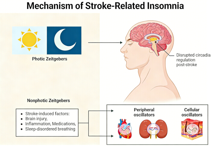
Figure 1: Schematic diagram of the pathophysiological mechanisms of stroke-related insomnia. Please click here to view a larger version of this figure.
| Types of interventions | Specific methods/examples | Main mechanism/role | Applicable Population/Precautions |
| Non-drug intervention | |||
| CBT‑I | Cognitive reconstruction, sleep restriction, stimulus control, relaxation training | Correction of irrational sleep beliefs, enhancement of bed sleep association, and reduction of over-awakening | Mild to moderate insomnia, accompanied by anxiety/depression; Remotely implementable |
| Light therapy | Scheduled exposure to natural light or simulated intense light in the morning (15–60 minutes) | Inhibiting melatonin secretion and resetting biological clock (suprachiasmatic nucleus) | Circadian rhythm disorder, sleep phase delay |
| Sleep health education | Get a regular routine, optimize your sleep environment and avoid caffeine/alcohol | Establish healthy sleep habit and reduce external interference | All patients with stroke as basic intervention |
| rTMS | Low frequency (1Hz) stimulation of the right frontal dorsolateral cortex | Regulate cortical excitability, improving sleep structure and cognitive function | Patients whose drug efficacy is unsatisfactory or who are not suitable for drug use; Need professional equipment and operation |
| Drug intervention | |||
| Non-benzodiazepines | Eszopiclone, zolpidem | It can selectively act on GABA‑A receptor, promote sleep and maintain sleep. | Short-term use (usually < 4 weeks); Monitoring Dependence and Cognitive Impacts |
| Melatonin receptor agonist | Ramelteon | Simulate endogenous melatonin to regulate sleep-waking rhythm | Circadian rhythm disorder, the elderly, co disease |
| Sedative antidepressants | Mianserin, paroxetine | Improve sleep and relieve depression/anxiety through 5‑HT and other systems | Insomnia patients with comorbid depression/anxiety |
| Integrated/Integrated Chinese and Western Medicine | |||
| Multimodal intervention | CBTI+light+moderate exercise | Multi-target regulation of sleep structure, emotion and physical fitness | Complex or refractory sleep disorder |
| Chinese patent medicine combined with western medicine | Tranquilizing pill/granule + Eszopiclone, etc | Holistic regulation (TCM syndrome differentiation)+rapid control of symptoms (western medicine) | Willing to accept the treatment of integrated traditional Chinese and western medicine |
Table 1: Comparison of sleep intervention strategies after stroke.
| Study (Year) | Population | Intervention | Key Outcome |
| Wu et al. (2017) | Post-ischemic stroke patients with insomnia | CBT-I + eszopiclone vs. eszopiclone alone | PSQI score lower in combined group (6.31 ± 0.76 vs. 9.36 ± 0.91); greater improvements in HAMA and HAMD scores |
| Weightman et al. (2023) | Stroke survivors with insomnia | Digital CBT-I (Sleepio) | Feasibility demonstrated; improved sleep quality and potential for enhanced motor learning |
| Weeks et al. (2024) | Post-stroke patients | Morning bright light therapy | Sleep onset latency reduced by 20–30 minutes; sleep quality improved by approximately 15% |
| Chen et al. (2023) | Post-stroke insomnia patients (network meta-analysis) | Chinese patent medicine + Western medicine | Highest clinical effective rate (SUCRA 0.999) and greatest PSQI improvement (SUCRA 0.982) compared to monotherapy |
| Bassetti et al. (2020) | Stroke patients with sleep disorders (consensus statement) | CPAP for OSA | Reduced risk of recurrent vascular events by 55%; improved neurocognitive recovery |
| Marshall et al. (2014) | Community-based cohort (20-year follow-up) | OSA identified by screening | OSA associated with increased all-cause mortality and stroke incidence |
| Li et al. (2018) | First-ever stroke patients (6-year follow-up) | Chronic insomnia | Mortality risk increased by 66% in patients with chronic insomnia |
Table 2: Summary of key findings from representative studies on sleep interventions in stroke patients.
Disclosures
The authors declare no conflicts of interest. The corresponding author affirms that the manuscript has been read and approved by all named authors.
Acknowledgements
The authors thank the staff of the Department of Neurology and Rehabilitation, Longhua Hospital Affiliated to Shanghai University of Traditional Chinese Medicine, for their clinical support and administrative assistance. This work was supported by the 2025 Oriental Talent Program – Health Platform Top‑notch Talent Project (Grant No. BJWS2025077) awarded to the corresponding author, Dr Qing Ye.