Sesquiterpene lactone XTT effectively attenuates lipopolysaccharide (LPS)-induced inflammatory responses in macrophages. XTT targets IKKα/β to inhibit NF-κB signaling without affecting MAPK activation, thereby exerting potent anti-inflammatory effects in vitro.
Research Article
1β-hydroxyl-5α-chloro-8-epi-xanthatin Suppresses Inflammatory Responses by Targeting IKKα/β and Inhibiting the NF-κB Signaling Pathway
1College of Pharmacy, Chinese Medicine Germplasm Resources Innovation and Effective Uses Key Laboratory of Sichuan Province, Chengdu University of Traditional Chinese Medicine, 2Center for Natural Products Research, Chengdu Institute of Biology, Chinese Academy of Sciences, 3Hospital of Chengdu University of Traditional Chinese Medicine, Chengdu University of Traditional Chinese Medicine
Opens in a new tab
In This Article
Summary
Loading...
$$\rightleftharpoonup{xx}$$
$$\longleftharp{xx}$$,
$$\longrightharp{xx}$$,
Abstract
Loading...
$$\rightleftharpoonup{xx}$$
$$\longleftharp{xx}$$,
$$\longrightharp{xx}$$,
Dysregulation of the nuclear factor-κB (NF-κB) cascade is tightly linked to the progression of inflammatory disorders. IκB kinases (IKKs) are well-established as central modulators of NF-κB activity, making them attractive therapeutic candidates for treating inflammatory pathologies. The sesquiterpene lactone 1β-hydroxyl-5α-chloro-8-epi-xanthatin (XTT) has been documented to exert robust anticancer effects. However, its anti-inflammatory activity and underlying molecular mechanisms remain unclear. In this work, XTT markedly suppressed the synthesis of nitric oxide (NO) and prostaglandin E2 (PGE2) in lipopolysaccharide (LPS)-activated RAW264.7 macrophages. XTT also substantially lowered LPS-triggered mRNA levels of inducible nitric oxide synthase (iNOS), cyclooxygenase-2 (COX2), tumor necrosis factor-α (TNF-α), interleukin-1β (IL-1β), and interleukin-6 (IL-6). Furthermore, XTT downregulated the protein levels of iNOS and COX2 elicited by LPS treatment. At the mechanistic level, XTT did not notably alter the activity of the mitogen-activated protein kinase (MAPK) cascade. Conversely, XTT strongly blocked LPS-triggered phosphorylation and breakdown of inhibitory κBα (IκBα) by interacting with IKKα/β, thus impairing NF-κB nuclear translocation. Taken together, these data show that XTT elicits robust anti-inflammatory effects in vitro via targeting IKKα/β and blocking NF-κB signaling. These results suggest that XTT may serve as a promising lead compound for the development of anti-inflammatory agents. Further biochemical and in vivo studies are required to validate direct IKKα/β inhibition and therapeutic potential.
Introduction
Loading...
$$\rightleftharpoonup{xx}$$
$$\longleftharp{xx}$$,
$$\longrightharp{xx}$$,
Inflammation is a tightly regulated biological process that enables organisms to respond to infection, tissue injury, and other environmental challenges1. Although acute inflammatory responses are generally protective, persistent activation of inflammatory pathways can disrupt tissue homeostasis and contribute to the onset and progression of chronic disorders, including inflammatory bowel disease (IBD) and rheumatoid arthritis (RA)2. Numerous intracellular signaling networks participate in inflammatory regulation, among which the nuclear factor-κB (NF-κB) pathway is one of the most extensively studied3.
Members of the NF-κB family, including p50, p52, p65 (RelA), RelB, and c-Rel, function as dimeric transcription factors that regulate the expression of genes involved in immunity and inflammation4,5. Under basal conditions, NF-κB dimers remain inactive in the cytoplasm through association with inhibitor of κB (IκB) proteins. Following stimulation by inflammatory mediators such as lipopolysaccharide (LPS) or tumor necrosis factor-α (TNF-α), the IκB kinase (IKK) complex becomes activated and phosphorylates IκBα, resulting in its ubiquitination and degradation. This process allows NF-κB complexes to accumulate in the nucleus and initiate transcription of inflammatory mediators and cytokines6. Because excessive or sustained NF-κB activation is closely associated with a variety of inflammatory and autoimmune diseases, pharmacological intervention at the level of IKK signaling has attracted considerable interest7,8,9.
Natural products have long been vital sources for drug development, including anti-inflammatory therapeutics10. 1β-hydroxyl-5α-chloro-8-epi-xanthatin (XTT) is a sesquiterpene lactone isolated from Xanthium sibiricum11,12. Prior research has shown that XTT suppresses cell proliferation and triggers apoptosis in human hepatocellular carcinoma cells via ROS-mediated ERK/p38 MAPK activation and JAK2/STAT3 inhibition caused by glutathione depletion13. However, the anti-inflammatory potential of XTT and its associated molecular mechanisms are still poorly understood.
The current findings indicate that XTT exhibits substantial anti-inflammatory effects in vitro. Mechanistic investigations indicate that XTT interacts with IKKα/β and suppresses NF-κB signaling, thereby inhibiting inflammatory responses in activated macrophages. These findings suggest that XTT may represent a promising lead compound for the development of anti-inflammatory agents.
Protocol
Loading...
$$\rightleftharpoonup{xx}$$
$$\longleftharp{xx}$$,
$$\longrightharp{xx}$$,
This study involved only established commercial cell lines and did not include human participants, clinical samples, or animal experiments. Therefore, institutional ethics approval and informed consent were not required. All experiments were conducted in accordance with institutional laboratory safety guidelines. Hazardous reagents were handled in designated fume hoods, with appropriate personal protective equipment (PPE) used. Chemical and biological waste was disposed of in accordance with institutional regulations. The chemicals, kits, and reagents used in the protocol are listed in the Table of Materials.
1. Cell culture
RAW264.7 murine macrophages, HeLa human cervical carcinoma cells and HEK293T human embryonic kidney cells were purchased from the Cell Bank of the Chinese Academy of Sciences (Shanghai, China). All cells were maintained in Dulbecco’s Modified Eagle Medium (DMEM) containing 10% fetal bovine serum (FBS) and 1% penicillin-streptomycin. The culture environment was set at 37 °C with 5% CO₂ and saturated humidity.
2. Cell viability assay
Cell viability was assessed via a Cell Counting Kit-8 (CCK-8) assay. RAW264.7 cells (2.5 × 10⁴ cells/well) were plated into 96-well plates and cultured overnight at 37 °C. Cells were then treated with varying concentrations of XTT for 24 h with or without LPS (0.5 µg/mL). The LPS concentration and treatment duration were optimized in preliminary experiments to induce moderate inflammatory responses without causing significant cytotoxicity. Subsequently, 10 µL of CCK-8 reagent was added to each well, and the plate was maintained at 37 °C for 2 h. Absorbance was measured at 540 nm using a microplate reader.
3. Determination of NO and PGE₂ levels in cell culture medium
RAW264.7 cells were plated into 24-well plates at a density of 2.5 × 10⁵ cells/well and cultured overnight at 37 °C. Cells were pre-treated with XTT for 2 h prior to LPS stimulation or TNF-α (15 ng/mL) for a further 24 h. The 2 h pre-treatment period was chosen to enable adequate interaction between XTT and cellular targets prior to inflammatory challenge. Levels of nitric oxide (NO) and prostaglandin E₂ (PGE₂) in the culture medium were quantified following the manufacturer’s protocols.
4. Quantitative real-time polymerase chain reaction (qRT-PCR)
RAW264.7 cells (2 × 10⁶ cells) were plated into 6-well plates overnight and pre-treated with various doses of XTT for 2 h before LPS challenge (0.5 µg/mL) for 24 h. Total RNA was isolated using TRIzol reagent according to the manufacturer’s instructions. RNA concentration and purity were evaluated using a spectrophotometer, and samples with A260/A280 ratios >1.8 were used for downstream analyses.
Total RNA (2 µg) was reverse-transcribed into cDNA in a final reaction volume of 20 µL using a cDNA synthesis kit. Quantitative real-time PCR was performed using SYBR Green chemistry on a real-time PCR detection system. Primer sequences were as follows:
iNOS forward: 5′-GGA TCT TCC CAG GCA ACC A-3′
iNOS reverse: 5′-AAT CCA CAA CTC GCT CCA AGA TT-3′
COX2 forward: 5′-CAA CAC CTG AGC GGT TAC-3′
COX2 reverse: 5′-GTT CCA GGA GGA TGG AGT-3′
IL-1β forward: 5′-GCC TTG GGC CTC AAA GGA AAG AAT C-3′
IL-1β reverse: 5′-GGA AGA CAC AGA TTC CAT GGT GAA G-3′
IL-6 forward: 5′-TGG AGT CAC AGA AGG AGT GGC TAA G-3′
IL-6 reverse: 5′-TCT GAC CAC AGT GAG GAA TGT CCA C-3′
TNF-α forward: 5′-CAC CAC GCT CTT CTG TCT-3′
TNF-α reverse: 5′-GGC TAC AGG CTT GTC ACT C-3′
GAPDH forward: 5′-TGC ACC ACC AAC TGC TTA GC-3′
GAPDH reverse: 5′-GGC ATG GAC TGT GGT CAT GAG-3′
The PCR amplification procedure was carried out as: initial denaturation at 95 °C for 5 min, followed by 39 cycles of 95 °C for 15 s and 60 °C for 30 s. Relative gene expression levels were calculated using the 2−ΔΔCt method with GAPDH as the internal control.
5. Western blotting
Following treatment, RAW264.7 cells were homogenized in RIPA buffer containing protease and phosphatase inhibitors. These inhibitors were prepared fresh immediately before use to prevent protein degradation and dephosphorylation. Total protein levels were quantified using a BCA assay kit. Equal amounts of protein were separated by 10% sodium dodecyl sulfate-polyacrylamide gel electrophoresis and transferred onto nitrocellulose membranes. After transfer, membranes were incubated with 5% skim milk prepared in TBST containing Tween-20 (TBST) for blocking, and then probed with primary antibodies (1:1,000 dilution) overnight at 4 °C. Membranes were next probed with horseradish peroxidase-conjugated secondary antibodies (1:5,000 dilution) for 1 h at room temperature. Protein bands were detected using a chemiluminescence detection system, and band intensities were analyzed via densitometric quantification.
6. Immunofluorescence assay
HeLa cells were seeded into 6-well plates and pre-treated with XTT (8 µM) for 4 h prior to TNF-α stimulation (15 ng/mL) for 30 min. This 4 h pre-incubation period was chosen to maximize target engagement prior to NF-κB activation. Cells were fixed with 4% paraformaldehyde for 15 min and permeabilized with 0.25% Triton X-100 for 15 min at room temperature. After blocking with 2% bovine serum albumin (BSA) and 10% goat serum diluted in phosphate-buffered saline (PBS), cells were incubated with an anti-p65 primary antibody (1:250 dilution) overnight at 4 °C, followed by probing with a TRITC-conjugated secondary antibody (1:1,000 dilution) for 1 h at room temperature. Nuclei were counterstained with DAPI (0.5 µg/mL) for 5 min in the dark. Fluorescence images were captured using a fluorescence microscope. HeLa cells were selected because their large cytoplasm-to-nucleus ratio facilitates visualization of p65 nuclear translocation.
7. Cytoplasmic and nuclear fractionation
HeLa cells were pre-incubated with XTT for 4 h and then stimulated with TNF-α (15 ng/mL) for 30 min. Cells were collected, rinsed with cold PBS, and processed for cytoplasmic and nuclear protein extraction using a commercial nuclear and cytoplasmic protein extraction kit per the manufacturer’s instructions. All procedures were performed on ice to preserve protein integrity.
8. Cellular thermal shift assay (CETSA)
CETSA was conducted as previously described14. Approximately 1 × 10⁷ HeLa cells were collected and resuspended in 500 µL of cold PBS containing protease inhibitors, then lysed via three freeze-thaw cycles in liquid nitrogen. Cell lysates were centrifuged at 20,000 × g for 15 min at 4 °C. The resulting supernatants were aliquoted equally into two groups and treated with either XTT (100 µM) or DMSO for 1 h at room temperature. XTT (100 µM) was applied to enhance detection sensitivity in CETSA experiments. Samples were then aliquoted into seven tubes and heated at the indicated temperatures for 3 min, followed by incubation at room temperature for another 3 min. Soluble protein fractions were collected for immunoblotting analysis.
9. Molecular docking
Molecular docking analysis was carried out as previously described14. Crystal structures of IKKβ (PDB IDs: 3BRT and 4KIK) were retrieved from the Protein Data Bank (PDB). Protein structures were preprocessed with PyMOL (version 2.3.4) to remove water molecules, original ligands, and impurities, followed by hydrogen atom addition. Gasteiger charges were computed, and active pockets were identified using AutoDock Tools (version 1.5.6). Protein structures were then stored in PDBQT format.
The XTT structure was generated in Chem3D (version 15.1) and optimized via energy minimization. Semi-flexible docking was performed using AutoDock Vina (version 1.1.2). Grid boxes were constructed to fully cover the active pockets, while all other parameters remained at default settings. Docking poses with the lowest binding energies and most stable conformations were selected for analysis. Binding energies below −5 kcal/mol were considered indicative of strong binding affinity according to published criteria. PyMOL was further employed to visualize binding modes and assess hydrogen bonding, hydrophobic interactions, van der Waals interactions, and potential binding sites.
10. Statistical analysis
All statistical analyses were conducted using GraphPad Prism 5.0 software. Data are presented as the mean ± standard error of the mean (SEM) from three independent experiments. Statistical significance was assessed using one-way analysis of variance (ANOVA) followed by Dunnett’s post hoc test. A value of p < 0.05 was considered statistically significant.
Results
Loading...
$$\rightleftharpoonup{xx}$$
$$\longleftharp{xx}$$,
$$\longrightharp{xx}$$,
XTT inhibited NO and PGE₂ production in LPS- or TNF-α-stimulated RAW264.7 macrophages
To determine whether the anti-inflammatory effects of XTT were independent of cytotoxicity, RAW264.7 cell viability was assessed following XTT treatment in the presence or absence of LPS. No appreciable reduction in cell viability was detected at concentrations ranging from 0.5 to 8 µM, indicating that the compound was well tolerated under the experimental conditions (Figure 1). Based on these findings, concentrations of 2–8 µM were selected for subsequent analyses.
Stimulation with LPS markedly increased the accumulation of NO and PGE₂ in the culture medium. Pretreatment with XTT attenuated both responses in a concentration-dependent manner, whereas it had little effect on unstimulated cells. A similar reduction in NO production was observed when RAW264.7 cells were challenged with TNF-α. In addition, morphological examination revealed a transition from the resting rounded phenotype to an activated morphology characterized by increased spreading and pseudopodia formation following LPS exposure15. These changes were noticeably reduced in cells treated with XTT (Figure 1), supporting its ability to suppress macrophage activation.
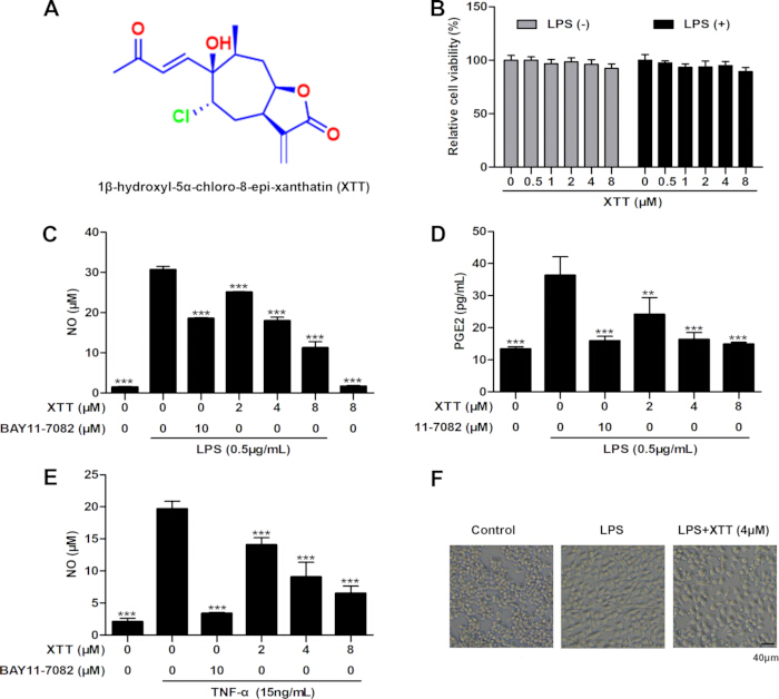
Figure 1: XTT suppressed NO and PGE₂ synthesis and attenuated morphological changes in LPS- or TNF-α-stimulated RAW264.7 cells. (A) Chemical structure of XTT. (B) RAW264.7 cells were incubated with the indicated concentrations of XTT for 24 h in the presence or absence of LPS (0.5 µg/mL), and cell viability was assessed using a CCK-8 assay. (C–E) RAW264.7 cells were pre-incubated with XTT (2, 4, and 8 µM) or BAY 11-7082 (10 µM) for 2 h prior to stimulation with LPS (0.5 µg/mL) or TNF-α (15 ng/mL) for 24 h. NO and PGE₂ levels in the culture medium were measured via the Griess assay and ELISA, respectively. (F) Morphological changes in RAW264.7 cells following incubation with XTT and LPS were visualized under an optical microscope. Scale bar = 40 µm. Values represent mean ± SEM (n = 3). *p < 0.05, **p < 0.01, ***p < 0.001 versusthe LPS-treated group. Please click here to view a larger version of this figure.
XTT suppressed the mRNA and protein expression of iNOS and COX2 in LPS-stimulated RAW264.7 cells
To further explore the molecular mechanism underlying the anti-inflammatory properties of XTT, the expression of inducible nitric oxide synthase (iNOS) and cyclooxygenase-2 (COX2) was examined in LPS-activated RAW264.7 cells. XTT treatment at 2, 4, and 8 µM significantly reduced LPS-triggered mRNA expression of iNOS and COX2 (Figure 2).
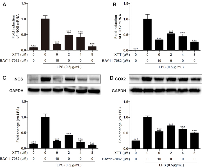
Figure 2: XTT downregulated the transcript and protein levels of iNOS and COX2 in LPS-activated RAW264.7 cells. RAW264.7 cells were pre-incubated with the indicated concentrations of XTT or BAY 11-7082 (10 µM) for 2 h prior to LPS stimulation (0.5 µg/mL) for 24 h. (A, B) Transcript levels of iNOS and COX2 were assessed via qRT-PCR using GAPDH as the internal reference. (C, D) Protein levels of iNOS and COX2 were detected viaWestern blotting. GAPDH served as the loading control. Representative blots from three independent experiments are shown, and densitometric analyses of iNOS and COX2 normalized to GAPDH are presented. Values represent mean ± SEM (n = 3). *p < 0.05, **p < 0.01, ***p < 0.001 versus the LPS-treated group. Please click here to view a larger version of this figure.
Treatment with XTT attenuated these increases in a concentration-dependent manner, with the strongest effect observed at 8 µM. The magnitude of inhibition was comparable to that produced by BAY 11-7082. These observations indicate that reduced NO and PGE₂ production is associated with suppression of iNOS and COX2 expression.
XTT decreased the mRNA expression of pro-inflammatory cytokines in LPS-stimulated RAW264.7 cells
To further characterize the anti-inflammatory profile of XTT, transcript levels of representative pro-inflammatory cytokines were evaluated. LPS stimulation strongly induced TNF-α, IL-1β, and IL-6 expression in RAW264.7 macrophages. Pretreatment with XTT significantly attenuated the induction of all three cytokines in a concentration-dependent manner (Figure 3).
At the highest concentration tested, the inhibitory activity of XTT was comparable to or greater than that produced by BAY 11-7082. The broad reduction in cytokine expression further supports the ability of XTT to limit inflammatory activation in macrophages.
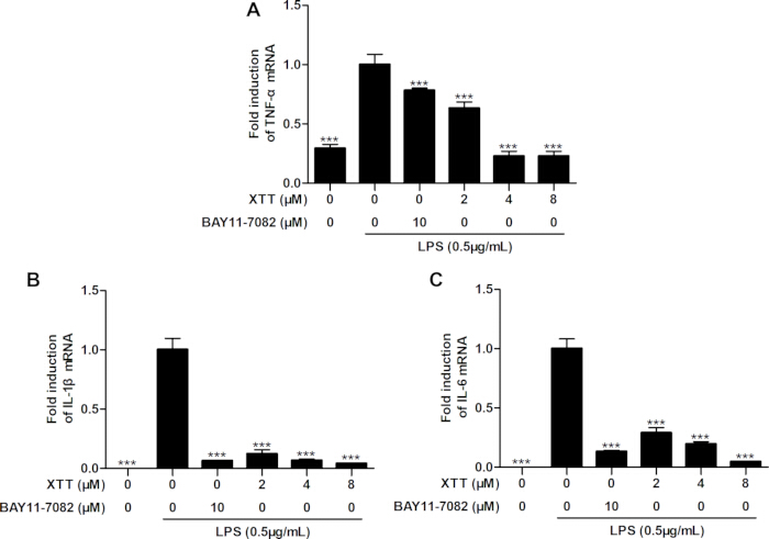
Figure 3: XTT downregulated the transcript levels of pro-inflammatory cytokines in LPS-stimulated RAW264.7 macrophages. Effects of XTT on TNF-α, IL-1β and IL-6 mRNA expression in LPS-stimulated RAW264.7 cells. (A–C) Transcript levels were determined by qRT-PCR. Data are mean ± SEM (n = 3). *p < 0.05, **p < 0.01, ***p < 0.001 versus LPS alone. Please click here to view a larger version of this figure.
XTT did not alter MAPK signaling activation in LPS-stimulated RAW264.7 cells
To clarify whether the mitogen-activated protein kinase (MAPK) signaling pathway contributes to the anti-inflammatory properties of XTT, the phosphorylation status of extracellular signal-regulated kinase (ERK), p38 MAPK, and c-Jun N-terminal kinase (JNK) was assessed via Western blotting.
LPS challenge markedly elevated the phosphorylation of ERK, p38 MAPK, and JNK in RAW264.7 cells. Specific inhibitors of these kinases effectively blunted their corresponding phosphorylation events. However, XTT treatment did not significantly alter LPS-triggered MAPK activation in RAW264.7 cells (Figure 4A–C), indicating that MAPK signaling is not a critical mediator of XTT’s anti-inflammatory actions.
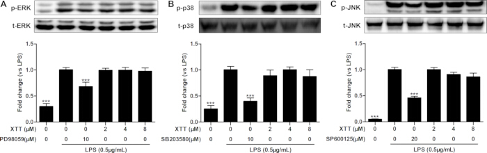
Figure 4: XTT did not significantly affect LPS-induced MAPK activation in RAW264.7 cells. RAW264.7 cells were pre-incubated with different concentrations of XTT or specific MAPK inhibitors for 4 h prior to LPS challenge for 30 min. (A–C) Phosphorylation status of ERK, p38 MAPK, and JNK was detected via Western blotting using specific antibodies. Total ERK, p38 MAPK, and JNK proteins served as internal references. Representative immunoblots and densitometric quantifications are shown. Data are presented as mean ± SEM from three independent experiments. *p < 0.05, **p < 0.01, ***p < 0.001 versus the LPS-treated group. Please click here to view a larger version of this figure.
XTT blocked activation of the NF-κB signaling pathway in activated macrophages
To explore the involvement of NF-κB signaling in the anti-inflammatory actions of XTT, the impacts of XTT on NF-κB pathway activation were examined in LPS-activated RAW264.7 cells and TNF-α-treated HeLa cells.
Western blot analysis showed that LPS challenge induced robust phosphorylation and degradation of IκBα in RAW264.7 cells. XTT treatment markedly mitigated these LPS-triggered changes in IκBα phosphorylation and degradation (Figure 5A and 5B).
Immunofluorescence analysis further confirmed that TNF-α stimulation evoked substantial nuclear translocation of NF-κB p65 in HeLa cells, whereas exposure to 8 µM XTT significantly curtailed p65 nuclear accumulation (Figure 5C). These observations were further validated by cytoplasmic and nuclear fractionation followed by Western blot analysis (Figure 5D). Collectively, these data indicate that NF-κB signaling suppression is a key mechanism underlying the anti-inflammatory properties of XTT.
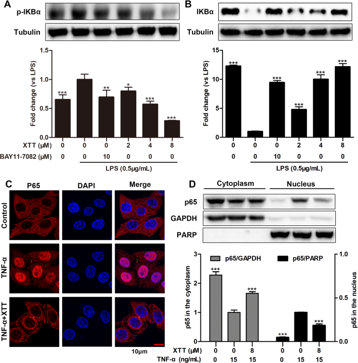
Figure 5: XTT inhibited activation of the NF-κB signaling pathway. RAW264.7 cells were pre-incubated with XTT (2, 4, and 8 µM) or BAY 11-7082 (10 µM) for 4 h prior to LPS challenge (0.5 µg/mL) for 5 min (A) or 10 min (B). (A, B) Phosphorylated and total IκBα levels were detected via Western blotting. Tubulin served as the loading control. Densitometric quantifications of IκBα normalized to tubulin are shown. HeLa cells were pre-incubated with XTT for 4 h followed by TNF-α stimulation (15 ng/mL) for 30 min. (C) Nuclear translocation of NF-κB p65 was examined by immunofluorescence staining. Scale bar = 10 µm. (D)Nuclear and cytoplasmic fractionation followed by Western blotting was conducted to assess p65 localization. GAPDH and PARP served as loading controls for cytoplasmic and nuclear fractions, respectively. Densitometric quantifications of p65 normalized to GAPDH or PARP are shown. Data are presented as mean ± SEM from three independent experiments. *p < 0.05, **p < 0.01, ***p < 0.001 versus the LPS-treated group. Please click here to view a larger version of this figure.
XTT interacted with IKKα/β and inhibited IKKα/β phosphorylation
To investigate whether XTT targets IKKα/β to suppress NF-κB signaling, cellular thermal shift assay (CETSA), molecular docking analysis, and phosphorylation studies were performed.
CETSA analysis demonstrated that XTT treatment reduced the thermal stability of IKKα/β proteins in HeLa cell lysates compared with the DMSO control, suggesting a direct interaction between XTT and IKKα/β (Figure 6A).
Molecular docking analysis was subsequently performed to evaluate the binding interactions between XTT and components of the IKK complex. XTT formed hydrogen bonding and hydrophobic interactions with residues within the NEMO/IKK interaction domain, including Leu80, Ala722, and Asn721, with a calculated binding energy of −5.5 kcal/mol (Figure 6B and 6C). In addition, XTT formed hydrogen bonds with Asn225 and Gly218 within the ATP-binding pocket of IKKβ, with a calculated binding energy of −7.3 kcal/mol (Figure 6D and 6E). Additional hydrophobic and van der Waals interactions involving residues such as Leu18, Tyr115, Phe155, and Arg154 further stabilized the XTT–IKKβ complex.
Western blot analysis demonstrated that LPS stimulation markedly increased IKKα/β phosphorylation in RAW264.7 cells, whereas XTT treatment effectively suppressed LPS-induced IKKα/β phosphorylation (Figure 6F). Collectively, these findings suggest that XTT interacts with IKKα/β and suppresses NF-κB pathway activation by inhibiting IKKα/β-mediated signaling.
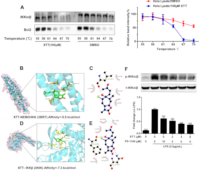
Figure 6: XTT interacted with IKKα/β and suppressed IKKα/β phosphorylation. (A) CETSA was conducted to assess the effect of XTT on the thermal stability of IKKα/β in HeLa cell lysates. Bcl2 served as the loading control. Densitometric quantifications of IKKα/β band intensities are shown. (B, C) Molecular docking analysis of XTT with the NEMO/IKK interaction domain was performed using AutoDock. (D, E) Molecular docking analysis of XTT with the ATP-binding pocket of IKKβ was performed using AutoDock. Calculated binding energies are indicated. (F) RAW264.7 cells were pre-incubated with XTT (2, 4, and 8 µM) or PS-1145 (10 µM) for 4 h prior to LPS challenge (0.5 µg/mL) for 10 min. Phosphorylated and total IKKα/β levels were detected via Western blotting. Densitometric quantifications of phospho-IKKα/β normalized to total IKKα/β are shown. Data are presented as mean ± SEM from three independent experiments. *p < 0.05, **p < 0.01, ***p < 0.001 versus the LPS-treated group. Please click here to view a larger version of this figure.
DATA AVAILABILITY:
Supplementary File 1: Supporting data for 1β-hydroxyl-5α-chloro-8-epi-xanthatin (XTT), including structural characterization (HRESIMS, 1H NMR, and 13C NMR spectra), HPLC purity analysis, and original experimental data supporting the reported results. Please click here to download this file.
Discussion
Loading...
$$\rightleftharpoonup{xx}$$
$$\longleftharp{xx}$$,
$$\longrightharp{xx}$$,
Sesquiterpene lactones are a class of natural products with diverse pharmacological activities, including antiseptic, anti-inflammatory, antipyretic, and detoxifying effects15. Previous studies have shown that XTT, a xanthanolide-type sesquiterpene lactone isolated from Xanthium sibiricum, displays potent antiproliferative and pro-apoptotic effects in human hepatocellular carcinoma cells13. This work assessed the anti-inflammatory properties of XTT in LPS-stimulated RAW264.7 macrophages and further probed the underlying molecular mechanisms. Activated macrophages produce inflammatory mediators and cytokines that amplify inflammatory signaling. Consistent with this biology, LPS stimulation increased NO and PGE₂ production, whereas XTT reduced both mediators together with iNOS and COX2 expression16,17,18,19. In this work, LPS stimulation markedly increased NO and PGE₂ production in RAW264.7 cells, whereas the known IκB inhibitor BAY 11-7082 significantly suppressed these responses, confirming the reliability of the inflammatory model20,21. XTT inhibited LPS-induced NO and PGE₂ production in a concentration-dependent manner and further downregulated iNOS and COX2 expression at both the transcriptional and translational levels. Notably, the inhibitory effects of 8 µM XTT were comparable to those of 10 µM BAY 11-7082, underscoring the robust anti-inflammatory activity of XTT in vitro.
In addition to inflammatory mediators, macrophage-derived cytokines such as TNF-α, IL-1β, and IL-6 are critical regulators of inflammatory progression22. These cytokines trigger intracellular signaling cascades that promote the expression of downstream inflammatory genes and contribute to the pathogenesis of inflammatory disorders, including inflammatory bowel disease (IBD) and rheumatoid arthritis (RA)23. Therapeutic targeting of these cytokines has demonstrated significant clinical benefits; for example, the anti-TNF-α antibody adalimumab exerts potent anti-inflammatory effects in RA patients by neutralizing TNF-α signaling24. In this study, XTT significantly decreased the mRNA expression of TNF-α, IL-1β, and IL-6 in LPS-stimulated RAW264.7 cells. The inhibitory effect of 8 µM XTT was greater than that observed with 10 µM BAY 11-7082 in vitro. These findings further confirm the potent anti-inflammatory activity of XTT and suggest its potential as a lead compound for anti-inflammatory drug development. Nevertheless, additional in vivo investigations are needed to evaluate its pharmacological efficacy and translational potential.
To clarify the molecular mechanisms underlying the anti-inflammatory effects of XTT, the involvement of the MAPK and NF-κB signaling pathways was investigated. The MAPK family, including extracellular signal-regulated kinase (ERK), p38 MAPK, and c-Jun N-terminal kinase (JNK), plays a critical role in governing inflammatory mediator production in macrophages and other immune cells25. Dysregulated MAPK activation is commonly associated with inflammatory diseases, and pharmacological inhibition of this pathway has emerged as a promising therapeutic strategy for disorders such as IBD and RA26. However, while LPS stimulation markedly activated MAPK signaling in RAW264.7 cells, XTT treatment had no significant impact on ERK, p38 MAPK, or JNK phosphorylation. These results indicate that the anti-inflammatory activity of XTT is independent of MAPK pathway regulation in macrophages.
Previous studies reported that XTT induces ERK and p38 MAPK activation in HepG2 cells through a reactive oxygen species (ROS)-dependent mechanism, thereby mediating antitumor activity13. The differential effects of XTT on MAPK signaling are therefore likely dependent on cellular context and biological microenvironment. XTT is structurally related to xanthatin, a multitarget anti-inflammatory compound that suppresses NF-κB, MAPK, and STAT3 signaling pathways27,28,29.
Similar to other molecules containing an α,β-unsaturated carbonyl group, XTT is chemically reactive and may form covalent interactions with cellular proteins. Although the present findings support IKKα/β as a major target of XTT, the precise binding mode, including whether the interaction is covalent or non-covalent, remains unclear. Electrophilic compounds with similar structural features are known to interact with multiple proteins30,31,32, and this potential promiscuity should be considered when interpreting the biological activity of XTT. Nevertheless, the present findings indicate that XTT selectively suppresses NF-κB signaling without significantly affecting MAPK activation, suggesting improved pathway selectivity relative to structurally related compounds.
NF-κB transcription factors are key modulators of immune and inflammatory reactions because they control the transcription of numerous inflammation-related genes33. Aberrant NF-κB activation is closely associated with the development of autoimmune and inflammatory conditions, and inhibition of this pathway has shown therapeutic benefit in inflammatory disorders such as IBD and RA25,34. NF-κB signaling activation is strictly controlled by IκBα. Upon inflammatory stimulation, IκBα is phosphorylated by the IKK complex and then broken down via the proteasomal pathway, thus enabling NF-κB dimers to translocate into the nucleus and initiate inflammatory gene transcription35,36. In this study, XTT markedly reduced LPS-induced IκBα phosphorylation and degradation in RAW264.7 cells. XTT also attenuated TNF-α-triggered nuclear translocation of NF-κB p65 in HeLa cells, as demonstrated by both immunofluorescence staining and cytoplasmic/nuclear fractionation assays37. HeLa cells were used to allow clear observation of p65 localization. These results indicate that inhibition of NF-κB signaling is a key mechanism driving the anti-inflammatory effects of XTT.
To further identify the upstream molecular target mediating NF-κB inhibition, the IKK complex was examined. The catalytic subunits IKKα and IKKβ are responsible for IκBα phosphorylation and are regarded as promising therapeutic targets for inflammatory diseases38. Several small-molecule IKK inhibitors have shown favorable anti-inflammatory actions in experimental models39. CETSA analysis demonstrated that XTT altered the thermal stability of IKKα/β proteins in HeLa cell lysates, confirming a direct interaction between XTT and IKKα/β40. Molecular docking analysis further suggested that XTT may interact with both the NEMO/IKK interaction domain and the ATP-binding pocket of IKKβ. XTT formed hydrogen bonds and hydrophobic interactions within the NEMO/IKK interaction region, potentially interfering with IKK complex assembly41,42,43. In addition, XTT showed a favorable predicted binding affinity for the ATP-binding pocket of IKKβ through interactions involving residues such as Asn225 and Gly21844. Western blot analysis further confirmed that XTT suppressed LPS-induced phosphorylation of IKKα/β in RAW264.7 cells. Collectively, these findings suggest that XTT suppresses NF-κB activation through interaction with IKKα/β and subsequent inhibition of IKK-mediated signaling.
Despite these findings, several limitations should be noted. First, the present study was limited to in vitro cellular models, and further in vivo studies are required to evaluate the pharmacological efficacy, safety, and translational relevance of XTT. Second, although CETSA, molecular docking, and phosphorylation analyses support interaction between XTT and IKKα/β, direct biochemical evidence demonstrating inhibition of intrinsic IKK kinase activity remains unavailable. Cellular phosphorylation changes may also be influenced by additional intracellular regulatory mechanisms. Future studies using purified active IKKα/β proteins and standardized in vitro kinase assays will therefore be necessary to validate XTT's direct kinase inhibition.
In summary, XTT showed robust anti-inflammatory activity in LPS- or TNF-α-stimulated cells by blocking NF-κB signaling. The anti-inflammatory effects of XTT are likely driven by interactions with IKKα/β and subsequent suppression of NF-κB pathway activation. Collectively, the data support the potential value of XTT as a candidate scaffold for anti-inflammatory drug development.
Disclosures
Loading...
$$\rightleftharpoonup{xx}$$
$$\longleftharp{xx}$$,
$$\longrightharp{xx}$$,
The authors declare no competing interests.
Acknowledgements
Loading...
$$\rightleftharpoonup{xx}$$
$$\longleftharp{xx}$$,
$$\longrightharp{xx}$$,
This work was supported by the National Natural Science Foundation of China (No. 82004031), the Sichuan Provincial Administration of Traditional Chinese Medicine Research Project (No. 2024MS566) and the National Key Research and Development Program of China (No. 2023YFC3504402).
Materials
| Name | Company | Catalog Number | Comments |
|---|---|---|---|
| Enhanced chemiluminescence reagent D20A2:D21A2:D22A2:D23A2: D24A2:D25A2:D26A2:D27A2:D28A2: D29A2:D30A2:D29 | Beyotime Biotechnology Co., Ltd | P90720 | luminescent signal detection |
| Horseradish peroxidase (HRP)-conjugated secondary antibodies | Signalway Antibody (Nanjing) | L30118 | specifically binds to the primary antibody |
| p65 primary antibody | Bioworld | BS4139 | specifically recognizes NF-κB p65 subunit protein |
| Spectrophotometer | Thermo Fisher Scientific | NanoDrop 2000 | SCR_018042 |
| 0.45 μM Ployvinylidence fluoride membrane | Millipore | PR05509 | adsorb protein molecules |
| 1β-hydroxyl-5α-chloro-8-epi-xanthatin | Chengdu Institute of Biology | natural monomeric compounds | |
| All-in-One cDNA Synthesis SuperMix | Bimake (Shanghai) | B24408 | reverse-transcribe |
| BAY11-7082 | Beyotime Biotechnology Co., Ltd | SF0011 | IκB/IKK inhibitor |
| Bcl2 antibody | Signalway Antibody (Nanjing) | 32012 | AB_3751037 |
| Bicinchoninic Acid (BCA) kit | Beyotime Biotechnology Co., Ltd | P0010S | total protein quantification |
| CCK-8 agent | Beyotime Biotechnology Co., Ltd | C0038 | cell viability measurement |
| Chemiluminescence detection system | Millipore, Bedford, USA | P90720 | acquire and process luminesence signals |
| COX-2 antibody | Signalway Antibody (Nanjing) | 33345 | AB_3676692 |
| DMSO | Sangon Biotech(Shanghai)Co.,Ltd. | A100231-0500 | solubilize compounds |
| GAPDH antibody | Signalway Antibody (Nanjing) | 41549 | AB_3751036 |
| Gel Imaging Instrument | E-BLOT | Touch Imager Pro | image acquisition and analysis |
| Gradient PCR instrument | Biometra TADVANCED | Biometra Tadvanced 96SG | gradient annealing temperature |
| Griess agent | Beyotime Biotechnology Co., Ltd | S0021S | NO quantification |
| Human cervical carcinoma HeLa cells | The Cell Bank of Chinese Academy of Sciences | SCSP-504 | Human-derived cells |
| Human TNF-α | Novus Biologicals | 210-TA | pro-inflammatory cytokines |
| iNOS antibody | Signalway Antibody (Nanjing) | 48309 | AB_2943634 |
| Laser Confocal Inverted Microscope | Olympus | FV1000 | SCR_016840 |
| LPS ( E.coli 055:B5) | Sigma-Aldrich (Shanghai) Trading Co., Ltd | L6529 | establish inflammatory models |
| Murine macrophage RAW264.7 cells | The Cell Bank of Chinese Academy of Sciences | SCSP-5036 | Mouse-derived cells |
| Nuclear and Cytoplasmic Protein Extraction Kit | Beyotime Biotechnology Co., Ltd | P0027 | separate cytoplasmic and nuclear proteins |
| PD98059 | Selleck Chemicals (Shanghai) | S1177 | MEK inhibitor |
| P-ERK (Thr202/Thr185) antibody | Signalway Antibody (Nanjing) | 12548 | detect the phosphorylation level of ERK protein |
| p-IKKα/β (Ser176/Ser177) antibody | Signalway Antibody (Nanjing) | 11931 | AB_3751034 |
| p-IκBα (phospho-Ser32)antibody | Signalway Antibody (Nanjing) | 13776 | AB_3751033 |
| p-JNK (T183/T221) antibody | Signalway Antibody (Nanjing) | 13371 | AB_3718662 |
| p-p38 MAPK (Thr180) antibody | Signalway Antibody (Nanjing) | 11581 | AB_3751032 |
| Prostaglandin E2 Parameter Assay Kit | R&D Systems | KGE004B | AB_2894942 |
| PS1145 | Selleck Chemicals (Shanghai) | S7691 | specific IKK inhibitor |
| Rapid SDS-PAGE Gel Preparation Kit | Beyotime Biotechnology Co., Ltd | P0018S | protein separation |
| Real-Time PCR System | Bio-Rad | CFX96 | SCR_018064 |
| RIPA buffer | Bimake (Shanghai) | P0013C | cell lysis |
| SB203580 | Selleck Chemicals (Shanghai) | S1076 | p38 MAPK inhibitor |
| SP600125 | Selleck Chemicals (Shanghai) | S1460 | JNK inhibitor |
| SYBR Green | Bimake (Shanghai) | B21202 | For real-time quantitative |
| TRITC-conjugated goat anti-mouse IgG | Beyotime Biotechnology Co., Ltd | A0568 | AB_2893016 |
| TRITC-conjugated second antibody | Beijing Baiao Leibo Technology Co., Ltd. | ZN1992 | AB_3751143 |
| TRIzol Reagent | Invitrogen | 15596026 | total RNA isolation |
| Vinculin antibody | Signalway Antibody (Nanjing) | 41534 | AB_3751035 |
Reprints and Permissions
Request permission to reuse the text or figures of this JoVE article
Request Permission