This non-invasive jacketed telemetry protocol enables simultaneous monitoring of cardiorespiratory and locomotor adaptation during exercise in rats. By avoiding surgical implantation, this approach allows repeated physiological measurements in the same animals, providing a reliable tool for exercise physiology research and teaching.
Method Article
A Jacketed Telemetry Method for Monitoring Cardiorespiratory Adaptation During Exercise in Rats
In This Article
Summary
Abstract
The assessment of cardiorespiratory adaptation during exercise is not only crucial for studying physiological adaptation to physical training, but also essential for preclinical research, as measurements at rest can mask underlying physiological phenomena. However, conventional preclinical methods, such as surgically implanted telemetry or hermetic chambers, come with important constraints. To overcome these limitations, we present a surgery-free methodology using a jacketed telemetry system, enabling the simultaneous monitoring of heart rate (HR), respiratory rate (RespR), and locomotor activity (AL) in rats during forced treadmill exercise. The protocol implements a two-phase acclimatization process designed to ensure both the appropriate running performance of the animals and data integrity: first, habituation to wearing the telemetry jacket, which integrates respiratory inductive plethysmography sensors and surface electrocardiogram electrodes; and second, a 2-week (five days per week) habituation to treadmill exercise. We detail equipment procedures and essential quality control steps, including real-time signal verification, to minimize animal stress and ensure reliable, low-noise physiological data. We also provide representative signal examples to help users achieve high data quality, especially during exercise protocols, but also in other experimental contexts. When implemented correctly, this protocol enables consistent monitoring at speeds up to 40 cm.s⁻1, allowing the quantification of changes in HR, RespR, and AL compared to baseline. By eliminating the need for surgical interventions, this protocol strongly adheres to the ethical principles of the 3Rs (reduce, refine, replace). While the provided techniques focus on exercise, they have potential applications across diverse fields, including comprehensive cardiorespiratory characterization, animal handling training, welfare monitoring, and physiology, pharmacology, or education.
Introduction
Developing models that provide valuable insights into the dynamic landscape of exercise physiology is of major importance in many fields, such as training and performance, or the prevention of diseases and rehabilitation. Among the physiological effects associated with exercise, understanding the adaptations of cardiac and respiratory functions is a crucial element in sports (training/performance) and clinical (prevention/rehabilitation) contexts to develop specific and tailored training/rehabilitation programs. It is therefore essential to continue deciphering the mechanisms of cardiorespiratory adaptations to exercise. To this end, animal models, including rodents, are now widely used in preclinical research designed to study the effect of exercise on cardiac and respiratory functions. Indeed, not only do they enable the evaluation of cardiorespiratory adaptations to exercise1, but they are also essential for studying pathophysiological phenomena potentially hidden at rest2,3. Nevertheless, acquiring relevant physiological data during exercise presents a significant technological challenge. For the collected data to be physiologically meaningful, the acquisition process must be as non-invasive as possible. Ideally, monitoring should be conducted without interfering with the activities of the patient or animal, and measurements should not be compromised by motion artifacts. Currently, in preclinical research, no single measuring device fully meets these essential requirements.
Furthermore, an ideal device should adhere to the ethical principles of the 3Rs (reduce, refine, replace), which guide preclinical animal research4. These principles, together with the current socio-economic context of animal use in research and teaching, underscore the need to develop tools and investigative models that enable more efficient and responsible use of animals. To this end, the development and use of wearable devices and sensors appear to be a priority.
Today, the main methods available for directly assessing cardiac and ventilatory functions in rodents cannot be used effectively in exercise protocols due to practical limitations. For example, unrestrained whole-body plethysmography chambers are recognized as an accurate and non-invasive approach for longitudinal, repeated measures of ventilatory function in awake animals5. Although they provide reliable measurements under resting conditions, active locomotion generates pressure artifacts and signal noise that can compromise the interpretation of respiratory data during exercise. Similarly, implanted radiotelemetry is established as the gold standard for studying cardiac function in freely moving rodents6 and can be used to monitor ECG and hemodynamic parameters2,7. However, it comes with important constraints, primarily related to the surgical implantation of sensors and transmitter. In particular, postoperative pain and impairment due to the surgical wound or the implanted device itself have to be managed with appropriate care protocols6,8,9. More recently, an increasing number of studies are focusing on non-invasive methods. For instance, studies have been conducted with wearable ECG radiotelemetry devices used externally in guinea-pigs10, contactless Doppler-based respiratory activity detection device11 or wearable biosensors12. However, these monitoring techniques have so far been limited to the proof-of-concept stage, and need to be further developed before they can be used on a larger scale. Jacketed telemetry, which is commonly used as a non-invasive solution for cardiorespiratory assessment in large animals such as dogs or primates13,14, has recently been adapted to laboratory rats15,16,17 and juvenile minipigs18. The system illustrated in Figure 1A–F consists of a non-invasive, low-constraint radio-device maintained in a jacket worn by the animal. It allows combined monitoring of cardiac activity by surface ECG, respiratory function by Respiratory Inductive Plethysmography (RIP), and locomotor activity by accelerometry. Previous studies have demonstrated the ability of this system to effectively monitor cardiorespiratory variables compared with invasive methods15,16. More recently, its use as a complementary non-surgical monitoring approach has been reported in various contexts, including pharmacological assessment in socialized conditions17,19 or cardiac disease models20.
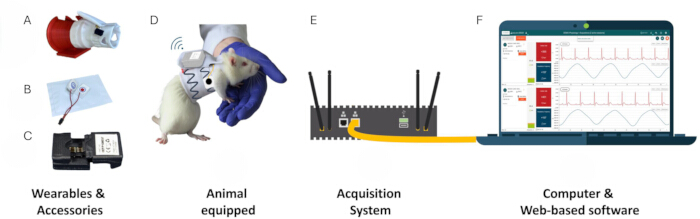
Figure 1: View of the main components of the jacketed telemetry system. (A) Instrumented jacket placed on its fitting tube, (B) Ag/AgCl patches for ECG monitoring, (C) Radio emitter for data collection and transmission, (D) Animal wearing the jacket and emitter, (E) Acquisition system for data storage and telemetry software execution, (F) Computer connected via an Ethernet cable, displaying the telemetry software through a web user interface (UI). Please click here to view a larger version of this figure.
Based on a previous evaluation study21 and habituation protocol22, the protocol proposed here presents a refined, non-invasive methodology using a jacketed telemetry system enabling the simultaneous monitoring of heart rate, respiratory rate and locomotor activity in rats during incremental exercise.
Protocol
All experiments were conducted in compliance with the European (L276-33 2010/63/EU) and French (AGRG1231951D) regulations concerning the protection and use of laboratory animals. The in vivo procedures of this study were approved by Ethics Committee #012 (Grenoble Alpes University), accredited by the French Ministry of Higher Education and Research, under the references APAFIS#31347-2021042914221915 v2 and APAFIS #52237-2024112821304952 v5.
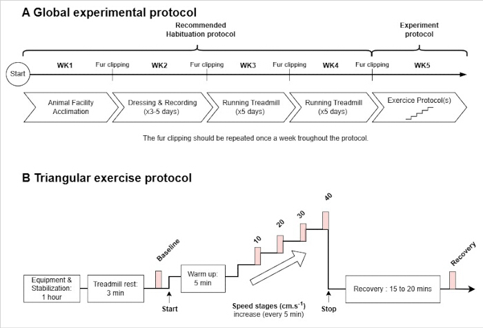
Figure 2: Experimental protocol overview. (A) Global experimental protocol including the habituation and exercise phases. The schedule is indicative and should be adjusted based on real-time observations and effective habituation during the experiment. Notably, animals can be habituated to handling during the “Animal Facility Acclimation". (B) Typical exercise protocol, including a post-equipment stabilization of 60 min, a baseline recorded after 3 min of rest on the treadmill, a warm-up period, incremental exercise and a recovery phase. Data analysis periods are indicated by light red squares. The specified speed stages are suggestions and should be tailored based on the specific animal model and experimental goals. Please click here to view a larger version of this figure.
1. Animal model
- Use rats sourced from approved suppliers and acclimated to housing conditions (1 bar; 24 °C; 40–70% humidity; 12 h light/dark cycle) for one week in pairs in conventional cages with free access to food and water, and provided with enrichment (nesting material, gnawing objects, cardboard tunnels).
- Mark the animals individually (ink on the tail).
2. Equipment procedure
- Fur removal: Timing 1–2 min per animal
- Clip the animal's hair once a week to ensure successful ECG recording.
- To minimize stress and anesthesia on the day of the experiment, perform this procedure the day before; it can also be done on the same day.
- Method A (Optional Anesthesia): Administer a gaseous anesthesia mix (1.5–5% isoflurane, 20% oxygen, and air) and position the rat in ventral recumbency (lying on its belly).
- Keep anesthesia duration to a minimum, typically 2–5 min, sufficient only for fur clipping.
- Carefully clip the hair at the dorsal and lateral chest (Figure 3A), using a clipper, and mark the tail with ink.
NOTE: Full recovery and ambulation typically occur within 5–10 min after discontinuation of anesthesia. - Ensure the animal has reached this state before returning it to its home cage.
- Method B (No Anesthesia): Alternatively, restrain the rat manually for the minimum time necessary to complete fur clipping, typically less than 2 min.
- Restrain the animal firmly but gently and avoid excessive pressure on the thorax.
- Carefully clip the hair at the dorsal and lateral chest (Figure 3A), using a clipper, and mark the tail with ink.
NOTE: If the animal shows signs of severe distress, release it immediately and allow a recovery period before attempting the procedure again. Assistance from another person is recommended when using this method. - Clean the clipped area with water to remove sebum produced by the rat’s skin and make sure the area is thoroughly dry. Dry the area with gauze.
- Animal equipment: Timing 3–5 min per animal
NOTE: The subsequent steps outline the process for fitting the animal with the jacket, sensors, and telemetric device, minimizing the need for physical restraint.- Choose the appropriate jacket size based on the animal's weight, sex, and the specific strain, following the manufacturer's selection charts and sizing tables23.
- Carefully place the instrumented jacket on the jacket fitting tool (be careful not to damage sensors and cables).
- Position the backpack over the notch located on the top of the fitting tool (Figure 4A). Set the fitting tool on its rectangular base (Figure 4B) and place the rat in front of the widest opening (funnel entrance side) (Figure 4C).
- If the rat does not want to go through the fitting tool, motivate the animal by placing an enrichment tunnel or its housing cage in front of the exit.
- As soon as the animal's head emerges from the other side and its front paws become visible: use one hand to hold the rat and the fitting tool together; use the other hand to guide the paws through each hole located on the front side of the jacket to properly position and secure it on the animal (Figure 4D).
- Gently slide the jacket down the animal, gradually removing the fitting tool as you go, and allow the animal to completely pass through the tool toward the exit.
- Ensure the jacket is properly positioned without any skin folds, and verify the sizing is appropriate according to the Jacket fit verification recommendations.
- If required, replace the jacket with the appropriate size. Animals that cannot be fitted safely or comfortably despite size adjustment should be excluded from the study.
- When the animal reaches the desired level of the fitting tool, remove the motivation element from its view if one has been used.
NOTE: This will prevent the rat from continuing to pass through the tool. - After placing the rat on the laboratory bench, unhook the backpack (Figure 4E) and unfold the jacket (Figure 4F).
- Insert the ECG Ag/AgCl electrode with the red (positive) sticker through the left opening of the jacket, marked with a red cross (Figure 4G), and insert the black (negative) electrode into the right opening.
- Position the electrodes in Lead II by placing the black (negative) electrode behind the right forelimb and the red (positive) electrode at the left lateral chest wall within the clipped area, under the RIP chest band (Figure 3B,C).
- Apply ECG gel (0.1 to 0.3 mL) to the black spot on each electrode using the syringe and the catheter for precise gel deposition. Press gently so that the electrodes adhere completely to the animal's skin (Figure 4H).
- Connect the sensors used (plethysmography/ECG electrodes) to the emitter, and place the telemetry transmitter and battery pack in the backpack on the animal's back (Figure 4I–J).
- Fold down the back of the jacket to protect the electronics/cables, reducing friction and keeping them protected. Fold the backpack towards the back of the rat (over the folded jacket) and attach it to the distal/posterior part of the jacket with the hook (Figure 4K).
NOTE: Gel is applied after electrode insertion to prevent the electrodes from slipping during placement through the jacket openings. Pre-application of gel is inadvisable, as it reduces friction and makes precise electrode positioning significantly more difficult.
- Signal acquisition initiation and signal quality verification
- Place the telemetry system in the same room as the recording (at a distance up to 10 meters) and in direct line-of-sight, ideally positioning the unit above the animal or on the same plane.
- Start the system and open the telemetry software.
- Start the acquisition according to the manufacturer's procedure24. Check the quality of the ECG and respiratory signals quality and implement any necessary adjustments (see Figure 5).
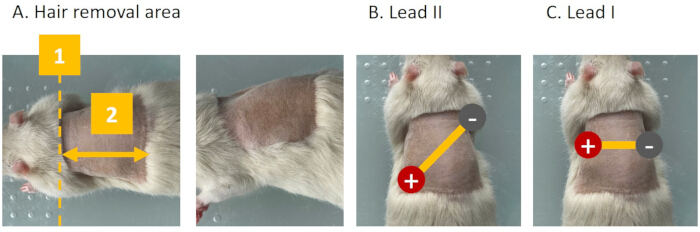
Figure 3: Views of an anesthetized rat. (A) Dorsal and lateral chest areas indicating where fur should be removed (step 2.1): 1) dashed line indicates the minimum upper boundary, and 2) double-headed arrows indicate the minimum length of the clipped area. (B) Recommended Lead II electrodes placement with the black (negative) electrode positioned behind the right forelimb and the red (positive) electrode on the left lateral chest wall within the clipped area, beneath the RIP chest band. (C) Lead I electrodes placement, showing electrodes positioned horizontally across the chest, the red (positive) behind the left forelimb and the black (negative) behind the right forelimb. Lead II configuration typically yields the highest QRS amplitude and clearest P-wave resolution in rats. Please click here to view a larger version of this figure.
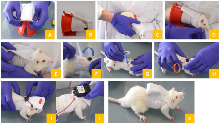
Figure 4: Step-by-Step procedure for equipping a rat with the jacket, electrodes, and electronic device (section 2). (A-B) Place the jacket on the fitting tube. (C-D) Guide the animal through the fitting tube and into the jacket. (E-F) Unhook the backpack and unfold the jacket. (G) Insert the ECG electrode with the red (positive) sticker through the left opening of the jacket, which is marked with a red cross and insert (0.1 to 0.3 mL of conductive gel). (H) Gently massage so that electrodes adhere completely to the animal's skin. (I-J) Connect the sensors used (plethysmography / ECG electrodes) to the emitter and insert it in the pouch. (K) Secure the backpack using the red hook. Please click here to view a larger version of this figure.
3. Equipment removal and storage procedure - Timing 1 min per animal
- Place the rat on the laboratory bench or hold it in the hand. Detach the hook from the backpack. Unfold the back of the jacket to access the notches and electrodes. Carefully remove them.
- Remove the transmitter-battery assembly from the backpack. Disconnect the cables from this assembly.
- Remove the front paws from the openings. Allow the rat to move forward while holding the jacket with the backpack.
- Once finished, replace the plastic covers on the electrodes to prevent the gel from drying out. Inspect cable sensors; replace jackets if damaged or worn. Let the jacket dry before storage in its individual pouch.
- Clean hardware using alcohol-free disinfectant and a wipe; ensure all parts are dry. Disconnect the emitter from the battery to prevent discharge.
NOTE: The jacket is reusable on the same animal if the size fits the animal's weight and sensors are not damaged (see corresponding section in the discussion for more information).
4. Acclimatization to wearing the telemetry Jacket
NOTE: The sessions were organized according to the following procedure.
- Equip the animal following the procedures detailed in the "Animal Equipment" section.
- Ensure the jacket fit is secure but comfortable. Start the recording and return the animal to its home cage.
- On the first habituation day, limit the time the animal wears the equipment to a few hours. Perform frequent clinical checks to monitor the animal’s state. Gradually increase the daily duration the animal wears the equipment.
- Progressively decrease observation frequency if no signs of discomfort are observed. Continue this progression until the habituation time matches the target recording duration of the study.
- Stop the recording and un-equip the animal according to the recommended procedure. Inspect the recorded data quality and evaluate the animal’s recovery and general well-being before returning it to the housing room.
NOTE: The steps previously described provide a standard framework, to be adapted to the specific needs of the study.
5. Habituation to treadmill exercise
- Equip the animal (according to the previously described "animal equipment" section). Start the recording.
- Return the animal to its cage for a post-equipment stabilization period of 60 min to ensure full physiological stabilization following jacket fitting in accordance with previously established recommendations17,18,19.
- Transfer the animal onto the stopped treadmill and wait for 3 min. Start a first slow speed stage at 5 or 10 cm.s-1 for 1.5 min.
- Subject the animal to incremental increases in running speed, rising by 5 cm.s-1 every 2 min, and motivate the animal to run during the early stages using the electric shocks from the treadmill at a typical intensity of 0.2 mA.
- At the end of the exercise, return the animal to its cage. Wait 30 min before unequipping the animal.
- Repeat this treadmill exercise habituation protocol over 2 weeks, 5 days per week with a gradual increase in running speed and exercise duration.
NOTE: The total duration of this process should range from 5 min (on the initial day of habituation) to progressively reach 25 min (on the final day of habituation). - Monitor the running speed reached as well as the number of shocks to adjust the intensity and duration of the exercise over the 2–week habituation.
- As a humane endpoint, terminate the session if the animal reaches 10 shocks or a cumulative shock duration of 10 s. Return the animal to its cage and monitor its behavior visually.
NOTE: A representative image of a rat running on the treadmill while wearing the jacketed telemetry system is shown in Figure 6.
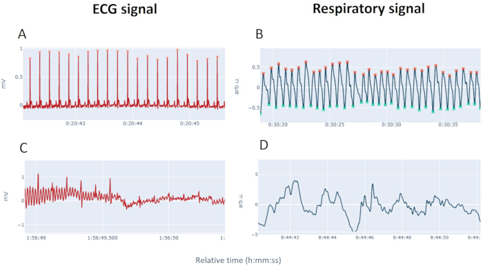
Figure 5: Typical ECG and respiratory traces during signal quality verification (step 2.3). ECGs were recorded at 500 Hz/24-bit and respiratory volumes at 200 Hz/32-bit, (A) The chart shows a high-quality ECG (resolved P, QRS, T waves, low noise, 3 s window with amplitude > 0.5 mV). (B) The chart shows a high-quality respiratory signal (resolved cycles at rest, 20 s windows, with amplitude > 0.5 arb.u.). (C) The chart shows a low-quality ECG (high interference like 50/60 Hz noise with amplitude > 0.5 mV, baseline wander, or small/flat QRS, 1.5 s windows). (D) The chart shows a low-quality respiratory signal (high baseline wander with amplitude > 5 arb.u., hindering identification, 12 s windows). The respiratory signal reflects relative changes in thoracic and abdominal circumference measured by respiratory inductance plethysmography (RIP); it is expressed in arbitrary units (arb.u.) as no volume calibration was performed in this protocol. Please click here to view a larger version of this figure.
6. Physiological monitoring during triangular Protocol
- Equip the animal (according to the previously described "animal equipment" section). Start the recording and wait for post-equipment stabilization in the home cage (recommended: 60 min), followed by a 3 min resting period in the treadmill prior to baseline (BL) measurements (Figure 2B).
- Allow the animal to adapt with a warm-up period at low speed (5 min at 5 cm.s-1) before the start of the exercise test.
- Subject the animal to successive speed levels, typically 10, 20, 30, and 40 cm.s-1 with a duration of 5 min per level.
- After exercise, return the animals to their cages while still equipped, allowing continuous monitoring of cardiac and respiratory functions during the recovery period.
NOTE: A typical exercise protocol for assessing maximal velocity is the triangular protocol. It consists of a progressive increase in speed (e.g., from 10 to 40 cm.s-1 max) every 5 min, adapted from Qin et al.25. The selection of speed levels, including both the number of stages and their specific ranges, must be tailored to the experimenter's objectives and the desired physiological stress.
7. Physiological variable recording and data analysis
- Equip the animal with the jacketed telemetry device and use telemetry software to monitor different variables including but not limited to: heart rate (HR), expressed in beats per min (bpm) that is calculated from the ECG recording, respiratory rate (RespR) in breaths per min (brpm) or the locomotor activity level (AL) in mG.
- Stop the recording at the end of the experiment after the recovery period.
NOTE: mG corresponds to and is defined as
  with
with  , the dynamic components of acceleration.
 - Follow the approach previously validated21, where an automated workflow has been shown to yield reliable physiological data without manual intervention.
- Check that exclusion thresholds for RR interval (85 ms to 6 s) and respiratory cycle duration (0.15 to 10 s) are set wide in the telemetry software to ensure a fully automated data-cleaning workflow, without manual editing, thereby limiting operator-dependent decisions.
- Review the signal quality and if required, adjust detection parameters within the software to optimize cycle detection for a given animal without manually editing individual cycles.
- Average variables every 10 s for each animal and report the results as mean ± SEM. Mark specific periods of interest within the telemetry software.
- Define the baseline as the last 30 s of the 3 min resting period after placing the animal on the treadmill, when locomotor activity level falls below 10 mG.
- For the Exercise Protocol, label the final 30 s of each successfully completed speed level. Exclude data from any failed level and all subsequent levels.
NOTE: Excluding failed levels results in a progressive reduction in sample size at higher speed stages.
Results
The results described in the “representative results” section were obtained in 20 adult Wistar rats (Males; 250–400 g).
Figure 5 illustrates the difference between typical high- and low-quality telemetric signals recorded from a resting animal. Figure 5A and Figure 5B present examples of high-quality data. The Electrocardiogram (ECG) in Figure 5A is of high quality, displaying well-resolved P, QRS, and T waves/complexes, with minimal baseline drift or powerline interference. Similarly, the Respiratory signal in Figure 5B exhibits the expected quality, with clear, well-resolved respiratory cycles. Conversely, Figure 5C and Figure 5D show examples of low-quality signals. The low-quality ECG in Figure 5C is characterised by high electrical interference, such as prominent 50/60 Hz powerline noise, or significant baseline wandering. The signal also shows attenuated or absent complexes (small or flat QRS complexes), making the accurate separation of cardiac cycles difficult. The poor-quality Respiratory signal in Figure 5D similarly suffers from high baseline wandering and attenuated cycles, which severely hinders the reliable identification of respiratory cycles, even when the animal is steady.
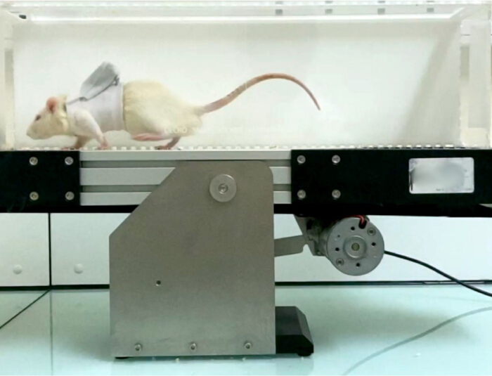
Figure 6: Picture of a Wistar rat exercising (step 6). The rat is exercising on the single-track treadmill (treadmill speed level = 20 cm.s-1) equipped with the telemetry jacket, electrodes, and electronic device. Please click here to view a larger version of this figure.
Data presented in Figure 7A–C have been obtained on four male Wistar rats, weighing between 330 and 340 g and aged 10 weeks upon arrival. The rats were fitted with the telemetry jacket for 5 consecutive days (Day 1 to Day 5). Each session consisted of equipping the animal followed by a 3 h recording in the home cage. Video analysis was manually performed to identify behaviors associated with physiological variations.
The first two habituation sessions induced more pronounced stress, characterized by increased heart rate (HR) (between 400 and 500 bpm) and respiratory rate (RespR) during the first hour post-fitting. These periods of high physiological activation coincided with intense exploratory activity (sniffing, frequent rearing) and grooming as observed via the camera. Conversely, when correctly performed, the 3–5 day habituation protocol described before is expected to attenuate this initial stimulation.
On the third day of habituation, HR and RespR returned to their baseline values more quickly, stabilizing at approximately 350 bpm and 100 brpm respectively, approximately 35 min after fitting.
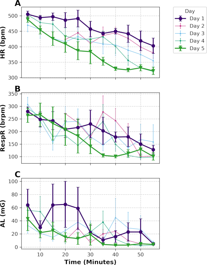
Figure 7: Physiological stabilization over consecutive habituation days (Step 4). Panels show (A) heart rate (HR; bpm), (B) respiratory rate (RespR; brpm), and (C) activity level (AL; mG) during the 1h post-fitting stabilization with the telemetry jacket. Data were collected across 5 consecutive habituation days (n=4; males, independent cohort). Data are represented as mean ± SEM calculated every 5 min. Bold lines highlight the first habituation day (purple circular markers) and the final habituation day (green triangle markers). Please click here to view a larger version of this figure.
The results were obtained in two experimental groups of eight rats that successfully completed the treadmill habituation procedure. These animals were included in two independent experiments conducted by different researchers. In the first group (Group 1; blue lines), all animals managed to run with the jacket up to 30 cm.s-1 speed, with 7 out of 8 animals completing the highest speed of 40 cm.s-1 without reaching the maximum number of electrical shocks. Conversely, in the second group (Group 2; orange lines), 7 out of 8 animals had to be stopped at 20 cm.s-1 because they reached the humane ethical endpoint criterion of 10 electric shocks.
Figure 8 shows individual examples from the two groups. The exercise protocol for Rat 0 was prematurely terminated. Its recovery period began when the treadmill was stopped, which occurred before the animal could complete the 20 cm.s-1 speed stage. Conversely, Rat 1 successfully completed the exercise protocol, demonstrating a gradual physiological adaptation after an initial increase at the onset of the treadmill exercise. This adaptation was characterized by a progressive increase in HR (Figure 8A), RespR (Figure 8B), and AL (Figure 8C) across the different speed stages, with elevated physiological activity sustained until the successful completion of the 40 cm.s-1 stage. Upon stopping the treadmill, the recovery phase began, marked by a decline in HR (Figure 8A), RespR (Figure 8B), and AL (Figure 8C), which progressively returned to baseline levels.
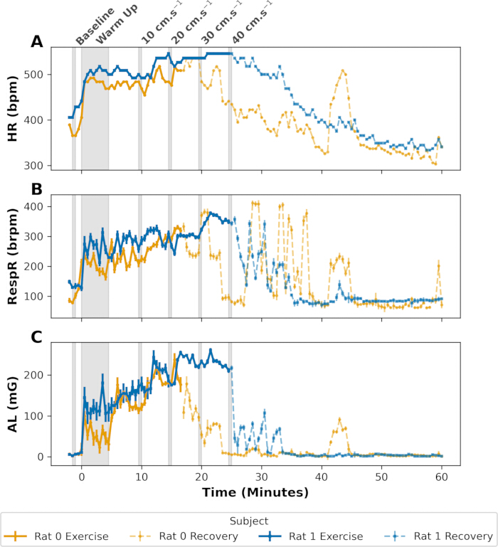
Figure 8: Individual physiological responses during the triangular exercise protocol (Step 6). Panels show (A) heart rate (HR; bpm), (B) respiratory rate (RespR; brpm), and (C) activity level (AL; mG) for two subjects: Rat 1 (male), which completed the maximum speed stage, demonstrating proper training and Rat 0 (male), which ceased activity at the 20 cm.s-1 speed stage. Solid lines indicate periods when the animal was running, while dotted lines represent the recovery period following the cessation of the treadmill activity. The warm-up refers to the 5 min period at 5 cm.s-1 after treadmill onset and before the actual start of the test. Data are represented as mean calculated every 30 s. Please click here to view a larger version of this figure.
As reported in Figure 9, the baseline measurements for group 1 were recorded in the treadmill as: HR at 395±14 bpm, RespR at 177±15 brpm, and AL at 15±2 mG. The monitoring device measured an initial physiological increase as: HR (+26%, +101 bpm) (Figure 9A), RespR (+46%, +82 brpm) (Figure 9B), and AL (+1210%, +189 mG) (Figure 9C) induced by the onset of the treadmill compared to baseline. A physiological increase induced by exercise at 40 cm.s-1 was measured for group 1: HR (+28%, +110 bpm) (Figure 9A), RespR (+71%, +126 brpm) (Figure 9B), and AL (+2839%, +443 mG) (Figure 9C) when compared to BL. Comparable BL values were recorded for the group 2: HR at 399±18 bpm, RespR at 150±15 brpm, and AL at 11±3 mG. The monitoring device measured a physiological increase induced by exercise up to 20 cm.s-1 as: HR (+23%, +90 bpm), RespR (+81%, +122 brpm), and AL (+1555%, +171 mG) when compared to Baseline. Values for higher speeds are not compared for this group given that only one animal reached those speed stages (Figure 9D).
Despite these differences, the two groups showed a large initial increase in HR, RespR, and AL at the onset of the treadmill use followed by a progressive and comparable increase in these parameters as the treadmill speed increased.
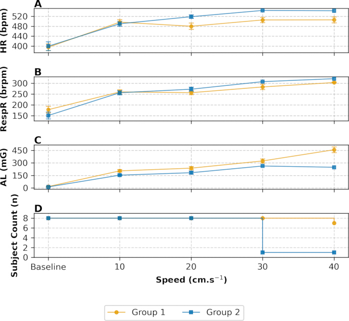
Figure 9: Physiological parameters and recovery profiles across independent experimental groups. Panels show (A) heart rate (HR; bpm), (B) respiratory rate (RespR; brpm), and (C) activity level (AL; mG) during a forced exercise speed stage of the triangular protocol (Step 6) and the subsequent recovery phase. Data are shown for Group 1 (blue; n = 8 males) and Group 2 (orange; n = 8 males), obtained from two independent trials conducted by different experimenters at different times. (D) The number of animals that completed the speed stage. Data are represented as mean ± SEM except for panel D, which represents animal counts. Please click here to view a larger version of this figure.
Discussion
This protocol presents a simple and non-invasive method for monitoring cardiorespiratory system adaptation in rodents during an incremental exercise test (IET)25. This approach is particularly relevant not only for studying exercise physiology, but also for protocols aimed at revealing functional alterations induced by pathological models that may not be apparent at rest2,3.
Although the protocol is designed for easy implementation, special attention must be paid to the selection of the appropriate jacket size. The habituation phases to the jacket and treadmill are the most critical steps. Furthermore, checking signal quality before recording begins is essential. To ensure data reliability, these various points may require adjustment or troubleshooting. These checkpoints are detailed below.
Jacket fit verification
If an animal manages to remove its jacket during the habituation period or appears uncomfortable, re-examine the fit to ensure the correct size has been selected, referring to the manufacturer's sizing recommendations23. If the animal can be equipped with the jacket but doubts remain about the fit, try passing a finger between the jacket and the animal’s shoulder to check that the thoracic bands are not excessively compressing the animal’s body. A finger should pass through easily, and the fabric should remain soft when the jacket is gently stretched. If inserting a finger is difficult or excessive pressure from the fabric is noticed, remove the jacket as the animal is likely being compressed. Conversely, a jacket that is too large will not remain in place for long after fitting and is easy for the animal to remove. Even if an oversized jacket can be fitted, it will tend to shift backward behind the shoulder blades. If the jacket moves even slightly, the animal may place a paw through the outer edge and eventually remove the entire device.
Acclimatization & Jacket fitting
The acclimatization phase is a critical step for minimizing stress on the day of the experiment. Indeed, even non-invasive procedures or routine handling can induce temporary stress responses26,27. The acclimatization period allows both the animals and the experimenters to become familiar with the procedure, and enables the animals to progressively adapt to the additional weight of the telemetry device (approximately 15 g), which represents less than 10% of the body weight of an adult rat within the recommended weight range.
While a five-day habituation period has been employed in this protocol, it represents an indicative duration and can be adjusted as needed. Indeed, the results in Figure 7A–C show that HR, RespR and AL values recorded after 1 h of stabilization plateaued after three days, corroborating earlier studies17,19,21 that successfully utilized a three-day protocol. In any case, the absence of normalization of cardiac and respiratory functions during a habituation session should be considered a strong indicator of animal discomfort, and therefore, insufficient habituation. If such a phenomenon occurs regardless of the fit of the jacket, it is strongly recommended to check the rats' environment and housing conditions to eliminate potential stressors28,29. Furthermore, it is essential to distinguish transient behaviors, such as attempts to gnaw the jackets or secretion of porphyrins (red tears), primarily induced by exposure to an unfamiliar situation26, from persistent reluctance to wear the jacket, which remains exceptional. In the former case, the frequency of these behaviors should decrease as habituation progresses, indicating the animal’s gradual acceptance of the procedure. In the latter case, the reluctance to wear the jacket does not diminish and repeated exposure may constitute negative reinforcement. In the experiments presented in this article, only one of the 16 rats attempted to gnaw the jacket on the first day of acclimatization and this behavior was not observed in subsequent sessions. While all animals ultimately tolerated the jacket, the possibility remains that a specific individual might consistently refuse to wear it, necessitating its exclusion.
Treadmill habituation and triangular protocol
Although the proposed habituation protocol is commonly used30,31; the contrasting outcomes obtained from the two separate experiments reflect the natural variability across individual and cohort responses. Despite undergoing the same standardized habituation, Group 2 reached the ethical shock limit at lower speed stages, suggesting insufficient adaptation to reach the targeted exercise speeds. Notably, the average heart rate was higher in Group 2 than in Group 1 at the 20 cm.s⁻1 stage despite similar baseline values, which may reflect a heightened stress response, lower cardiovascular fitness, or individual variability; however, the limited sample size (n=1 in Group 2 beyond this speed) precludes any definitive interpretation. This underscores the necessity of continuous monitoring during habituation to adjust the protocol dynamically and account for environmental factors or cohort-specific characteristics that drive inter-experiment variability. To ensure adequate preparation, each animal's advancement and number of shocks received over the ten-day period should be tracked throughout the habituation period, allowing for protocol adjustments based on regular reassessments of individual progress toward the target maximum speeds of the incremental test. As a general indication, animals typically require the first two to three sessions to adapt to treadmill running at low speeds (below 10–20 cm.s⁻1), before progressively tolerating higher speeds (20–30 cm.s⁻1) over the intermediate sessions (day 3 to 6). By the final habituation sessions, well-adapted animals should be capable of sustaining the target speed (around 40 cm.s⁻1) before day 10 for the full session duration without reaching the shock limit. A pre-test incremental session is recommended to verify each animal's capability to reach the target speed. This step serves as a final check to allow extension of the habituation period for animals that fail to complete it satisfactorily. Ultimately, it is critical to ensure that habituation sessions remain sub-maximal; they should not constitute physical training or influence performance during the subsequent IET30,31. Rather than representing a methodological failure, the inclusion of both outcomes here is intentional, as it provides users with a realistic illustration of the range of responses that can be expected in practice. This protocol serves a dual purpose: acclimatizing the animals to the equipment and identifying those unable to perform the required exercise. While 'non-runners' are frequently not mentioned in published reports, the literature suggests that more than 10% of rats may fail to complete habituation32. Consequently, these individuals should be identified early and excluded from the study. Given the potential for animal exclusion due to both treadmill non-compliance and jacket refusal, including one or two additional animals per cohort to maintain a statistically robust sample size is recommended.
Signal quality
Another frequent troubleshooting scenario involves addressing poor recorded signal quality (as illustrated in Figure 6, "representative results"). If poor ECG quality persists after 2–3 min (the time required for the gel to establish proper contact between the skin and the electrodes), the following corrective actions can be taken: 1. Check for interference: Ensure that no skin folds, jacket folds or fur regrowth are interfering with electrode contact. 2. Improve conductivity: Gently massage the skin over the electrodes. If necessary, apply additional conductive gel directly to the black dots in the center of the adhesive side of the electrodes. 3. Inspect equipment: Carefully examine the electrodes and wires for any signs of damage, and consider replacing the electrodes if signal quality remains poor or if damage is apparent. If the RIP signal is noisy, first verify the jacket size and the integrity of the internal sewn sensors. If the size is inappropriate or the sensors are damaged, the jacket must be replaced, as this is the only effective solution for restoring signal quality. Overall signal quality may decrease at higher speeds compared to baseline recordings, due to motion-induced mechanical noise manifesting as increased baseline wander and motion artifacts. The influence of these artifacts and the corresponding mitigation strategies has been thoroughly described elsewhere21.
This study uses a rodent-specific jacket for multimodal physiological assessment (cardiac, ventilatory, and actigraphy) during incremental exercise on a treadmill. Previous studies have demonstrated that this device yields results comparable with invasive methods for assessing cardiac16,33,34 and ventilatory15 functions, which are established as the gold standard. However, most of these invasive methods are generally incompatible with exercise protocols performed in freely moving animals. Only the use of implantable telemetry sensors (for a review see35) makes this type of experimentation possible. However, implanting these sensors requires extensive surgery, which may compromise animal welfare through postoperative pain, location of surgical wounds, or discomfort related to the device bulk, and may ultimately alter the animal's phenotype or ability to perform exercise8,9,36. In this context, preserving the animal’s physical integrity during the investigation represents a major advantage of this method. Furthermore, while specialized metabolic treadmills allow for the measurement of oxygen consumption ( ) and carbon dioxide production (
 ), they do not provide direct functional indices of cardiac or respiratory performance. The external telemetry system used in this protocol is compatible with this equipment, addressing this gap by effectively complementing the endpoints accessible through these methodologies. Finally, although treadmill speed is often used as a proxy for mechanical work performed during IET, the integration of an accelerometer enables direct measurement of actigraphy, thereby providing richer data on animal activity and allowing movement analysis beyond simple locomotion speed.
Beyond exercise physiology, this jacketed telemetry system may have broader applications in experimental settings requiring non-surgical ambulatory cardiorespiratory monitoring. Following appropriate acclimatization, prolonged recordings have been implemented up to 48 h33, thereby opening perspectives for circadian or sleep-related physiology studies, as well as longitudinal monitoring protocols. In addition to the exercise protocol presented here, this technology has already been applied in other contexts, including cardiovascular phenotyping in disease models, where ECG-derived parameters and heart rate variability have recently been reported in rat models of ischemic cardiomyopathy20 and of chronic kidney disease-associated cardiovascular dysfunction37, illustrating its applicability beyond acute exercise settings. Such an approach may also be of interest for other preclinical applications involving spontaneous or challenge-induced activity, including seizure or epilepsy models, safety pharmacology studies17,19, and post-surgical recovery monitoring38. Although these applications were not specifically evaluated in the present study, they illustrate the versatility and translational potential of this non-surgical monitoring platform. The current method presents several limitations that require consideration. First, the proposed protocol is generic and designed for healthy animals. Therefore, the fit and tolerance of the jacket may vary depending on animal size, body composition, strain, sex, and physiological condition, potentially limiting its applicability in very small, obese, or diseased animals. Moreover, researchers working with specific models (e.g., obese animals) must adapt the exercise program independently of the measurement system.
Conducting a preliminary test on a small cohort of animals to thoroughly evaluate potential constraints before performing the main experiment is highly recommended. It should also be noted that this protocol was developed using a proprietary jacketed telemetry system. While the general principles described here are broadly applicable, performance characteristics such as signal quality, jacket fit, and data output format may differ from those of other telemetry platforms. Researchers are therefore encouraged to validate their specific system before performing the main experiment. In addition, considering the ethical and societal aspects of preclinical research, the use of a mild aversive stimulus (low-intensity electrical stimulation) to control exercise, as presented here, may be questioned. Despite increasing regulatory scrutiny, this approach remains the most widely used method. As recently reviewed by Okamoto et al.39, it is still the preferred option when precise control of exercise intensity and duration is required. Conversely, when strict control of exercise is not essential, some studies propose replacing electrical stimulation with potentially less aversive stimuli, including compressed air, tactile cues, or noise (for a review see40). Similarly, the use of reward-based paradigms as an alternative to aversive stimuli has been explored for more than a decade41; however, standardized exercise protocols relying solely on positive reinforcement have not yet been established. The potential applications of this method are considerable, spanning both specific research contexts and educational settings. In research settings, this approach is broadly applicable to studies requiring a controlled exercise protocol to assess cardiorespiratory function. Its main applications include: i) Performing exercise tests comparable to those used in humans to accurately evaluate the adaptive capacity of the cardiorespiratory system in healthy and pathological conditions; ii) Standardizing training or rehabilitation protocols across various animal models of experimental disease; iii) Facilitating the longitudinal monitoring of the physiological effects induced by training or rehabilitation over extended periods; iv) detecting physiological alterations, such as those associated with heart failure, that may not be apparent in animals at rest. From a pedagogical perspective, this model offers substantial advantages for university practical training, thereby directly or indirectly adhering to the 3Rs principles4 : “Refinement”, “Reduction” and “Replacement” in the use of animals. It promotes “Refinement” through its completely non-invasive nature, thereby preserving the animal’s physical integrity, unlike traditional methods for studying cardiorespiratory physiology. Furthermore, such a multi-modal approach allows students to study cardiac and respiratory physiology using a single system during one session, providing an integrated illustration of physiological homeostasis. This indirectly contributes to a “Reduction” in animal numbers. In an educational context, this system could also support “Replacement” by enabling the creation of realistic physiological databases to complement practical training and simulation-based learning in physiology education to illustrate a wide range of physiological scenarios42. Indeed, for educational purposes, the troubleshooting scenarios described in this protocol are particularly valuable, as they provide examples derived from real-world experimental conditions in which inter-individual variability and technical challenges can be illustrated. As universities increasingly complement or replace animal use with simulation tools, it is relevant to have access to such examples. Additionally, enriching the physiological recordings with video data obtained during the present protocol can provide students with visual context, including examples of unresponsive animals, thereby enhancing interpretation and learning.
Disclosures
Timothé FLENET and Charles EYNARD are employees and shareholders of ETISENSE SAS. Agathe CAMBIER is an employee of ETISENSE SAS. ETISENSE SAS is the company that is manufacturing the jacketed telemetry system used in this protocol.
Acknowledgements
These results were obtained as part of the LEARN project (23FC3R-001), which was supported by a FC3R “Digital Tools” grant42. This financial support from the GIS FC3R using funds managed by Inserm, supported the development of a digital platform for higher education dedicated to experimental physiology.
Generative AI was utilized for language editing and manuscript optimization to improve clarity and readability. The authors reviewed and edited the output as needed and take full responsibility for the final content of the manuscript.
Materials
| Name | Company | Catalog Number | Comments |
|---|---|---|---|
| Camera fisheye 6 MPX | Reolink, Hong Kong, China | FE-W | Used to observe the animals during habituation. |
| Clipper | Aesculap Schermaschinen, Suhl, Germany | Clipper Aesculap Exacta GT416 | Used for hair removal. An alternate corded or battery-powered clipper with 0.1 mm clipping capability is suitable. |
| Computer with a web browser | Any supplier | N/A | Used to open the telemetry software and record video. Any computer with a recent web browser can be used. |
| Disposable adhesive surface ECG electrodes | ETISENSE, Lyon, France | ELEC-R-1PAIR | Surface Ag/AgCl Electrodes to be inserted beneath the jacket for ECG recording. |
| ECG Contact gel | ASEPT INMED, QUINT-FONSEGRIVES, France | Uni Gel ECG, 251005 | Used to improve contact and adherence of ECG electrodes. Any ECG contact gel can be used. |
| Gas anesthesia station | TEM SEGA, Saint-Médard-en-Jalles, France | Portico veterinary gas anesthesia station with ISOTEC 3 evaporator | Used for animal anesthesia during hair clipping. Any equivalent rodent anesthesia station can be used. |
| Gauze | Any supplier | N/A | Used to dry the clipped area after cleaning. Any sterile or non-sterile absorbent gauze is suitable. |
| Instrumented jacket | ETISENSE, Lyon, France | JKT-RAT-T1 | Telemetry jacket with respiratory sensors used to hold the electrodes and the transmitter in place. Available in different sizes depending on animals weight and strains, refer to manufacturer selection tables for adequate size choice23. |
| Intravenous catheter without needle | TERUMO, Laguna, Philippines | SURFLO 22G 0,85x25 SR+OX2225C1 | Used for precise gel application. Can be reused. Any equivalent syringe and catheter combination can be used. |
| Isoflurane | Virbac, Santé Animale, France | Vetflurane, 401346 | Veterinary formulation of Isoflurane used for gaseous anesthesia during hair clipping (1.5–5% in 20% oxygen and air). Any other equivalent veterinary isoflurane can be used. Use according to local regulations. |
| Jacket fitting-tool | ETISENSE, Lyon, France | FTUB-R-BD | Optional tool, used to facilitate fitting of the jacket onto the animal with minimal restraint. |
| Motorized treadmill | BIOSEB, Vitrolles, France | BX-TM | Single or multi-lane motorized treadmill delivering electric shocks of 0.2 mA. |
| Permanent ink marker | Any supplier | N/A | Used to mark the tail for individual animal identification. Any non-toxic, waterproof permanent marker is suitable. |
| Surface disinfectant | Franklab, Montigny le Bretonneux, France | FB Spray, 10515 | Used to clean the hardware after use. Any alcohol-free surface disinfectant can be used. |
| Syringe without needle (1 or 5 mL) | BD, Pont-de-Claix, France | BD Plastipak,303172 | Used for precise gel application. Can be reused. Any equivalent syringe and catheter combination can be used. |
| Telemetry software | ETISENSE, Lyon, France | LASA software v2.17 or later version | Web-based physiological data acquisition and analysis software. |
| Telemetry system for physiological data acquisition | ETISENSE, Lyon, France | DECRO systems : SYS-NANOACQ4, SYS-S2, SYS-S1 or equivalent | Available in different configurations depending on the number of animals. |
| Telemetry transmitter, and battery pack for physiological data acquisition | ETISENSE, Lyon, France | EMET-HD-ACQ-REVG | Telemetry transmitter recording ECG, Respiratory Signals and 3D acceleration, with a rechargeable battery (12 hours continuous battery life). |
| Video recording software | Reolink, Hong Kong, China | Reolink Client v8.17.6 or later version | Used to record the video. Windows or Mac version available |
References
- Yan Z, et al. Cardiac effects of treadmill running at different intensities in a rat model. Front Physiol. 2021;12:774681.
- Kuczmarski JM, Unrath K, Thomas GD. Exaggerated cardiovascular responses to treadmill running in rats with peripheral arterial insufficiency. Am J Physiol Heart Circ Physiol. 2018;314(1).
- Dellavechia-De-Carvalho C, Rebelo MA, De-Moraes C, Papoti M. Aerobic capacity evaluation of Sprague Dawley rats in treadmill running: comparison between protocols. Braz J Med Biol Res. 2025;58.
- Russell W, Burch R. The principles of humane experimental technique. Methuen & Co. Ltd.; London; 1959.
- Hoymann HG. Lung function measurements in rodents in safety pharmacology studies. Front Pharmacol. 2012;3:156.
- Kramer K, Kinter LB. Evaluation and applications of radiotelemetry in small laboratory animals. Physiol Genomics. 2003;13(3):197-205.
- Brown MB, et al. Novel assessment of haemodynamic kinetics with acute exercise in a rat model of pulmonary arterial hypertension. Exp Physiol. 2015;100(6):742-754.
- Hale E, et al. The rat telemetry assay and venous catheter access buttons for use in cardiovascular safety pharmacology assessments - surgical methods, refinements and colony maintenance. J Pharmacol Toxicol Methods. 2024;127:107517.
- Schuler B, et al. Optimized surgical techniques and postoperative care improve survival rates and permit accurate telemetric recording in exercising mice. BMC Vet Res. 2009;5:28.
- Amrani A-IE, et al. Jacketed external telemetry in the guinea-pig. Toxicol Lett. 2012;211.
- Zeng T, Mott C, Mollicone D, Sanford LD. Automated determination of wakefulness and sleep in rats based on non-invasively acquired measures of movement and respiratory activity. J Neurosci Methods. 2012;204(2):276-287.
- Cotur Y, et al. Bioinspired stretchable transducer for wearable continuous monitoring of respiratory patterns in humans and animals. Adv Mater. 2022;34(33):2203310.
- Chui RW, et al. Assessment of two external telemetry systems (PhysioJacketTM and JETTM) in beagle dogs with telemetry implants. J Pharmacol Toxicol Methods. 2009;60(1):58-68.
- Authier S, et al. Safety pharmacology investigations in toxicology studies: an industry survey. J Pharmacol Toxicol Methods. 2013;68(1):44-51.
- Flenet T, et al. High-resolution respiratory inductive plethysmography in rats: validation in anesthetized conditions. Physiol Meas. 2017;38(7):1362-1372.
- Fontecave-Jallon J, et al. Inductive plethysmography in rats: towards a new standard for longitudinal non-invasive cardiac output monitoring in preclinical studies. Physiol Meas. 2018;39(9):095006.
- Fares R, et al. Non-invasive jacketed telemetry in socially housed rats for a combined assessment of respiratory system, electrocardiogram and activity using the DECRO system. J Pharmacol Toxicol Methods. 2022;117:107195.
- Penard L, et al. Implementation of cardiorespiratory monitoring in the juvenile minipig using a noninvasive jacketed telemetry system. Birth Defects Res. 2021;113(10):806.
- Fares R, Champeroux P. Simultaneous assessment of central nervous and respiratory systems using jacketed telemetry in socially housed rats: application of the 3Rs principles in core battery safety pharmacology studies. J Pharmacol Toxicol Methods. 2023;121:107268.
- Detrait M, et al. Cardiac sympathetic denervation mitigated ischemic cardiomyopathy progression in a rat model of sleep apnea. J Am Heart Assoc. 2025;14(11).
- Tanguy S, et al. Jacketed telemetry in rats: a novel non-invasive method for cardiorespiratory phenotyping during treadmill exercise. Lab Anim. 2025;59(2):215-225.
- Fares RP, Boire A, Eynard C, Flenet T. Simultaneous non-invasive telemetric electrocardiogram and respiratory measurement with a connected jacket (DECRO system) in rats. Research Square. 2020:10.21203/rs.3.pex-860/v1.
- ETISENSE. DECRO - Rat jacket specification and size selection. Lyon: ETISENSE; 2026:https://support.etisense.com/portal/en/kb/articles/rat-table-selection-of-jackets.
- ETISENSE. DECRO - How to start and stop a data acquisition. Lyon: ETISENSE; 2026:https://support.etisense.com/portal/en/kb/articles/make-and-stop-a-data-acquisition.
- Qin F, et al. Maximum oxygen consumption and quantification of exercise intensity in untrained male Wistar rats. Sci Rep. 2020;10(1):11520.
- Mason G, et al. Non-invasively assessing disturbance and stress in laboratory rats by scoring chromodacryorrhoea. Altern Lab Anim. 2004;32(1 Suppl):153-159.
- Delaunois A, Dedoncker P, Hanon E, Guyaux M. Repeated assessment of cardiovascular and respiratory functions using combined telemetry and whole-body plethysmography in the rat. J Pharmacol Toxicol Methods. 2009;60(2):117-129.
- National Research Council Committee on Recognition and Alleviation of Distress in Laboratory Animals. Recognition and alleviation of distress in laboratory animals. National Academies Press; Washington, DC; 2008.
- Conour LA, Murray KA, Brown MJ. Preparation of animals for research--issues to consider for rodents and rabbits. ILAR J. 2006;47(4):283-293.
- Reboul C, et al. Cardiac remodeling and functional adaptations consecutive to altitude training in rats: implications for sea level aerobic performance. J Appl Physiol. 2005;98(1):83-92.
- Goret L, et al. Training does not affect the alteration in pulmonary artery vasoreactivity in pulmonary hypertensive rats. Eur J Pharmacol. 2005;527(1-3):121-128.
- Garrigos D, et al. A handful of details to ensure the experimental reproducibility on the forced running wheel in rodents: a systematic review. Front Endocrinol. 2021;12:638261.
- Scorrano F, Stuber N, Dreessen K, Cordier V. Evaluation of a new telemetry jacket system for rats. Lab Anim. 2022;56(1):155.
- Fontana-Pires L, et al. Continuous non-invasive extraction of hemodynamic variables from thoracocardiographic signals using the ensemble averaging technique: validation in anesthetized rats without ventilatory support. Physiol Meas. 2025;46(12):125002.
- Laske TG, Garshelis DL, Iles TL, Iaizzo PA. An engineering perspective on the development and evolution of implantable cardiac monitors in free-living animals. Philos Trans R Soc Lond B Biol Sci. 2021;376(1830):20200217.
- Helwig BG, Ward JA, Blaha MD, Leon LR. Effect of intraperitoneal radiotelemetry instrumentation on voluntary wheel running and surgical recovery in mice. J Am Assoc Lab Anim Sci. 2012;51(5):600-608.
- Soulie M, et al. Altered ECG parameters with loss of cardiac variability and prolonged QTc in a non-diabetic chronic kidney disease rat model. Arch Cardiovasc Dis. 2024;117(6-7).
- Cambier A, et al. Refining the implementation of surgical protocols: benefits of non-invasive cardiorespiratory monitoring in rat. Lab Anim. 2022;56(1):152.
- Okamoto M, Hata T, Hiraga T, Soya H. Treadmill running model for rodents as a translational approach based on human physiological responses. In: Soya H, editor. Exercise Brain Stimulation for Cognitive Function and Mental Health. Singapore: Springer Nature Singapore; 2025.
- Poole DC, et al. Guidelines for animal exercise and training protocols for cardiovascular studies. Am J Physiol Heart Circ Physiol. 2020;318(5).
- Fantegrossi WE, Xiao WR, Zimmerman SM. Novel technology for modulating locomotor activity as an operant response in the mouse: implications for neuroscience studies involving exercise in rodents. J Neurosci Methods. 2013;212(2):338-343.
- FC3R. Digital tools - Call for project FC3R - LEARN (L3Rn) (23FC3R-001): development of an innovative platform for teaching experimental physiology in higher education. France: FC3R; 2024:https://www.fc3r.com/en/digital-tools-3-2023.html.
Reprints and Permissions
This article has been published
Video Coming Soon