This protocol standardizes fixation, staining, and confocal imaging methods for visualizing tunneling nanotubes and mitochondrial transfer between astrocytes and neurons in vitro.
Method Article
Standardized Immunofluorescence Imaging of Tunneling Nanotubes and Mitochondrial Transfer Between Astrocytes and Neurons
In This Article
Summary
Abstract
Mitochondria are essential organelles that regulate energy production, cellular signaling, and metabolic homeostasis in neural cells. Tunneling nanotubes (TNTs) are thin membranous structures that mediate long-distance intercellular communication and facilitate the transfer of cellular components, including mitochondria, between connected cells. Reliable visualization of TNTs and mitochondrial transfer requires careful sample handling because these structures are highly fragile and sensitive to fixation, washing, and imaging conditions. This protocol describes standardized procedures for the fixation, staining, and confocal imaging of TNTs in astrocytes and astrocyte–neuron coculture systems. The workflow includes membrane and cytoskeletal staining for TNT visualization, mitochondrial labeling for tracking mitochondrial localization, and immunofluorescence staining for Miro1 colocalization analysis. Critical steps for preserving TNT morphology, including gentle washing and light-protected handling, are emphasized throughout the procedure. The protocol also outlines imaging approaches for the characterization of TNTs and mitochondria in fixed-cell preparations. These methods provide a reproducible experimental framework for studying TNT formation and mitochondrial transfer between neural cells in vitro.
Introduction
Mitochondria serve as the energy factories of the cell, providing the energy required for cellular activities through the production of adenosine triphosphate while also playing important roles in cellular signal transduction and redox balance1. In the nervous system, mitochondrial metabolites can increase the complexity and excitability of neuronal processes, thereby supporting neuronal growth and development2. Mitochondria are not static organelles; they continuously alter their morphology, location, and distribution to adapt to various intracellular and extracellular stresses and metabolic demands3. Intercellular mitochondrial transfer contributes to the maintenance of tissue homeostasis and promotes development under physiological conditions, while under pathological conditions, it participates in damage repair, immune regulation, and disease progression4,5.
Intercellular communication of materials and energy is essential for various biological processes, including cellular homeostasis, tissue repair, and neural network regulation. Current studies have revealed multiple modes of intercellular communication, among which tunneling nanotubes (TNTs) represent one of the most extensively studied mechanisms associated with intercellular mitochondrial transfer6. TNTs are F-actin-based, bridge-like membrane structures with a tubular morphology, ranging from 50 to 200 nm in diameter and extending up to several cell diameters in length7. Research has shown that TNTs can serve as physical connections between neural cells and are closely associated with the onset and progression of neurodegenerative diseases, brain cancer, and other disorders. For instance, mitochondrial transfer from microglia to neurons via TNTs has been reported to rescue neuronal cells8,9.
Current research on TNTs commonly faces several challenges, including the structural fragility of TNTs, which makes them prone to breakage and may consequently affect experimental outcomes; difficulty distinguishing TNTs from filopodia or other membrane protrusions; and challenges associated with accurately quantifying intercellular TNTs. To improve the reproducibility and consistency of TNT-related studies, standardized methods for the washing, fixation, staining, and imaging of TNTs are required to enable accurate assessment of their structure and distribution while minimizing experimental variability. Factors such as cell density prior to fixation, protection from light, washing intensity, fixation duration, and post-fixation staining conditions can all influence the structure and stability of TNTs. In this study, we describe procedures for the pretreatment, staining, and confocal imaging of TNTs and mitochondria-associated TNT structures in astrocyte and astrocyte-neuron culture systems.
This research protocol can be generally applied to most laboratory settings. Validation in primary astrocytes and a mouse hippocampal neuronal cell line confirmed that the protocol is suitable for both primary neural cells and immortalized cell lines. However, this protocol still has several limitations. Confocal microscopy provides lower spatial resolution than super-resolution imaging approaches for TNT visualization, and MitoTracker-based assessment of mitochondrial transfer may be affected by dye leakage and non-mitochondrial signal interference10.
Protocol
This study did not involve human subjects or live vertebrate animals; only commercially obtained or established cell lines were used, and no ethical approval was required.
1. Staining for TNTs in Astrocytes
- Cell Seeding
- Seed isolated primary rat astrocytes at passage 3 (P3) into 35 mm glass-bottom culture dishes at a density of 5 × 10⁴ cells per dish. Add 2 mL of Dulbecco’s Modified Eagle Medium/Nutrient Mixture F-12 (DMEM/F-12) complete medium supplemented with 10% fetal bovine serum (FBS) and 1% triple antibiotic mixture (penicillin G, streptomycin, and amphotericin B). Culture the cells at 37°C in a humidified incubator containing 5% CO₂.
- Incubate the cells for approximately 24 h until attachment occurs.
- Pretreatment and Fixation
- Transfer the culture dishes to a clean bench under light-protected conditions after cell attachment.
NOTE: Minimize unnecessary movement and vibration during handling to preserve TNT integrity. - Remove the culture medium gently under dim light conditions.
NOTE: Avoid complete drying of the cell surface during medium removal because TNTs may collapse and attach to the culture substrate. - Gently wash the cells once with 500 µL of phosphate-buffered saline (PBS) without calcium and magnesium ions (pH 7.3–7.5). Add and remove the PBS slowly along the wall of the culture dish to avoid disrupting intercellular TNTs during washing.
- Remove the PBS carefully.
- Fix the cells with a commercial 4% tissue cell fixative solution containing paraformaldehyde (pH 7.2–7.4) for 30 min at room temperature (RT; 20°C–25°C) under dim light conditions.
CAUTION: Handle fixative solutions using appropriate personal protective equipment and perform all procedures in accordance with institutional chemical safety guidelines. - Remove the fixative solution.
- Wash the cells once with PBS.
- Transfer the culture dishes to a clean bench under light-protected conditions after cell attachment.
- Cell Permeabilization and Fluorescent Staining
- Permeabilize the cells with 500 µL of ready-to-use Triton X-100 permeabilization solution for 20 min at RT.
CAUTION: Triton X-100 may disrupt fragile TNT structures. Minimize excessive agitation during permeabilization. - Remove the permeabilization solution.
- Wash the cells once with PBS.
- Prepare the staining solution immediately before use. Dilute the F-actin staining dye in PBS to a final concentration of 1 µM and use 4′,6-diamidino-2-phenylindole (DAPI) as a ready-to-use nuclear stain. Mix the two staining solutions thoroughly before use.
NOTE: Phalloidin-based staining reagents may be used as alternatives for F-actin visualization. - Mix the F-actin and nuclear staining solutions at a 1:1 ratio (equal volumes) under light-protected conditions.
- Add the prepared staining solution to the culture dishes.
- Incubate the cells for 30 min at 37°C in the dark.
- Remove the staining solution.
- Wash the cells three times with PBS using 500 µL per wash for 1 min each wash.
- Add 1 mL of PBS to prevent the cells from drying during imaging preparation.
- Permeabilize the cells with 500 µL of ready-to-use Triton X-100 permeabilization solution for 20 min at RT.
- Confocal Imaging
- Acquire images using a Nikon inverted confocal microscope equipped with a ×20 objective lens. Use 405 nm excitation for DAPI and 488 nm excitation for F-actin staining. Capture images at a resolution of 1024 × 1024 pixels under light-protected conditions at RT.
- Perform qualitative assessment and manual quantification using at least five randomly selected fields of view per sample. Quantify the number of TNTs and the number of TNT-positive cells containing one or more TNTs in each field of view (Figure 1A and 1B).
NOTE: Acquire images immediately after staining to minimize fluorescence loss and structural disruption of TNTs.
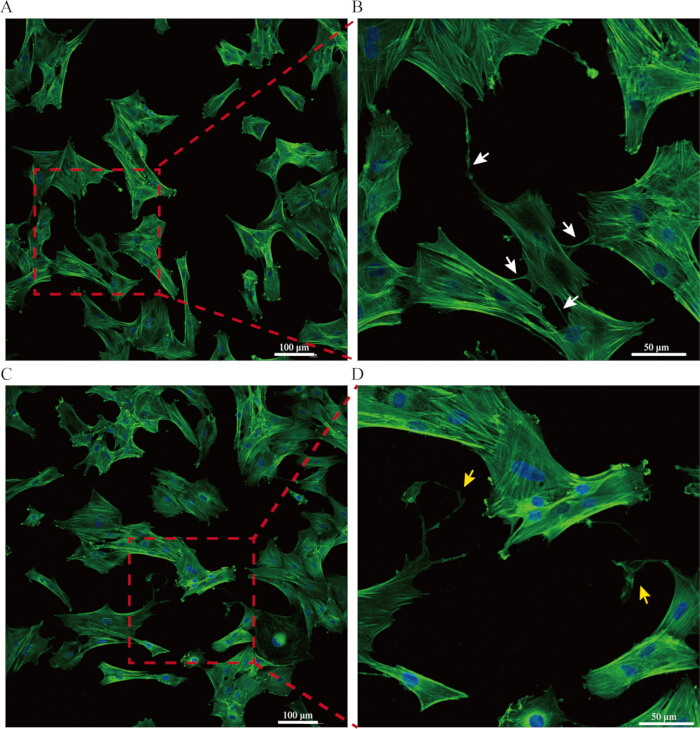
Figure 1: Fluorescence staining of tunneling nanotubes (TNTs) in astrocytes. (A) Representative confocal microscopy images showing nuclear and F-actin staining in astrocytes cultured in glass-bottom culture dishes. (B) Enlarged view of the boxed region in panel A. White arrows indicate TNT-like structures between adjacent cells. (C) Representative images showing TNT-like structures disrupted following improper sample handling. (D) Enlarged view of the boxed region in panel C. Yellow arrows indicate disrupted TNT-like structures. Please click here to view a larger version of this figure.
2. Staining for Mitochondrial Transfer from Astrocytes to Neurons via TNTs
- Astrocyte Seeding and Mitochondrial Labeling
- Seed isolated primary rat astrocytes at passage 3 (P3) into 35 mm glass-bottom culture dishes at a density of 3 × 10⁴ cells per dish. Add 2 mL of DMEM/F-12 complete medium supplemented with 10% FBS and 1% triple antibiotic mixture (penicillin G, streptomycin, and amphotericin B). Culture the cells at 37°C in a humidified incubator containing 5% CO₂.
- Incubate the cells for approximately 24 h until attachment occurs.
- Transfer the culture dishes to a clean bench under dim light conditions after cell attachment.
NOTE: Minimize unnecessary movement and vibration during handling to preserve TNT integrity. - Remove the culture medium gently under light-protected conditions. Avoid disrupting TNTs during medium removal.
NOTE: Avoid complete drying of the cell surface during medium removal because TNTs may collapse and attach to the culture substrate. - Reconstitute 50 µg of lyophilized MitoTracker dye in dimethyl sulfoxide (DMSO) to prepare a 1 mM stock solution and store the stock solution at −20°C. Before use, dilute the stock solution in 400 µL of complete DMEM/F-12 medium supplemented with 10% FBS and 1% triple antibiotic mixture to obtain a final MitoTracker concentration of 50 µM.
CAUTION: Protect fluorescent dyes from direct light exposure during preparation and incubation procedures. - Add the mitochondrial staining working solution to the culture dishes.
- Incubate the cells at 37°C in a humidified incubator containing 5% CO₂ for 30 min under light-protected conditions.
- Remove the mitochondrial staining solution under light-protected conditions.
- Gently wash the cells once with 500 µL of PBS without calcium and magnesium ions (pH 7.3–7.5). Add and remove the PBS slowly along the wall of the culture dish to avoid disrupting intercellular TNTs during washing.
- Remove the PBS carefully.
- Astrocyte-Neuron Coculture
- Seed immortalized mouse hippocampal neuronal cells at passage 3 (P3) into the astrocyte-containing culture dishes at a density of 3 × 10⁴ cells per dish. Add cells in a total volume of 2 mL consisting of a 1:1 mixture of DMEM and DMEM/F-12 complete medium supplemented with 10% FBS and 1% triple antibiotic mixture. Establish the coculture using a direct-contact method without a Transwell insert. Culture the cells at 37°C in a humidified incubator containing 5% CO₂.
NOTE: Add neuronal cells gently to minimize disruption of pre-existing TNT structures. - Maintain the coculture for 24 h under standard incubator conditions while protecting samples from unnecessary light exposure during handling procedures.
- Seed immortalized mouse hippocampal neuronal cells at passage 3 (P3) into the astrocyte-containing culture dishes at a density of 3 × 10⁴ cells per dish. Add cells in a total volume of 2 mL consisting of a 1:1 mixture of DMEM and DMEM/F-12 complete medium supplemented with 10% FBS and 1% triple antibiotic mixture. Establish the coculture using a direct-contact method without a Transwell insert. Culture the cells at 37°C in a humidified incubator containing 5% CO₂.
- Fixation and Permeabilization
- Remove the culture medium under light-protected conditions.
- Wash the cells once with PBS.
- Fix the cells with 4% tissue cell fixative solution containing paraformaldehyde for 30 min at RT under dim light conditions.
CAUTION: Handle fixative solutions using appropriate personal protective equipment and perform all procedures in accordance with institutional chemical safety guidelines. - Remove the fixative solution.
- Wash the cells once with PBS.
- Permeabilize the cells with 500 µL of ready-to-use Triton X-100 permeabilization solution for 20 min at RT under dim light conditions.
CAUTION: Triton X-100 may disrupt fragile TNT structures. Minimize excessive agitation during permeabilization. - Remove the permeabilization solution.
- Wash the cells once with PBS.
- Fluorescent Staining and Imaging
- Prepare the staining solution immediately before use. Dilute the F-actin staining dye in PBS to a final concentration of 1 µM and use DAPI as a ready-to-use nuclear stain. Mix the staining solutions thoroughly before use.
NOTE: Phalloidin-based staining reagents may be used as alternatives for F-actin visualization. - Mix the F-actin and nuclear staining solutions at a 1:1 ratio (equal volumes) under light-protected conditions immediately before use.
- Add the prepared staining solution to the culture dishes.
- Incubate the cells for 30 min at 37°C in the dark.
- Remove the staining solution.
- Wash the cells three times with PBS using 500 µL per wash for 1 min each wash.
- Add 1 mL of PBS to prevent the cells from drying during imaging preparation.
- Acquire images using a Nikon inverted confocal microscope equipped with a ×20 objective lens. Use 405 nm excitation for DAPI, 488 nm excitation for F-actin staining, and 665 nm excitation for MitoTracker staining. Capture images at a resolution of 1024 × 1024 pixels under light-protected conditions at RT.
- Perform qualitative assessment using at least five randomly selected fields of view per sample. Evaluate the presence of TNT-like structures and mitochondria-associated fluorescence signals within intercellular membrane connections (Figure 2A and 2B).
NOTE: Acquire images immediately after staining to minimize fluorescence loss and structural disruption of TNTs.
- Prepare the staining solution immediately before use. Dilute the F-actin staining dye in PBS to a final concentration of 1 µM and use DAPI as a ready-to-use nuclear stain. Mix the staining solutions thoroughly before use.
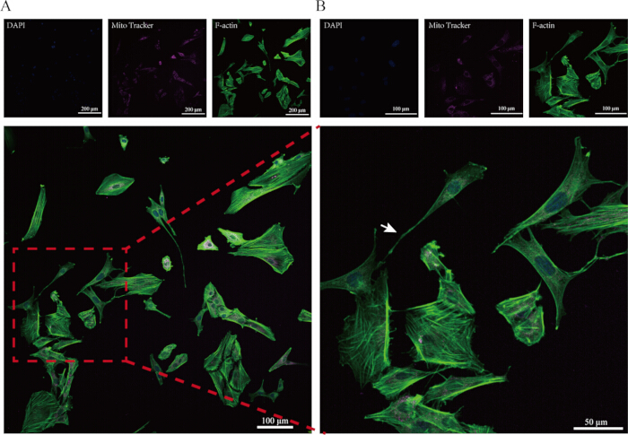
Figure 2: Fluorescence staining of mitochondria and TNTs in an astrocyte-neuron coculture system. (A) Representative confocal microscopy images showing nuclear, mitochondrial, and F-actin staining in astrocytes and neurons cultured in glass-bottom culture dishes. Astrocytes exhibited irregular star-like or polygonal morphologies with extended cellular processes, whereas neuronal cells appeared round or oval with slender neurite-like extensions. (B) Enlarged view of the boxed region in panel A. White arrows indicate mitochondria-associated TNT-like structures between cells. The two cell populations were distinguished primarily based on their characteristic cellular morphology. Please click here to view a larger version of this figure.
3. Immunofluorescence Colocalization Staining for Mitochondria and Miro1 in TNTs
- Astrocyte Seeding and Mitochondrial Labeling
- Seed isolated primary rat astrocytes at passage 3 (P3) into 35 mm glass-bottom culture dishes at a density of 3 × 10⁴ cells per dish. Add 2 mL of DMEM/F-12 complete medium supplemented with 10% FBS and 1% triple antibiotic mixture (penicillin G, streptomycin, and amphotericin B). Culture the cells at 37°C in a humidified incubator containing 5% CO₂.
- Incubate the cells for approximately 24 h until attachment occurs.
- Transfer the culture dishes to a clean bench under dim light conditions after cell attachment.
NOTE: Minimize unnecessary movement and vibration during handling to preserve TNT integrity. - Remove the culture medium gently under light-protected conditions. Avoid disrupting TNTs during medium removal.
NOTE: Avoid complete drying of the cell surface during medium removal because TNTs may collapse and attach to the culture substrate. - Reconstitute 50 µg of lyophilized MitoTracker dye in DMSO to prepare a 1 mM stock solution and store the stock solution at −20°C. Before use, dilute the stock solution in 400 µL of complete DMEM/F-12 medium supplemented with 10% FBS and 1% triple antibiotic mixture to obtain a final MitoTracker concentration of 50 µM.
CAUTION: Protect fluorescent dyes from direct light exposure during preparation and incubation procedures. - Add the mitochondrial staining working solution to the culture dishes.
- Incubate the cells at 37°C in a humidified incubator containing 5% CO₂ for 30 min under light-protected conditions.
- Remove the mitochondrial staining solution under light-protected conditions.
- Gently wash the cells once with 500 µL of PBS without calcium and magnesium ions (pH 7.3–7.5). Add and remove the PBS slowly along the wall of the culture dish to avoid disrupting intercellular TNTs during washing.
- Remove the PBS carefully.
- Astrocyte-Neuron Coculture
- Seed immortalized mouse hippocampal neuronal cells at passage 3 (P3) into the astrocyte-containing culture dishes at a density of 3 × 10⁴ cells per dish. Add cells in a total volume of 2 mL consisting of a 1:1 mixture of DMEM and DMEM/F-12 complete medium supplemented with 10% FBS and 1% triple antibiotic mixture. Establish the coculture using a direct-contact method without a Transwell insert. Culture the cells at 37°C in a humidified incubator containing 5% CO₂.
NOTE: Add neuronal cells gently to minimize disruption of pre-existing TNT structures. - Maintain the coculture for 24 h under standard incubator conditions while protecting samples from unnecessary light exposure during handling procedures.
- Seed immortalized mouse hippocampal neuronal cells at passage 3 (P3) into the astrocyte-containing culture dishes at a density of 3 × 10⁴ cells per dish. Add cells in a total volume of 2 mL consisting of a 1:1 mixture of DMEM and DMEM/F-12 complete medium supplemented with 10% FBS and 1% triple antibiotic mixture. Establish the coculture using a direct-contact method without a Transwell insert. Culture the cells at 37°C in a humidified incubator containing 5% CO₂.
- Fixation and Permeabilization
- Remove the culture medium under light-protected conditions.
- Wash the cells once with PBS.
- Fix the cells with a commercial 4% tissue cell fixative solution containing paraformaldehyde (pH 7.2–7.4) for 30 min at RT under dim light conditions.
CAUTION: Handle fixative solutions using appropriate personal protective equipment and perform all procedures in accordance with institutional chemical safety guidelines. - Remove the fixative solution.
- Wash the cells once with PBS.
- Permeabilize the cells with 500 µL of ready-to-use Triton X-100 permeabilization solution for 20 min at RT under dim light conditions.
CAUTION: Triton X-100 may disrupt fragile TNT structures. Minimize excessive agitation during permeabilization. - Remove the permeabilization solution.
- Wash the cells once with PBS.
- Blocking and Immunofluorescence Staining
- Block the cells with 5% goat serum for 1 h at RT under dim light conditions. Prepare the blocking solution by diluting goat serum 1:20 in PBS.
- Remove the blocking solution.
- Dilute rabbit monoclonal anti-Miro1 antibody in PBS at a dilution ratio of 1:100. Add the diluted primary antibody solution to the culture dishes and incubate overnight (12–16 h) at 4°C.
NOTE: Protect fluorescently labeled samples from direct light exposure throughout the staining procedure. - Remove the primary antibody solution.
- Wash the cells three times with PBS using 500 µL per wash for 1 min each wash.
- Dilute the fluorescent goat anti-rabbit IgG secondary antibody in PBS at a dilution ratio of 1:100. Add 500 µL of diluted secondary antibody solution to each culture dish and incubate for 1 h under light-protected conditions.
NOTE: A no-primary-antibody control was not included in the present study. Future experiments should incorporate appropriate immunofluorescence controls to further validate staining specificity. - Remove the fluorescent secondary antibody solution.
- Fluorescent Counterstaining and Imaging
- Prepare the staining solution immediately before use. Dilute the F-actin staining dye in PBS to a final concentration of 1 µM and use DAPI as a ready-to-use nuclear stain. Mix the staining solutions thoroughly before use.
NOTE: Phalloidin-based staining reagents may be used as alternatives for F-actin visualization. - Add the staining solution to the culture dishes.
- Incubate the cells for 30 min at 37°C in the dark.
- Remove the staining solution.
- Wash the cells three times with PBS using 500 µL per wash for 1 min each wash.
- Add 1 mL of PBS to prevent the cells from drying during imaging preparation.
- Acquire images using a Nikon inverted confocal microscope equipped with a ×20 objective lens. Collect Z-stack images using a step size of 5 µm over a total imaging range of 40 µm (8 optical sections). Use 405 nm excitation for DAPI, 488 nm excitation for F-actin staining, 594 nm excitation for Miro1 immunostaining, and 665 nm excitation for MitoTracker staining. Capture images at a resolution of 1024 × 1024 pixels under light-protected conditions at RT.
- Perform qualitative assessment using at least five randomly selected fields of view per sample. Evaluate TNT-like structures, mitochondria-associated fluorescence signals, and apparent colocalization between Miro1 and mitochondria-associated staining within intercellular membrane connections (Figure 3A–3C).
NOTE: Acquire images immediately after staining to minimize fluorescence loss and structural disruption of TNTs.
- Prepare the staining solution immediately before use. Dilute the F-actin staining dye in PBS to a final concentration of 1 µM and use DAPI as a ready-to-use nuclear stain. Mix the staining solutions thoroughly before use.
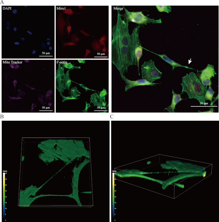
Figure 3: Colocalization staining of mitochondria and Miro1 in TNTs in an astrocyte-neuron coculture system. (A) Representative confocal microscopy images showing nuclear, mitochondrial, Miro1, and F-actin staining in astrocytes and neurons cultured in glass-bottom culture dishes. White arrows indicate regions of apparent colocalization between mitochondria-associated signals and Miro1 within TNT-like structures. (B) Three-dimensional reconstructed Z-stack image showing TNT-like structures formed between adjacent cells. (C) Side-view reconstruction of the same Z-stack image showing TNT-like structures extending between adjacent cells. Please click here to view a larger version of this figure.
Results
In astrocyte monoculture preparations, thin membrane protrusions connecting adjacent cells were observed following F-actin and nuclear staining (Figure 1A and 1B). Multiple TNT-like structures were detected extending between neighboring astrocytes, and individual astrocytes frequently formed connections with several surrounding cells. Successful preservation of TNT-like structures was associated with gentle handling during washing and fixation procedures. Excessive mechanical disturbance during medium removal or washing resulted in reduced detectability and structural disruption of these membrane connections, as shown in disrupted TNT-like structures following improper sample handling (Figure 1C and 1D).
In astrocyte-neuron coculture preparations, mitochondria-associated fluorescence signals were observed within TNT-like structures connecting adjacent cells following MitoTracker staining and confocal imaging (Figure 2A and 2B). These structures were visualized together with F-actin and nuclear staining to support morphological assessment of TNT-like connections. Proper light protection and minimized washing intensity improved preservation of fluorescence signal and TNT morphology during imaging preparation.
In immunofluorescence colocalization experiments, overlapping fluorescence signals corresponding to mitochondria-associated staining and Miro1 immunostaining were observed within TNT-like structures in astrocyte-neuron coculture preparations (Figure 3A). These observations suggest a potential association between Miro1 and mitochondria localized within TNT-like structures. Three-dimensional reconstructed Z-stack imaging further supported the presence of TNT-like structures extending between adjacent cells (Figure 3B and 3C). TNT preservation, fluorescence signal intensity, and image quality were influenced by fixation, permeabilization, and imaging conditions.
Discussion
The first critical step in the experimental protocol described above is the light-protected pretreatment before fixation. TNT-like structures are highly fragile, and maintaining light-protected conditions after removal from the incubator is important for preserving fluorescence signal intensity and structural integrity during sample preparation. The second critical step is to perform PBS washing with gentle manipulation to minimize disruption of TNT-like structures caused by fluid flow. Adherence to these key handling procedures helps preserve the morphology and stability of TNT-like structures and improves the consistency of subsequent imaging results. Excessive aspiration, prolonged washing, or strong fluid agitation may lead to collapse or fragmentation of TNT-like structures during sample processing. Due to their highly dynamic nature, TNTs are sensitive to light. Phototoxicity induced by light exposure can cause TNT disruption, making these structures difficult to visualize11. Therefore, light-protected conditions should be maintained during TNT-related experiments.
Intracellular mitochondrial movement contributes to cellular energy distribution, particularly in neurons. Studies have shown that mitochondria can move along actin filaments and microtubules12. Recent studies have also demonstrated that TNTs can function as long-distance intercellular communication structures associated with mitochondrial transfer between neural cells13. The methodological approach described in this study enables visualization of TNT-like structures and mitochondria-associated fluorescence signals in neural cell culture systems. Previous studies have shown that intercellular mitochondrial transfer between neural cells may contribute to tissue repair and neuroprotective processes in neurological disorders such as Parkinson’s disease and cerebral ischemic injury14,15. Using the methods described in this study, TNT-like structures were observed between astrocytes and between astrocytes and neurons, together with mitochondria-associated fluorescence signals localized within these structures.
As noted in previous studies10, MitoTracker staining is subject to dye leakage and non-mitochondrial signal transfer, which may reduce data reliability. To address this limitation, future investigations should employ more robust methods, such as genetically encoded mitochondrial labeling, to obtain more conclusive results. Under the current experimental conditions, the two cell types were distinguished primarily based on cellular morphology, an approach that may reduce cell-type identification specificity. Future studies should incorporate cell type-specific fluorescent labeling to improve the accuracy of coculture analysis. Miro1 is an adaptor protein localized to the outer mitochondrial membrane and has been implicated in mitochondrial transport processes in neurons16,17. Using the immunofluorescence colocalization protocol described in this study, overlapping fluorescence signals corresponding to mitochondria-associated staining and Miro1 immunostaining were observed within TNT-like structures. These observations suggest a potential association between Miro1 and mitochondria localized within TNT-like structures. However, the spatial resolution of conventional confocal microscopy may limit definitive assessment of molecular colocalization within thin membrane protrusions.
We performed Z-stack imaging to confirm the presence of TNT-like structures between cells, thereby supporting structural validation of these membrane connections. While the present protocol is suitable for most experimental environments, confocal imaging of fixed cells may result in loss of fragile TNT-like structures during sample processing compared with live-cell imaging, thereby limiting accurate TNT quantification18. Furthermore, imaging of fixed cells does not allow unambiguous determination of mitochondrial transfer directionality, which warrants further investigation in future studies. This protocol provides a standardized experimental workflow for staining TNT-like structures, mitochondria-associated signals, and related proteins in neural cell culture systems. Compared with uncontrolled washing and fixation procedures, the protocol emphasizes gentle sample handling and fluorescence preservation during imaging preparation. These methods may help improve experimental consistency and reproducibility in TNT-related imaging studies.
In summary, this study provides a practical and broadly applicable laboratory protocol for the qualitative assessment of TNTs and investigation of mitochondrial transport via TNT-like structures. However, sample processing steps such as fixation and washing may introduce quantification variability compared with live-cell imaging approaches. Furthermore, confocal microscopy provides lower spatial resolution than super-resolution microscopy, which limits detailed structural characterization of TNTs. In addition, MitoTracker-based mitochondrial staining is affected by dye leakage and nonspecific signal transfer, which should be addressed in future studies to improve experimental accuracy.
Disclosures
The authors declare that they have no competing financial interests.
Acknowledgements
This study was funded by the National Natural Science Foundation of China (82260650); Natural Science Foundation of Inner Mongolia Autonomous Region Project (2025MS08067); The College Students' Innovation and Entrepreneurship Training Program of Baotou Medical College (S202510130008); Baotou Medical College Flower Bud Program Project (HLJH202529) and Baotou Medical College Research Projects for Cultivation of Public Health and Preventive Medicine First-class Discipline (GWGG-20250807).
Materials
| Name | Company | Catalog Number | Comments |
|---|---|---|---|
| 1% triple antibiotic mixture | Biosharp | BL142A | Prevents bacterial and fungal contamination during cell culture |
| 4% Tissue Cell Fixative | Solarbio | P1110 | Cell fixation reagent |
| Anti-Miro1 antibody | Abcam | AB319154 | Primary antibody against Miro1 |
| Astrocytes | Laboratory-isolated primary cells | NA | Astrocytes were isolated from the cerebral cortex of neonatal SD rat pups aged 1–3 days. The purity of the isolated cells was confirmed to be >95% by GFAP staining, and passage 3 (P3) cells were used for all experiments |
| Astrocyte cell culture medium medium (DMEM/F-12) | Gibco | 6125011 | DMEM and F-12 were mixed at a 1:1 ratio (v/v), supplemented with 10% fetal bovine serum (FBS) and 1% triple antibiotics |
| Confocal microscope | Nikon AX | ECLIPSE TI2-E | Images were acquired using a Nikon AX confocal microscope (Nikon Corporation, Japan) controlled by NIS-Elements (version 5.4) |
| DAPI | Servicebio | G1012-100ML | Nuclear staining dye |
| DMSO | BioFroxx | 1084ML100 | Solvent used for preparation of fluorescent dye stock solutions |
| F-actin staining dye | Thermo Fisher Scientific | A57243 | F-actin staining reagent for visualization of cytoskeletal structures and tunneling nanotubes |
| Fetal bovine serum | ExCell Bio | FSD500 | Provides essential nutrients for cell culture |
| Fluorescent secondary antibody (Goat Anti-Rabbit IgG) | ABclonal | AS039 | Secondary antibody for immunofluorescence staining |
| Glass-bottom cell culture dish | NEST | 801001 | Imaging dish for confocal microscopy |
| Goat serum | Solarbio | 210C051 | Blocking reagent for immunofluorescence staining |
| MitoTracker Deep Red | Thermo Fisher Scientific | M22426 | Mitochondrial staining dye |
| Neurons | Cell Bank of CAS | HT22-GNM47 | Immortalized mouse hippocampal neuronal cells were maintained in DMEM complete medium. Cells at passage 3 (P3) were collected and used for subsequent experiments. |
| Neuron cell culture medium (DMEM) | Servicebio | G4511-500ML | DMEM supplemented with GlutaPlus and sodium pyruvate, without HEPES, supplemented with 10% fetal bovine serum (FBS) and 1% triple antibiotics |
| Phosphate-buffered saline (PBS) | Servicebio | G4203-500ML | Washing buffer used during fixation and staining |
| Triton X-100 | Solarbio | P1080 | Cell permeabilization reagent |