This case details a robot-assisted radical prostatectomy performed on a renal transplant recipient and provides a step-by-step guide to safely replicate the procedure in controlled settings, minimizing the risk of injury to the transplant or the patient.
Case Report
The Surgical Management of Prostate Cancer in Renal Transplant Recipients: The Minimally Invasive Approach
In This Article
Summary
Abstract
Patients with a renal transplant present unique anatomical challenges during pelvic surgery. The graft is typically located in the iliac fossa with vascular anastomoses to the iliac vessels and a short ureter implanted into the bladder. These features increase the risk of graft injury during radical prostatectomy, especially when lymph node dissection is planned. This case describes the experience in one of the few cases performed in our department. A 66-year-old man with localized prostate cancer (Gleason 3 + 4 = 7a) and a functioning renal transplant in the right iliac fossa. His history included end-stage renal disease due to IgA nephropathy (retroperitoneal transplants in 1990 on the left side and 2009 on the right side), coronary artery disease with stenting, and atrial fibrillation on new oral anticoagulation (NOAC). He underwent nerve-sparing robot-assisted radical prostatectomy (RARP) with bilateral pelvic lymphadenectomy. Histopathology confirmed organ-confined acinar adenocarcinoma (pT2c) with negative margins and no lymph node metastases. Postoperative cystogram confirmed a satisfactory vesicourethral anastomosis. When the intraoperatively inserted suprapubic catheter was clamped to enable natural urination, renal parameters temporarily increased. This was resolved after reopening the catheter for a few days. Ultrasound showed preserved graft perfusion without hydronephrosis or lymphocele. This case supports the feasibility of RARP in renal transplant recipients when meticulous technique is used to protect graft function.
Introduction
Renal transplantation remains the preferred treatment for end-stage renal disease, offering superior survival and quality of life compared with long-term dialysis. As graft survival improves, the incidence of malignancies due to immunosuppressive drugs such as prostate cancer is rising in this population1. Prostate cancer in renal transplant recipients is generally not considered high risk or locally advanced at diagnosis, and it is associated with good control and low mortality rates during follow-up. Additionally, renal transplant recipients are at a considerable risk of death from other causes, e.g., cardiovascular disease, infections, etc.
Solid organ transplant recipients face a higher overall absolute risk of cancer (5-year cumulative incidence of 4.4%) compared to the general population. While certain malignancies, such as kidney and colorectal cancers, show significantly higher incidence rates in recipients over age 50, the 5-year cumulative incidence of prostate cancer remains similar to or even lower than that of the general population at recommended screening ages2.
Conversely, recent data indicate a 3-year cumulative incidence of prostate cancer reaching 1.74% in renal transplant recipients, approximately twofold higher than in age-matched controls in the general population3. This highlights that while prostate cancer is not disproportionately increased by transplantation, the aging transplant population still requires standardized screening and management.
Open radical prostatectomy has been successfully performed in renal transplant patients for many years. However, it might carry higher risks, including increased blood loss (usually 400–600 mL), longer hospital stays, more wound-related complications, and a greater need for retraction near the graft, which can threaten the ureter or the anastomosis4.
In contrast, RARP provides three-dimensional magnification, tremor filtration, and precise dissection, allowing better visualization and reduced manipulation of the graft structures5. Studies show that RARP is associated with lower estimated blood loss (often 100–200 mL), fewer major complications, shorter length of stay, and comparable or better functional recovery, while maintaining similar oncological outcomes6. Zeng et al. reported low graft injury rates and stable renal function after RARP, with overall complication rates of about 17% and infrequent major complications7.
While existing literature has established the general feasibility of RARP in renal transplant recipients and identified common technical modifications such as adjusted port placement and modified lymphadenectomy, the granular, step-by-step technical execution of these modifications in a complex clinical scenario remains underreported. This case report provides a detailed procedural guide for surgeons encountering this challenging anatomy for the first time. By illustrating these technical nuances in a real-world case with a right-sided iliac fossa transplant, concurrent anticoagulation, and multiple comorbidities, the case complements the existing aggregate literature with practical, actionable guidance for the safe replication of this procedure.
Case Presentation:
A 66-year-old man presented with biopsy-confirmed prostate adenocarcinoma (Gleason 3+4=7a). His medical history included end-stage renal disease secondary to IgA nephropathy, a first renal transplant in the left iliac fossa in 1990 (later explanted), and a second functioning transplant in the right iliac fossa in 2009. The patient is on everolimus 1 mg twice daily, tacrolimus 3 mg twice daily, and prednisone 5 mg once daily. Additional comorbidities were coronary artery disease with prior stent placement and atrial fibrillation managed with long-term apixaban. He reported lower urinary tract symptoms with a post-void residual volume of approximately 200 mL. He also complained about long-standing erectile dysfunction.
Diagnosis, Assessment, and Plan:
Physical examination showed good general condition with stable cardiopulmonary status. Digital rectal examination revealed a prostate of about 50 mL without nodules. Transrectal ultrasound confirmed a 49 mL prostate with homogeneous parenchyma and intact capsule; pelvic doppler ultrasound verified a well-perfused right iliac transplant. Laboratory findings included creatinine 1.25 mg/dL and eGFR 61 mL/min/1.73 m2.
After a multidisciplinary discussion involving urology, nephrology, and cardiology, the patient was deemed suitable for surgical treatment. The plan was nerve-sparing RARP with bilateral pelvic lymphadenectomy, chosen for its precision in altered anatomy and low risk profile. Potential complications discussed included transient renal impairment, lymphocele, and graft injury; anticoagulation was managed per institutional protocol.
Protocol
The patient consented in writing to all treatment steps and anonymous publication. The study complies with the Declaration of Helsinki and was approved by the ethics committees of the Westfalen-Lippe Medical Association and the University of Muenster (2023–500-f-S) for retrospective data management. Please refer to the Table of Materials for details of the materials and equipment used in this procedure.
1. Preoperative preparation
- A detailed multidisciplinary discussion involving urology, nephrology, cardiology, and anesthesiology was conducted. The team reviewed the patient’s renal function (creatinine 1.25 mg/dL, eGFR 61 mL/min/1.73 m2), cardiac status, and anticoagulation.
- The immunosuppression was continued without interruption.
- Preoperative staging included pelvic magnetic resonance imaging (MRI) of the prostate to evaluate tumor location, volume, extracapsular extension, and proximity to the neurovascular bundles. This imaging also helped assess the transplanted kidney's location in the right iliac fossa (Figure 1).
- Transrectal ultrasound complemented the MRI, showing a prostate volume of 49 mL with homogeneous parenchyma and an intact capsule.
- Pelvic ultrasound confirmed good perfusion of the transplanted kidney.
- Lower urinary tract symptoms were assessed (notable post-void residual of approximately 200 mL), and the risks and benefits of surgery versus radiotherapy were discussed with the patient.
- Preoperatively, the patient was allowed a free diet until the evening of the surgery, without any restrictions.
- The anticoagulation therapy with apixaban was paused 2 days before surgery and resumed on the second POD. Low-molecular-weight heparin was administered during the pause: enoxaparin 0.4 mL s.c. once daily.
- Prophylactic cefuroxime (1.5 g i.v.) was administered 30 min before skin incision.
- Throughout the procedure, continuous intraoperative monitoring was performed, including electrocardiography, non-invasive blood pressure (cycled every 3–5 min), heart rate, and peripheral oxygen saturation (maintained at ≥95%).
- The trocar placement was adjusted and shifted away from the graft location to maintain a safe distance from the right iliac fossa graft.
2. The RARP with pelvic lymphadenectomy
- After induction of general anesthesia, the patient was carefully secured on the operating table with adequate padding at all pressure points (shoulders, arms, sacrum, heels, and calves) to prevent neuropraxia.
- The table was placed into a Trendelenburg position (30° head-down) to improve pelvic exposure and was maintained throughout the robotic phase.
- The transperitoneal access was established using a modified 6-port fan configuration for the da Vinci robotic system to accommodate the right-sided transplant. An 8-mm camera port was placed at the umbilicus, with the right-sided 8-mm robotic ports shifted 2–3 cm cephalad and slightly more medial than standard to avoid proximity to the graft and its vascular anastomoses.
- Insufflation with CO2 maintained a pneumoperitoneum pressure of 8 mmHg.
- After docking the robotic system, we performed stepwise dissection with constant awareness of the transplant anatomy.
- The umbilical ligaments were dissected to drop the bladder and enter the space of Retzius, taking great care to avoid tension on the short transplant ureter and its bladder implantation.
- The transplanted kidney, its artery and vein, as well as the implanted ureter, were identified before proceeding with the planned prostatectomy.
- The endopelvic fascia was incised bilaterally and the prostate was released laterally from the levator ani muscles. The dorsal venous complex was ligated with sutures.
- The bladder neck was divided anteriorly, and the posterior plane was developed carefully.
- The posterior dissection included mobilization of the seminal vesicles and vasa deferentia, incision of Denonvilliers’ fascia, and development of the plane between the prostate and rectum toward the apex.
- The maximum length of the urethra was preserved before transecting it proximal to the prostate.
- On the left side, a standard dissection was performed using the bipolar robotic instrument and clips to close the lymphatic vessels.
- On the right side, the dissection was limited, staying lateral to the graft vessels and using clips rather than energy to minimize thermal or traction injury.
- A tension-free vesicourethral anastomosis was fashioned over a 20 Fr Foley catheter using running barbed suture.
- Saline leak test was performed. A suprapubic catheter was also placed per institute standards. The pneumoperitoneum was gradually desufflated, the table was returned to neutral supine position, the robot was undocked, and all port sites were closed in layers.
- A final inspection confirmed excellent graft perfusion and no inadvertent injury to the transplant structures.
3. Postoperative management and monitoring
- The estimated blood loss was under 100 mL. There were no episodes of intraoperative hypotension, vasopressor requirement, or ventilatory compromise.
- The patient was transferred to the intermediate care unit for close hemodynamic and renal function monitoring. Serial laboratory checks focused on creatinine, urea, and electrolytes.
- Anticoagulation (apixaban) was resumed on the second postoperative day according to institutional protocol.
- Postoperative abdominal ultrasound on days 1, 3, and 7 was performed to assess graft perfusion, rule out hydronephrosis, and exclude lymphocele formation.
- The suprapubic catheter (14 Fr) was clamped for a trial of natural urination on postoperative day 3; a transient rise in renal retention parameters was noted but resolved promptly after the suprapubic catheter was reopened.
- The patient was discharged with stable kidney function, and the suprapubic catheter was removed on the 10th postoperative day after successful natural urination with low post-void residual urine.
Results
The procedure was completed without intraoperative complications and with minimal blood loss. Histopathology confirmed acinar adenocarcinoma, Gleason 3 + 4 = 7a, pT2c stage, negative margins (R0), tumor volume ≈1.0 cm3, and six tumor-free lymph nodes. Postoperatively, renal retention parameters transiently increased after suprapubic catheter clamping but normalized rapidly after reopening the catheter. Serial ultrasounds demonstrated preserved graft perfusion, no hydronephrosis, and no lymphocele formation. The patient was discharged on postoperative day 7 with stable renal function.
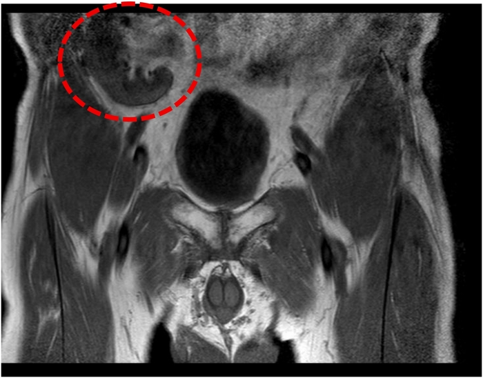
Figure 1: The transplanted kidney in the right iliac fossa. Coronal MRI showing the position of the kidney in the right iliac fossa and lateral to the bladder. Please click here to view a larger version of this figure.
Discussion
Radical prostatectomy in renal transplant recipients is challenging because the graft occupies the surgical field. Open radical prostatectomy has traditionally been performed, but is associated with higher blood loss and a greater need for retraction near the iliac vessels8.
Furthermore, RARP in renal transplant recipients has been established as a feasible and safe approach by multiple systematic reviews and case series. Zeng et al. synthesized data from 13 studies and reported low graft injury rates, stable renal function, and an overall complication rate of approximately 17%7. Similarly, Piana et al. provided a comprehensive overview of technical modifications, including adjusted port placement (described in 54% of patients), modified lymphadenectomy, and careful Retzius space development (performed in 60% of cases)9. Collectively, these reports confirm that RARP can be performed successfully in this challenging population.
However, systematic reviews and aggregated data cannot provide the granular, step-by-step technical detail surgeons need when encountering this complex anatomy for the first time. Our case report contributes precisely this: a detailed procedural guide grounded in a real-world patient with a right-sided iliac fossa transplant, multiple comorbidities, and concurrent anticoagulation. Several technical nuances from our approach merit specific emphasis. First, we modified port placement by shifting the right-sided robotic ports 2–3 cm cephalad and slightly more medial than standard. This specific measurement, while consistent with the general principle of "shifted ports" reported in the literature, provides a practical reference point for preoperative planning. Second, we prioritized early identification of the transplant kidney, its vascular anastomoses, and the short ureter—particularly its implantation into the bladder—before proceeding with the prostatectomy. This step, while conceptually straightforward, is critical to avoid inadvertent traction or injury during bladder mobilization and Retzius space development.
A key technical challenge was the presence of extensive adhesions from the patient's two prior retroperitoneal transplant procedures (1990 and 2009). This required meticulous, athermal dissection using clips rather than bipolar energy adjacent to the graft vessels on the right side, a strategy we recommend to minimize thermal spread and vascular injury. The decision to perform bilateral pelvic lymphadenectomy—often waived in this population—was made due to the patient's intermediate-risk prostate cancer (Gleason 3 + 4 = 7a). We performed a limited dissection on the right side, staying lateral to the graft vessels, which successfully yielded six tumor-free nodes without compromising graft perfusion. This demonstrates that PLND can be safely performed when oncologically indicated, provided the dissection is carefully tailored.
Our perioperative management also offers instructive lessons. The transient rise in renal retention parameters after suprapubic catheter clamping on postoperative day 3 underscores the delicate balance between encouraging early voiding and protecting transplanted kidney function. This phenomenon, which resolved promptly upon reopening the catheter, highlights the importance of close renal function monitoring and a low threshold for catheter re-institution in this population. While routine suprapubic catheter use is not standard in all centers, it was valuable for postoperative management and urinary drainage, consistent with our institutional protocol and prior evidence suggesting that a significant proportion of patients may develop acute urinary retention after RARP10.
This case report is inherently limited by its single-patient nature and, importantly, the loss of the patient to long-term follow-up beyond the early postoperative period (the surgery was performed in 2020). Therefore, we cannot draw conclusions about functional outcomes, such as long-term continence, or oncological durability, such as biochemical recurrence-free survival. Potency was out of scope since the patient had preoperative erectile dysfunction. The primary value of this report lies in demonstrating the technical feasibility and perioperative safety of the robot-assisted approach in this complex anatomical setting. The favorable results presented here—including successful graft preservation, uncomplicated early recovery, and negative surgical margins—are confined to the immediate postoperative course. While the short-term outcomes are encouraging, they should be interpreted with caution, and further prospective series with complete follow-up are necessary to confirm the long-term benefits and oncological control of RARP in renal transplant recipients.
Disclosures
The authors have nothing to disclose. The authors used a language model (Gemini) exclusively for language polishing and grammatical refinement. All scientific content was controlled, verified, and approved by the authors, who assume full responsibility for the final manuscript.
Acknowledgements
The authors have no acknowledgments.
Materials
| Name | Company | Catalog Number | Comments |
|---|---|---|---|
| Apixaban | Bristol-Myers Squibb | Eliquis (NDC B. 0003-0894 ) | NOAC (Factor-Xa-Inhibitor); 2 Tage präoperativ pausiert, ab POD 2 fortgesetzt |
| Bipolar Robotic Instrument | Intuitive Surgical | Maryland Bipolar 471172 | Zur Dissektion und Hämostase mit bipolarer Energie |
| Cefuroxime | Hikma | 1,5 g Vial NDC 0143-9977-22 | 1,5 g i.v. 30 Min. vor Hautinzision (perioperative Antibiotikaprophylaxe) |
| Da Vinci Surgical System (X) | Intuitive Surgical | Da Vinci X Surgical System | Roboterplattform für radikale Prostatektomie |
| Electrocardiography (ECG) Monitor | Philips / GE Healthcare u. a. | Krankenhaus-spezifisches Modell Philips IntelliVue | Kontinuierliches Herzmonitoring intra- und postoperativ |
| Enoxaparin | Sanofi | Lovenox 40 mg/0,4 mL NDC 0075-0620-40 | 0,4 mL s.c. 1× täglich zur Thromboseprophylaxe |
| Everolimus | Novartis | Zortress 1 mg(NDC 0078-0422) | 1 mg 2× täglich (Immunsuppressivum; Transplantationssetting) |
| Foley Catheter | Bard | 20 Fr three-way Bard 0167V20S | 20 Fr Dreiwege-Katheter zur Blasendrainage/Spülung |
| Large Needle Driver | Intuitive Surgical | 471006 | EndoWrist Large Needle Driver zum Nähen |
| Laparoscopic Clip Applier | Teleflex | Hem-o-lok Medium-Large | Zum Verschluss von Lymphgefäßen und Vermeidung thermischer Verletzungen |
| Monopolar Scissors | Intuitive Surgical | 470179 Monopolar Curved Scissors, X | Zur scharfen Dissektion und monopolaren Kauterisation |
| Non-invasive Blood Pressure (NIBP) Monitor | Philips | Krankenhaus-spezifisches Modell | Messung alle 3–5 Minuten zur hämodynamischen Überwachung |
| Prednisone | Verschiedene | 5 mg Tabletten | 5 mg 1× täglich (Immunsuppressivum / Kortikosteroid) |
| ProGrasp Retractor | Intuitive Surgical | 471093 | Starke Greifzange zur Geweberetraktion und -manipulation |
| Running Barbed Suture | (J&J) | STRATAFIX Spiral PDS 3-0 SXPP1B422 | 3-0 Widerhakennähte für spannungsfreie vesikourethrale Anastomose |
| Suprapubic Catheter | Bard | 14 Fr | 14 Fr Katheter intraoperativ gelegt zur Harnableitung |
| Tacrolimus | Astellas Pharma | Prograf (NDC 0469-0617 für 1 mg Kapseln) | 3 mg 2× täglich (Immunsuppressivum; Calcineurin-Inhibitor) |
References
- Sherer BA, et al. Prostate cancer in renal transplant recipients. Int Braz J Urol. 2017;43(6):1021-32.
- Hall EC, Pfeiffer RM, Segev DL, Engels EA. Cumulative incidence of cancer after solid organ transplantation. Cancer. 2013;119(12):2300-8.
- Bratt O, et al. Prostate cancer in kidney transplant recipients - a nationwide register study. BJU Int. 2020;125(5):679-85.
- Schmidt J, et al. Radical prostatectomy in kidney transplant recipients - a multicenter experience. Eur Urol Open Sci. 2024;67:45-53.
- Slota JM, et al. Robotic radical prostatectomy in kidney transplant recipients: A propensity-matched cohort study. BJUI Compass. 2025;6(12):e70128.
- Shahait M, et al. Oncological and functional outcomes of robot-assisted radical prostatectomy in kidney transplant recipients. JSLS. 2021;25(3):e2021.00045.
- Zeng J, et al. Safety and clinical outcomes of robot-assisted radical prostatectomy in kidney transplant patients: A systematic review. J Endourol. 2018;32(10):935-43.
- Waeckel T, Doerfler A, Bechade C, Tillou X. Comparative study of open versus robotic radical prostatectomy in renal transplant recipients and review of the literature. Prostate Int. 2026;14(1):75-80.
- Piana A, et al. Robot-assisted radical prostatectomy in renal transplant recipients: A systematic review. J Clin Med. 2023;12(21):6754.
- Farzat M, et al. BMI impact on readmissions for patients undergoing robot-assisted radical prostatectomy: A monocentric, single-surgeon serial analysis of 500 cases. J Clin Med. 2023;12(12):3908.