This study describes an in vitro. model to evaluate the protective effects of Sanqi Baiji San against ethanol-induced gastric epithelial cell injury and its involvement in PI3K/AKT-related signaling.
Method Article
Role of Sanqi Baiji San in Mitigating Ethanol-Induced Gastric Epithelial Cell Injury via PI3K/AKT-related Signaling
Opens in a new tab
In This Article
Summary
Loading...
$$\rightleftharpoonup{xx}$$
$$\longleftharp{xx}$$,
$$\longrightharp{xx}$$,
Abstract
Loading...
$$\rightleftharpoonup{xx}$$
$$\longleftharp{xx}$$,
$$\longrightharp{xx}$$,
This work aimed to clarify the protective mechanism of Sanqi Baiji San (SQBJ) against ethanol-induced gastric epithelial cell injury and to explore its potential relevance to gastric ulcer (GU). Network pharmacology was used to screen SQBJ’s active components (Traditional Chinese Medicine Systems Pharmacology Database and Analysis Platform [TCMSP], Oral Bioavailability [OB] ≥ 20%, Drug-Likeness [DL] ≥ 0.1), map their targets (Universal Protein Resource [UniProt]), collect GU-related targets (GeneCards/OMIM/DrugBank), and analyze overlapping targets via Gene Ontology (GO)/Kyoto Encyclopedia of Genes and Genomes (KEGG) enrichment and molecular docking. Ethanol-injured Gastric Epithelial Cell Line-1 (GES-1) cells were treated with SQBJ or the PI3K inhibitor LY294002. CCK-8 was used to determine the optimal SQBJ concentration. Network pharmacology analyses identified 126 common targets enriched within PI3K/AKT/MAPK-related cascades and suggested potential interactions between principal SQBJ constituents and PI3K/AKT-related proteins. In ethanol-challenged cells, SQBJ alleviated cell injury by reducing inflammatory mediator release and oxidative stress, as evidenced by decreased intracellular reactive oxygen species and malondialdehyde levels. SQBJ restored mitochondrial membrane potential and ATP content and reduced apoptosis-associated changes in B-cell lymphoma-2, cleaved caspase-3, and Bcl-2-associated X protein levels. SQBJ also modulated PI3K/AKT- and MAPK-associated signaling markers. These protective effects were largely weakened by LY294002, suggesting that PI3K/AKT-related signaling is involved in SQBJ-mediated cytoprotection. These findings provide an in vitro mechanistic basis for the potential application of SQBJ in GU, although further validation in animal models of ethanol-induced gastric ulcer is required.
Introduction
Loading...
$$\rightleftharpoonup{xx}$$
$$\longleftharp{xx}$$,
$$\longrightharp{xx}$$,
Gastric ulcer (GU) is a highly prevalent gastrointestinal disorder worldwide, characterized by localized inflammation, erosion, and ulceration of the gastric mucosa. According to estimates yielded by the 2021 Global Burden of Disease (GBD 2021) analysis, peptic ulcer disease (PUD) continues to impose a substantial clinical and economic burden on millions of patients globally. Although the overall prevalence of PUD has declined with the widespread and extensive clinical reliance on anti-infective regimens and acid-suppressing proton pump inhibitors (PPIs), this downward trend has plateaued, with a rebound even observed in some regions1. Notably, alcohol (ethanol) consumption is a critical pathogenic factor independent of Helicobacter pylori (H. pylori.) infection and non-steroidal anti-inflammatory drug (NSAID) use. Long-term excessive alcohol intake damages the gastric mucosal barrier, accelerates ulcer development, and even elevates susceptibility to gastric malignancies—thereby rendering this condition a critical international healthcare crisis2. In developing countries, the incidence of alcohol-related gastric mucosal injury is on the rise due to multiple factors, including increasing alcohol consumption and lifestyle changes, creating an urgent need for effective and safe prevention and treatment strategies.
The pathological mechanism underlying ethanol-induced gastric mucosal injury is multifactorial and multi-layered. Upon direct contact with the gastric mucosa, ethanol rapidly disrupts the mucus-bicarbonate barrier and triggers an uncontrolled surge of intracellular reactive oxygen species (ROS). This oxidative burst subsequently instigates lipid peroxidation (marked by elevated malondialdehyde, MDA), depletes endogenous antioxidants within the stomach lining, including superoxide dismutase (SOD), along with glutathione (GSH), and ultimately induces mitochondrial dysfunction3. Accumulating evidence indicates that intracellular ROS induced by ethanol is mainly derived from mitochondria and is closely linked to the collapse of the inner mitochondrial membrane potential (Δψm) and the inhibition of adenosine triphosphate (ATP) synthesis4. Meanwhile, oxidative stress further activates the pro-inflammatory cascades under the command of nuclear factor-kappa B (NF-κB). This activation subsequently promotes the massive release of inflammatory mediators chiefly tumor necrosis factor-α (TNF-α), interleukin-6 (IL-6), and C-reactive protein (CRP), thus forming a vicious cycle of "oxidative stress-inflammatory response"5. On this basis, the intrinsic apoptotic cascade driven by mitochondria is triggered, as evidenced by a disrupted ratio of Bcl-2 to Bax alongside the sequential enzymatic proteolytic cleavage of cysteinyl aspartate-specific proteinase 3 (Caspase-3), which eventually leads to massive death of gastric mucosal epithelial cells and ulcer formation6. Accordingly, simultaneous targeting of the three core pathological links of oxidative stress, inflammatory response, and apoptosis represents a key strategy for the effective intervention of alcoholic gastric mucosal injury.
The phosphoinositide 3-kinase/protein kinase B (PI3K/AKT) signaling pathway constitutes a central regulatory axis governing cellular longevity, proliferation, and programmed death, executing a vital function in preserving gastric mucosal homeostasis. Following activation, PI3K/AKT signaling phosphorylates multiple downstream substrates, inhibits the activity of the pro-apoptotic factor Bad, thereby impeding the mitochondrial escape of cell-destructive cytochrome c, maintaining ΔΨm homeostasis, and promoting cell survival7. Furthermore, there is a complex antagonistic crosstalk between the PI3K/AKT cascade and mitogen-activated protein kinase (MAPK) networks: the up-regulated activation of PI3K/AKT can attenuate inflammatory signal transduction and apoptotic cascade reactions by inhibiting the activation of stress kinases, including p38, c-Jun N-terminal kinase (JNK), and extracellular regulated protein kinases (ERK)8. Multiple studies have indicated that reduced activity of the PI3K/AKT pathway is closely associated with exacerbated inflammation and increased apoptosis in gastric mucosal injury models, while restoration of this pathway’s activity can significantly ameliorate mucosal injury9. However, there is currently a lack of systematic causal experimental verification regarding the precise regulatory mechanism of PI3K/AKT in ethanol-induced gastric mucosal injury, particularly its synergistic relationship with the MAPK pathway.
In terms of modern pharmacotherapy, PPIs are the first-line option for the clinical management of GU, yet their long-term administration has raised growing safety concerns. Accumulating studies have linked chronic PPI use to a variety of adverse outcomes, including renal disease, cardiovascular events, osteoporotic fractures, nutrient deficiencies (iron, magnesium, and vitamin B12), Clostridioides difficile. infection, and even an elevated risk of gastric cancer10. Furthermore, the phenomenon of rebound gastric acid hypersecretion after PPI discontinuation has been confirmed by large-scale pharmacovigilance data11. These safety risks have driven both clinical and basic research communities to actively explore alternative therapies with multi-target protective mechanisms, minimal side effects, and suitability for long-term use. In particular, natural compound preparations from traditional Chinese medicine (TCM) have attracted extensive attention due to their holistic therapeutic concept of "multi-component, multi-target, and multi-pathway" action12.
Sanqi Baiji Powder (SQBJ) is a classic TCM compound formula composed of Panax notoginseng and Bletilla striata at a 1:1 mass ratio. Panax notoginseng is well known for its effects of promoting blood circulation and arresting bleeding, as well as alleviating painful swelling. Its primary bioactive constituents, including notoginsenoside R1 along with ginsenosides (Rg1, Rb1), exert significant anti-inflammatory, antioxidant, and mucosa-repairing effects. Bletilla striata is characterized by its astringent effects that arrest bleeding, reduce swelling, and promote the regeneration of damaged tissues. Concurrently, Bletilla striata polysaccharide (BSP), serving as its primary active extract, has documented efficacy in suppressing the JNK/p38 MAPK cascade, thereby decreasing the expression of interleukin-1β (IL-1β), TNF-α, and IL-6, and enhancing the antioxidant defense capacity of the gastric mucosa13. Accumulated literature evidence has verified that co-administration of Panax notoginseng. saponins (PNS) and BSP exerts a synergistic effect in alcoholic gastric ulcer models14, and SQBJ-related preparations have shown significant mucosal protective effects in preclinical studies of reflux esophagitis and peptic ulcer15. However, there is currently no systematic in vitro experimental verification of the signaling pathway mechanism underlying the gastric mucosal protective effect of SQBJ, especially the core role of the PI3K/AKT pathway and its regulatory relationship with MAPK. Meanwhile, the causal association between the pivotal constituent chemicals and functional receptors of SQBJ has yet to be fully elucidated.
To address these gaps, the present study adopted an integrated research strategy combining network pharmacology and in vitro experimental verification, employing the ethanol-challenged GES-1 cell line as the experimental platform to investigate the potential molecular mechanism underlying the gastric epithelial protective effects of SQBJ, with emphasis on oxidative stress, inflammatory responses, apoptotic processes, PI3K/AKT-related signaling, and MAPK-associated changes. First, we used the PI3K inhibitor LY294002 to preliminarily evaluate the involvement of PI3K/AKT signaling and found that inhibiting this pathway attenuated the protective effect of SQBJ, suggesting that PI3K/AKT-related signaling contributes to SQBJ-mediated cytoprotection. Second, we explored the association between PI3K/AKT-related changes and MAPK pathway regulation, providing preliminary evidence that MAPK signaling changes may be linked to SQBJ modulation of the PI3K/AKT axis's functional response. Collectively, this work establishes an in vitro molecular framework for evaluating how SQBJ mitigates ethanol-induced gastric epithelial injury and provides preliminary evidence for further exploration of TCM compound formulas in alcoholic gastric mucosal injury. However, animal experiments are still required to confirm whether these cellular protective effects can be translated into in vivo. therapeutic efficacy within experimental models of alcohol-driven gastric ulceration (Supplemental Figure S1).
Protocol
Loading...
$$\rightleftharpoonup{xx}$$
$$\longleftharp{xx}$$,
$$\longrightharp{xx}$$,
This study did not involve human participants, human tissue samples, clinical specimens, or animal experiments. Publicly available and anonymized database resources were used for network pharmacology analysis, and commercially available GES-1 cells were used for in vitro experiments. Therefore, formal ethical approval and informed consent were not required. Detailed information on the main reagents, antibodies, assay kits, instruments, manufacturers, catalog numbers, and Research Resource Identifiers (RRIDs), where applicable, is summarized in the Table of Materials.
Preparation and quality control of SQBJ aqueous extract
Herbal materials
The raw materials of Panax notoginseng (Burk.) F. H. Chen (root and rhizome) and Bletilla striata .(Thunb. ex A. Murray) Rchb. f. (tuber) were obtained from a commercial traditional Chinese medicine supplier and subsequently authenticated by a senior TCM pharmacist following the Chinese Pharmacopoeia (2025 edition). Voucher specimens (No. SQBJ-20240301, No. PN-20240301, No. BS-20240301) were deposited in the herbarium of our institution. The two herbs were crushed and mixed at a mass ratio of 1:1 to prepare SQBJ powder.
Preparation of SQBJ aqueous extract
SQBJ powder underwent two successive reflux extractions with deionized water at a solid-to-liquid ratio of 1:10 (g/mL) for 1 h per session. Thereafter, the collected extracts were combined and passed through four layers of medical gauze to remove residual material. The resulting filtrate was concentrated under reduced pressure at 60 °C until reaching a relative density of 1.05–1.10, followed by vacuum freeze-drying to obtain SQBJ aqueous extract powder, with a final extraction yield of 18.2 % ± 0.6 %. The extract powder was sealed and stored at −20 °C, dissolved in serum-free DMEM medium, and subsequently passed through a 0.22 µm membrane filter for sterilization prior to cell experiments.
Quality control of SQBJ aqueous extract
To evaluate batch-to-batch consistency and standardization of the SQBJ aqueous extract, we conducted quantitative determination alongside high-performance liquid chromatography (HPLC) fingerprint profiling. The Table of Materials lists the comprehensive technical parameters regarding the HPLC system. Based on the chemical profiles of both constituent herbs, we targeted four representative markers: notoginsenoside R1, ginsenoside Rg1, ginsenoside Rb1, and militarine. However, this HPLC-based quality control strategy mainly reflected several representative constituents and did not provide a comprehensive chemical profile of all active compounds in SQBJ.
Chromatographic separation was achieved via a reversed-phase C18 column (4.6 mm × 250 mm, 5 µm). The operation was maintained at a column temperature of 30 °C, with a flow rate of 1.0 mL/min, an injection volume of 10 µL, and a detection wavelength of 203 nm. The composition of the mobile phase consisted of acetonitrile (A) and 0.1% phosphoric acid aqueous solution (B), utilizing the following gradient elution run: 0–15 min, 19% A; 15–35 min, 19%–35% A; 35–55 min, 35%–55% A; and 55–60 min, 55%–19% A. Detailed instrument and column information is provided in the Table of Materials.
Reference substance preparation
To perform quantitative analysis, we procured commercial standards for notoginsenoside R1, ginsenoside Rg1, ginsenoside Rb1, and militarine, all with purity ≥ 98%. The individual reference chemicals were precisely measured and dissolved into a single methanol stock to achieve a uniform mixed reference solution with a final concentration of 50 µg/mL for each component.
Sample preparation:
SQBJ aqueous extract dry powder (50 mg) was precisely measured, followed by dissolution using 25 mL of analytical-grade methanol. This mixture underwent an ultrasonic bath treatment for 30 min under controlled instrumentation parameters (power: 250 W; frequency: 40 kHz). Following equilibration back to ambient room temperature, the liquid was passed through a 0.22 µm organic filter membrane to yield the final analytical test solution.
Content determination results:
The average contents of core characteristic components in three consecutive batches of SQBJ aqueous extract were as follows: notoginsenoside R1 3.12 ± 0.11 mg/g, ginsenoside Rg1 8.45 ± 0.23 mg/g, ginsenoside Rb1 5.26 ± 0.18 mg/g, and militarine 2.78 ± 0.09 mg/g, with the RSD of each component content < 5 % between batches, confirming good batch-to-batch consistency of the extract.
Ethics and compliance statement
Neither animal testing nor the inclusion of human clinical specimens, tissue biopsies, and live subjects was involved throughout this study. The network pharmacology analysis was performed using publicly available databases, including TCMSP, UniProt, GeneCards, OMIM, DrugBank, STRING, Metascape, PubChem, and RCSB PDB. All data retrieved from these databases were publicly accessible and anonymized; therefore, the bioinformatics profiling was entirely exempt from requiring formal informed consent or additional ethical oversight.
The present in vitro. experiments were conducted using the commercially available human gastric mucosal epithelial cell line GES-1. No primary human cells or patient-derived biological materials were used. The cell line was obtained from an authenticated cell repository and handled in accordance with institutional biosafety and cell culture guidelines. Because this study involved only established cell lines and publicly available databases, it was exempt from formal institutional ethics committee approval. All experimental procedures were performed in accordance with relevant institutional laboratory safety and research compliance requirements.
Network pharmacology studies
Identification and screening of active ingredient targets
The active components of Panax notoginseng and Bletilla striata., the two constituent herbs of SQBJ, were retrieved from publicly available databases. All database-derived information used in this study was publicly accessible and did not contain identifiable personal information. Using botanical names as queries, we identified candidate phytochemicals from the Traditional Chinese Medicine Systems Pharmacology Database and Analysis Platform (TCMSP). To identify eligible bioactive molecules, the screening protocol applied thresholds of oral bioavailability (OB) ≥ 20% and drug-likeness (DL) ≥ 0.1. Next, we identified the potential downstream gene targets associated with these candidate components, omitting any constituents without matched valid gene targets. All target genes were annotated and standardized via the Universal Protein Resource (UniProt), restricting the reference database search exclusively to Homo sapiens, thereby generating the finalized target gene list for the functional SQBJ active components.
Retrieval of pathology-related targets
To compile genes linked to the condition, we queried three public databases: GeneCards, Online Mendelian Inheritance in Man (OMIM), and DrugBank, with "gastric ulcer" as the search term. The cross-platform dataset was subsequently merged, followed by the elimination of redundant entries to establish a standardized list of gastric ulcer-associated target genes.
Network construction for ingredients, targets, and disease
We determined the overlapping target genes between SQBJ active components and gastric ulcer. Subsequently, a multi-layer interaction network was constructed using network visualization software. To evaluate node importance, the integrated topological evaluation algorithm computed each node's topological parameters, including degree and betweenness centrality. Based on these calculated values, we identified the active components and key functional targets of SQBJ in the treatment of gastric ulcer.
Mapping of protein-protein interaction (PPI) networks
The identified consensus targets shared between SQBJ and gastric ulcer were cross-referenced against the STRING database. Our query specified the multi-protein analysis template while restricting the analytical background to Homo sapiens. PPIs were filtered with a minimum interaction confidence threshold of medium confidence (> 0.4).
The resulting functional connectivity records were exported as a TSV file, which was subsequently integrated into dedicated network visualization software for PPI analysis. Based on the calculated connection topography, we evaluated the core functional nodes in the network by ranking the specific degree value of each node.
Downstream functional annotation and pathway mapping
To elucidate the biological roles of the shared intersection nodes, we conducted Gene Ontology (GO) functional enrichment analysis and Kyoto Encyclopedia of Genes and Genomes (KEGG) pathway enrichment analysis utilizing a web-accessible bioinformatic annotation tool. The functional classification categorizes genes into three classic categories: biological process (BP), cellular component (CC), and molecular function (MF). Bubble plots and bar charts for enrichment results were generated using an online bioinformatics plotting platform to map out the primary biological processes and signaling pathways associated with the therapeutic effect of SQBJ on gastric ulcer.
Computational molecular docking simulation
The spatial geometry files of the core active components of SQBJ were downloaded from public compound databases and subsequently saved as MOL2 files using file-processing software. We also obtained the three-dimensional crystal structures of the core target proteins by querying the Protein Data Bank. These macromolecular structures were preprocessed by removing original ligands and water molecules, and the resulting clean models were saved in PDB format. To simulate receptor-ligand binding energetics, the docking verification involving core active components and target proteins was executed via standard computational molecular docking software. The docking results were optimized and rendered using molecular visualization software.
Cell culture
GES-1 human-derived gastric epithelial cells were cultivated utilizing RPMI-1640 medium integrated with 10% fetal bovine serum and 1% penicillin–streptomycin at 37 °C under a humidified atmosphere containing 5% CO₂. We identified the source culture as an established cell line, obtaining the initial stock from an authenticated cell repository. At an observed confluence of 80–90%, the monolayers were rinsed 2x with PBS followed by detachment using 0.25% trypsin-EDTA for 2 min at 37 °C. To terminate the enzymatic cleavage reaction, we introduced an equal volume of complete medium. The resulting cellular suspension was centrifuged at 1,000 × g. for 5 min, after which the pellet was resuspended in fresh complete medium, and reseeded at the appropriate density. Cultures were returned to identical incubation conditions. Throughout the propagation period, an inverted phase-contrast microscope was used to assess cell viability, morphology, and growth status, and cells in their logarithmic growth phase were routinely collected for downstream analyses.
Establishment and treatment of ethanol-induced GES-1 cell injury model
Logarithmic-phase GES-1 cells were seeded at 5 × 10⁵ cells/well in 6-well plates or 1 × 10⁴ cells/well in 96-well plates and incubated overnight to adhere. Ethanol-induced injury was established by exposing cells to 500 mmol/L ethanol in unsupplemented RPMI-1640 for 4 h. This condition was selected based on preliminary concentration- and time-response experiments. In the preliminary assay, 250 mmol/L ethanol for 4 h caused only mild injury, with cell viability remaining at approximately 78.6 ± 5.4%, whereas 750 mmol/L ethanol reduced viability to 32.8 ± 4.7%, indicating excessive cytotoxicity. Exposure to 500 mmol/L ethanol for 4 h reduced cell viability to 54.7 ± 4.9%, which produced reproducible moderate-to-severe injury while retaining sufficient viable cells for subsequent intervention and mechanistic assays. Therefore, 500 mmol/L ethanol for 4 h was selected for model establishment. This condition was used to simulate acute ethanol-induced epithelial injury in vitro and does not fully reproduce the physiological exposure pattern of the gastric mucosa in vivo.
For pharmacological intervention, three concentrations of SQBJ aqueous extract (5, 10, and 50 µg/mL) were selected and administered immediately after ethanol challenge. In the PI3K inhibition arm, cells were preincubated with 20 µmol/L LY294002 for 30 min prior to co-treatment with ethanol and the optimal SQBJ concentration. A positive control cohort received 20 µmol/L omeprazole immediately after ethanol exposure. Omeprazole was used as a reference gastroprotective agent rather than a dose-equivalent comparator. The concentration was selected based on a preliminary cytotoxicity evaluation and on commonly used in vitro intervention ranges, and was intended to provide a pharmacological reference for cellular protection. Control cells were maintained in complete medium without ethanol or additional agents. All incubations were conducted in a humidified atmosphere containing 5% CO₂ at 37 °C. Each experimental group comprised at least three replicate wells, and the entire protocol was executed independently 3x.
Determination of optimal SQBJ concentration via CCK-8 method
Log-phase HGE/GES-1 elements were plated into 96-well vectors at a density of 1 × 10⁴ cells/well and incubated overnight in complete RPMI-1640 medium under standard conditions (37 °C, 5% CO₂) to ensure adherence. The cultures were randomized into four groups: vehicle control (complete medium only), low-dose SQBJ intervention (5 µg/mL), medium-dose SQBJ intervention (10 µg/mL), and high-dose SQBJ intervention (50 µg/mL); these three concentrations were utilized for therapeutic screening and prepared by dissolving SQBJ in PBS. All conditions were assayed in at least three parallel wells, and the total protocol was repeated in three distinct experimental batches. After the designated treatment period, each well received 10 µL of CCK-8 reagent, followed by a 2 h incubation period at 37 °C. The optical density (OD) at 450 nm was measured using a microplate reader. The percentage of cell viability was calculated as (absorbance_treatment / absorbance_control) × 100%, allowing identification of the most effective SQBJ concentration for subsequent experiments.
qPCR
Log-phase GES-1 elements were cultured in 6-well plates (5 × 10⁵ cells/well) and maintained overnight at 37 °C in a humidified incubator containing 5% CO₂ to allow adherence. Thereafter, cells were exposed to the predetermined optimal concentration of SQBJ and harvested at baseline (0 h) and at 6, 12, 24, and 48 h post-treatment to determine the dynamic response of signaling molecules. Based on the time-course results, the 24 h interval was chosen for subsequent mechanistic assays because it showed the strongest signaling response while maintaining stable cell viability. Each time point was represented by three triplicate wells, and the entire experiment was independently repeated 3x.
We isolated total RNA using a specialized extraction reagent, assessing the concentration and quality of the yield using a spectrophotometer. All samples exhibited an A₂₆₀/A₂₈₀ absorbance ratio within the range of 1.8 and 2.0. A reverse transcription kit with gDNA wiper was used to reverse-transcribe cDNA from 1 µg of total RNA. Real-time PCR analysis was executed via a quantitative PCR system with primers for PI3K, AKT, p38, JNK, ERK, and the reference gene GAPDH. Details of the PCR system, reagents, and primer synthesis service are detailed in the Table of Materials. Primers were designed to specific regions of the corresponding human genes. qPCR protocol: hot-start activation at 95 °C for 30 s, followed by 40 cycles of denaturation at 95 °C for 5 s and annealing/extension at 60 °C for 30 s. Target gene mRNA expression calculation relied on the relative quantitative PCR method with the 2-ΔΔCt method.
ELISA
Supernatants were harvested and cleared via centrifugation at 1,200 × g. for 10 min at 4 °C for cellular debris removal. Levels of inflammatory mediators (CRP, IL-6, TNF-α) and oxidative stress indices (SOD, MDA, GSH) were evaluated using commercial ELISA kits following the manufacturers' specific directions. The concentration profiles of these targets were verified with commercial assay formulations per the suppliers' instructions. Each experimental group comprised three replicate wells, and the procedure was executed across three separate cell culture batches to confirm experimental data reproducibility. Briefly, 100 µL of either standards or clarified supernatants were dispensed into pre-coated reaction plates and incubated at 37 °C for 90 min. Following three successive wash cycles, each cavity received 100 µL of the specific detection antibody for a 60 min incubation at 37 °C, followed by another three wash cycles. Next, substrate solution (100 µL) was introduced, and the samples were protected from light for 15 min before blocking the enzymatic reaction with 50 µL of stop solution. Optical density at 450 nm was detected via a microplate reader. Concentrations were interpolated from the corresponding standard curves. To minimize bias caused by differences in cell number or treatment-induced changes in cell viability, MDA, SOD, GSH, CRP, TNF-α, and IL-6 profiles were calibrated against the protein contents of each analytical sample, as determined by the BCA assay, and expressed as relative values compared with the control group.
Detection of intracellular ROS in GES-1 cells using DCFH-DA fluorescent probe
To quantify intracellular reactive oxygen species (ROS) levels, we utilized the membrane-permeable fluorogenic indicator DCFH-DA. Harvested cells were resuspended in unsupplemented RPMI-1640 containing 10 µmol/L DCFH-DA and maintained for 20 min under standard culture conditions (37 °C, 5% CO₂) in the dark. Following three successive washes with serum-free medium to clear unbound probe, fluorescence intensity was monitored by flow cytometry with 488 nm excitation and 525 nm emission. Relative ROS levels were reported as the mean fluorescence intensity for each treatment group, normalized to the viable cell number determined in parallel wells using the CCK-8 assay, and expressed relative to the corresponding control group.
Determination of mitochondrial membrane potential (ΔΨm)
We evaluated changes in mitochondrial membrane potential (ΔΨm) using a commercial JC-1 assay kit. In brief, harvested cells were resuspended in a freshly prepared JC-1 working solution—an equal-volume mixture of JC-1 dye and staining buffer supplied by the kit—to a final cell concentration of 1 × 10⁶ cells/mL. After incubation for 20 min at 37 °C in a humidified environment containing 5% CO₂, protected from light to avoid bleaching, the sample was harvested by centrifugation (800 × g., 5 min) and rinsed once with 1 mL of ice-chilled JC-1 staining buffer. Fluorescence was subsequently acquired by flow cytometry.
JC-1 aggregates (indicative of intact ΔΨm) were monitored via the FL2 channel using 488 nm excitation and 590 nm emission wavelengths, whereas JC-1 monomers (indicative of ΔΨm dissipation) were tracked via the FL1 channel using 488 nm excitation and 525 nm emission settings. The ΔΨm was expressed as the red-to-green fluorescence signal proportion; dropping values indicate mitochondrial depolarization.
Determination of intracellular ATP level
Mitochondrial functionality was assessed via intracellular ATP quantification using an ATP assay kit. A luciferin–luciferase bioluminescent reaction was employed to determine ATP content, which is directly proportional to mitochondrial activity. Briefly, harvested cells were re-suspended in ice-cold ATP lysis buffer and kept for a 5 min lysis window on ice. Centrifugation was then performed at 12,000 × g. for 5 min at 4 °C, after which 20 µL of the obtained cleared supernatant was transferred to 96-well white opaque microplates. Subsequently, 100 µL of freshly prepared ATP detection working solution—an admixture of luciferase reagent and luciferin substrate prepared following the provided protocol—was introduced into each cavity. The resulting luminescence signal was promptly captured with a luminescence microplate reader. To obtain relative ATP levels, raw luminescence values were normalized to the individual supernatant total protein concentrations quantified with a commercial BCA protein assay kit, thereby correcting for variations in cell number.
Detection of cell apoptosis by Annexin V/PI double staining
Apoptotic cell detection was performed with a commercial Annexin V-FITC/PI cell death assay configuration as outlined by the manufacturer. After washing, the pellets were resuspended in Annexin V-FITC binding buffer to a cell density of 1 × 10⁶ cells/mL (195 µL per sample). Subsequently, each independent tube received 5 µL of Annexin V-FITC followed by 10 µL of PI staining solution. The sample was vortexed gently and incubated in the dark at room temperature for 15 min. Following this process, apoptotic cells were monitored via a flow cytometer. The respective fluorescent channels facilitated the differentiation between viable cells (Annexin V⁻/PI⁻), early apoptotic cells (Annexin V⁺/PI⁻), and late apoptotic/necrotic cells (Annexin V⁺/PI⁺). The percentage of apoptotic cells in each group was quantified following these gating parameters.
Western blot
Membranes were incubated overnight at 4 °C with primary antibodies against Bcl-2, Bax, cleaved Caspase-3, AKT, p38 MAPK, JNK, ERK1/2, and GAPDH. Antibodies were selected according to validation information for human samples and western blot applications. RRIDs were added for all antibodies with available records in the Antibody Registry or RRID Portal. Target protein levels were normalized to GAPDH. After standard washing protocols, the blots were exposed to HRP-conjugated secondary antibodies for 1 h at room temperature. Bands were detected using an ECL substrate solution. Western blot band grey values were evaluated using digital image analysis software.
Immunofluorescence staining
GES-1 monolayers were established on glass coverslips within 24-well plates prior to the indicated treatments. After treatment, fixation was executed utilizing 4% paraformaldehyde for 15 min at room temperature, followed by membrane permeabilization using 0.3% Triton X-100 in PBS for 10 min, and blocking via 5% bovine serum albumin for 30 min at 37 °C. Coverslips were then transferred for overnight incubation at 4 °C with primary antibodies targeted to AKT, p38, JNK, and ERK. Following three successive PBS washes, specimens were incubated for 1 h at room temperature with a fluorescence-conjugated anti-rabbit secondary antibody protected from light. DAPI counterstaining was applied for 5 min to visualize nuclei. Coverslips were subsequently mounted with anti-fade reagent, and data collection utilized a confocal laser scanning microscope to determine the subcellular distribution of the target proteins. All immunofluorescence images were captured under the same imaging parameters using a 20× objective lens. Scale bars corresponding to 50 µm were added to all representative immunofluorescence images.
Statistical analysis
Quantitative values are illustrated as the mean ± standard deviation (SD), originating from at least three independent experimental runs with each sample verified in triplicate. Statistical analyses were conducted via specialized numerical analysis tools. To determine distinct variations across multiple cohorts, we utilized one-way analysis of variance (ANOVA) alongside Tukey’s post hoc test for inter-group comparisons. A two-tailed threshold of P < 0.05 designated statistical significance.
Results
Loading...
$$\rightleftharpoonup{xx}$$
$$\longleftharp{xx}$$,
$$\longrightharp{xx}$$,
Bioinformatic dissection of SQBJ-mediated mechanisms in gastric ulcer
To explore how SQBJ may exert its pharmacological actions on GU, we initially predicted the targets corresponding to its primary bioactive constituents. Using R programming, we extracted and cleared redundant entries among these bioactive components, ultimately yielding 411 candidate targets. Subsequently, GU-linked pathological targets were harvested by querying GeneCards and OMIM platforms, isolating 1,643 distinct condition-specific loci (Supplemental File 1). Intersecting the disease targets with those of SQBJ’s components identified 126 shared key targets (Figure 1A). Using these shared targets, evaluation of PPI topology (Figure 1B) and its graphical representation (Figure 1C) identified a hub module of densely interconnected proteins, implicating central regulatory nodes associated with the SQBJ-GU target network. Functional annotation via GO enrichment profiling (Figure 1D) further classified these targets into core biological processes (e.g., cellular response to peptide stimuli, positive regulation of peptidyl-tyrosine phosphorylation), cellular components (e.g., membrane microdomain, phosphorus-containing molecular complexes), and molecular actions, including protein tyrosine kinase activity and protein serine kinase activity. KEGG pathway analysis (Figure 1E) extended these observations to canonical signaling cascades, revealing prominent enrichment in pathways including PI3K-AKT signaling, prostate cancer, and focal adhesion. Molecular docking simulations (Figure 1F) visualized the predicted binding conformations of key molecules within these pathways. Collectively, this computational dissection suggested that SQBJ may act through multi-component, multi-target, and multi-pathway mechanisms, particularly by regulating key targets such as PIK3CA, with quercetin and DFV identified as the primary predicted bioactive components.
Dose- and time-dependent modulation of GES-1 cell viability and signaling pathways by SQBJ fractions
To define the optimal SQBJ dose, we first conducted dose-response assays. GES-1 cells were treated with three SQBJ fractions (SJD: 5 µg/mL, SJM: 10 µg/mL, and SJH: 50 µg/mL) for 24 h, with triplicate wells per group and three independent experiments. Cell viability analysis (Figure 2A) revealed a dose-dependent protective effect: SJM (10 µg/mL) induced the greatest increase in viability, making it the optimal dose for subsequent time-course investigations. At this dose, we evaluated time-dependent changes in critical signaling markers (PI3K, AKT, p38, JNK, and ERK) over 48 h. As shown in Figure 2B-F, the relative abundance of total JNK, PI3K, p38, AKT, and ERK showed a time-dependent trend. The expression of PI3K, AKT, p38, JNK, and ERK began increasing at 6 h, reached a further peak at 12 h, peaked at 24 h, and declined at 48 h. Meanwhile, the CCK-8 assay showed that 10 µg/mL SQBJ produced the most stable improvement in cell viability at 24 h without obvious cytotoxicity. Therefore, 24 h served as the most appropriate duration for follow-up pathway exploration because it represented the time point with maximal signaling response and stable cellular protection. In summary, these results indicate that SQBJ affects GES-1 cell viability in a dose- and time-dependent manner and is associated with PI3K/AKT- and MAPK-related signaling markers.
Attenuation of ethanol-induced inflammation and oxidative stress in GES-1 cells with involvement of PI3K/AKT-related signaling
Gastric mucosal injury results mainly from key pathological conditions such as inflammation and oxidative stress. To ascertain whether SQBJ modulates cytoprotection in association with PI3K/AKT signaling, we established six experimental cohorts: Control, Model (500 mmol/L ethanol for 4 h), SQBJ (10 µg/mL), positive drug (20 µmol/L omeprazole after ethanol exposure), PI3K inhibitor (25 µmol/L LY294002 prior to ethanol challenge), and a combined group pre-treated with 25 µmol/L LY294002 before ethanol intervention followed by SQBJ treatment, with three wells per group and three independent experiments. ELISA and fluorescence assays showed that the Model group exhibited significantly increased inflammatory cytokines (CRP, TNF-α, IL-6; Figure 3A-C), elevated oxidative stress markers (MDA, ROS; Figure 3D,E), and reduced antioxidant levels (SOD, GSH; Figure 3F,G). SQBJ and the positive drug markedly reversed ethanol-induced inflammatory and oxidative stress responses. However, co-treatment involving LY294002 blunted these beneficial outcomes; the combination group demonstrated negligible reductions in CRP, TNF-α, IL-6, MDA, and ROS readings compared with the PI3K inhibitor group, while SOD and GSH levels showed only limited recovery. These changes were not sufficient to indicate a clear protective effect of SQBJ under PI3K inhibition, suggesting that the observed suppression of inflammation and oxidative damage by SQBJ is closely linked to PI3K/AKT-related signaling. When PI3K activity was inhibited by LY294002, the protective effect of SQBJ was largely weakened, indicating that PI3K/AKT-related signaling contributes to SQBJ-mediated cytoprotection.
Mitochondrial function in ethanol-injured GES-1 cells with involvement of PI3K/AKT-related signaling
Maintenance of mitochondrial function is an important feature of protection against ethanol-induced damage in GES-1 cells. Flow cytometric plots (Figure 4A) revealed that ethanol exposure (Model group) reduced the red/green ratio (reflecting ΔΨm dissipation), while SQBJ and positive drug treatments restored this ratio. Quantitative analysis (Figure 4B) verified a prominent reduction of this fluorescence proportion within the Model group, which was reversed by SQBJ; co-treatment with a PI3K inhibitor attenuated this protective effect. For ATP levels (Figure 4C), the Model group showed marked reduction, and SQBJ partially rescued ATP content, whereas PI3K inhibition blunted this protection. These findings indicate that SQBJ preserves mitochondrial function in ethanol-injured GES-1 cells with involvement of PI3K/AKT-related signaling.
PI3K/AKT-mediated regulation of GES-1 cell apoptosis and proliferation by SQBJ
To investigate SQBJ’s effects on GES-1 growth profile and cell death responses in relation to the PI3K/AKT cascade, we performed proliferation assays using the CCK-8 method. We also determined programmed cell death patterns through the Annexin V/PI flow cytometry protocol; apoptotic protein expression was analyzed by western blot. CCK-8 results (Figure 5A) showed that the ethanol-induced Model group exhibited reduced cell viability, which was rescued by SQBJ and positive drug treatment; co-treatment with a PI3K inhibitor diminished this protective effect. Annexin V/PI staining (Figure 5B) revealed that the Model group had increased apoptosis, while SQBJ and positive drug decreased apoptosis; PI3K inhibition reversed this trend. Western blot analysis (Figure 5C) showed that in the Model group, Bcl-2 (anti-apoptotic) expression decreased, while Bax (pro-apoptotic) and cleaved Caspase-3 levels increased. SQBJ reversed these changes, whereas PI3K inhibitor co-treatment blunted SQBJ’s effects. Collectively, SQBJ regulates GES-1 cell proliferation and apoptosis in association with PI3K/AKT-related apoptotic signaling.
Modulation of PI3K/AKT and MAPK signaling in ethanol-injured GES-1 cells
To clarify whether SQBJ modulates the PI3K/AKT network and alters MAPK-related signaling in ethanol-damaged GES-1 cells, we analyzed transcriptional levels via qPCR (Figure 6A), protein abundance via western blot (Figure 6B), and intracellular spatial distribution via immunofluorescence confocal microscopy (Figure 6C). qPCR revealed that ethanol exposure (Model group) downregulated AKT mRNA while upregulating p38, JNK, and ERK. mRNAs; SQBJ treatment reversed these changes, whereas PI3K inhibitor co-treatment blunted this modulation (Figure 6A). Western blot analysis showed that ethanol exposure reduced AKT-related protein levels while increasing p38, JNK, and ERK levels; SQBJ treatment reversed these ethanol-associated changes, while LY294002 weakened the regulatory effect of SQBJ on these signaling markers (Figure 6B). Immunofluorescence further showed diminished AKT fluorescence intensity and enhanced p38, JNK, and ERK signals in the Model group; SQBJ restored AKT localization and suppressed p38/JNK/ERK signals, with PI3K inhibition reversing these effects (Figure 6C). Collectively, these results suggest that SQBJ modulates PI3K/AKT- and MAPK-associated signaling markers in ethanol-injured GES-1 cells. Because direct pathway interaction experiments were not performed, these findings should be interpreted as evidence of pathway involvement rather than proof of a strict upstream-downstream signaling hierarchy.
Data Availability Statement
The raw data supporting the findings of this study, including the original quantitative data for cell viability, ELISA assays, ROS detection, mitochondrial membrane potential analysis, ATP measurement, apoptosis analysis, qPCR, western blot densitometry, and immunofluorescence quantification, have been uploaded as Supplemental File 2.
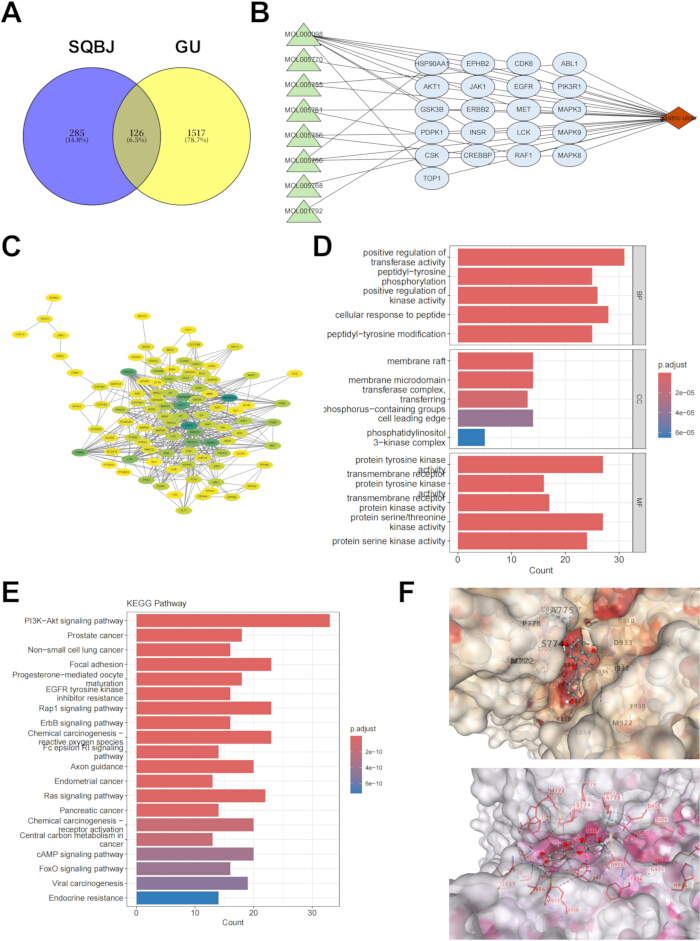
Figure 1. Network pharmacology analysis of SQBJ's potential mechanisms in treating GU. (A) Venn diagram showing overlapping key targets between SQBJ’s bioactive components and GU-associated targets. (B) Protein–protein interaction network analysis of the shared key targets between SQBJ and GU. (C) Visualization of the PPI network constructed from shared key targets between SQBJ and GU. (D) Gene Ontology enrichment analysis (covering biological process, cellular component, and molecular function categories) of shared key targets between SQBJ and GU. (E) Kyoto Encyclopedia of Genes and Genomes pathway analysis of shared key targets between SQBJ and GU. (F) Molecular docking simulations illustrating binding conformations of key bioactive components of SQBJ with core targets. Please click here to view a larger version of this figure.
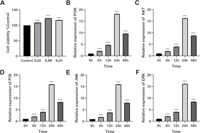
Figure 2. Dose- and time-dependent modulation of GES-1 cell viability and signaling pathways by SQBJ fractions. (A) Cell viability of GES-1 cells treated with SJD, SJM, SJH for 24 h, evaluated by CCK-8 assay. (B) Temporal expression of PI3K in GES-1 cells following SQBJ treatment. (C) Temporal expression of AKT in GES-1 cells following SQBJ treatment. (D) Temporal expression of p38 in GES-1 cells following SQBJ treatment. (E) Temporal expression of JNK in GES-1 cells following SQBJ treatment. (F) Temporal expression of ERK in GES-1 cells following SQBJ treatment. Data are presented as mean ± SD from three independent experiments. n=3. *** p. < 0.001. Please click here to view a larger version of this figure.
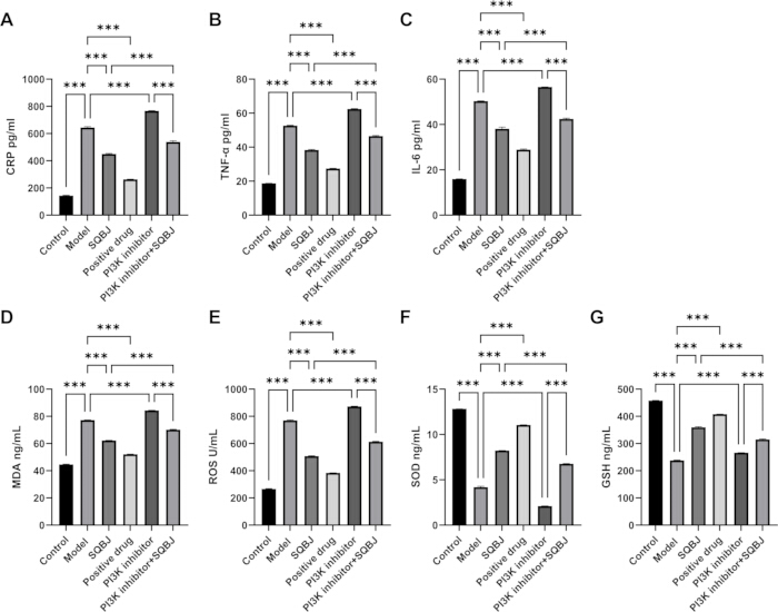
Figure 3. Attenuation of ethanol-induced inflammation and oxidative stress in GES-1 cells with involvement of PI3K/AKT-related signaling. (A) ELISA assessment of CRP levels; (B) ELISA assessment of TNF-α levels; (C) ELISA assessment of IL-6 levels; (D) ELISA assessment of MDA levels; (E) Fluorometric detection of intracellular ROS using DCFH-DA probe; (F) ELISA assessment of SOD levels; (G) ELISA assessment of GSH levels. Data are presented as mean ± SD from three independent experiments. n=3. *** p < 0.001. Abbreviations: CRP = C-reactive protein; TNF-α = tumor necrosis factor-α; IL-6 = interleukin-6; MDA = malondialdehyde; ROS = reactive oxygen species; SOD = superoxide dismutase; GSH = glutathione. Please click here to view a larger version of this figure.
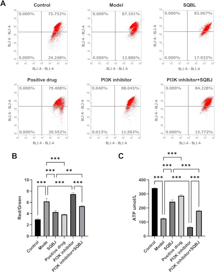
Figure 4. Mitochondrial function in ethanol-injured GES-1 cells with involvement of PI3K/AKT-related signaling. (A) Flow cytometric profiles of mitochondrial membrane potential (ΔΨm) detected via JC-1 staining across experimental groups. (B) Quantitative analysis of JC-1 red/green fluorescence ratio, reflecting ΔΨm status. (C) Intracellular ATP levels measured by luciferase assay in different treatment groups. Data are presented as mean ± SD from three independent experiments; n = 3. ** p < 0.01, *** p < 0.001. Please click here to view a larger version of this figure.
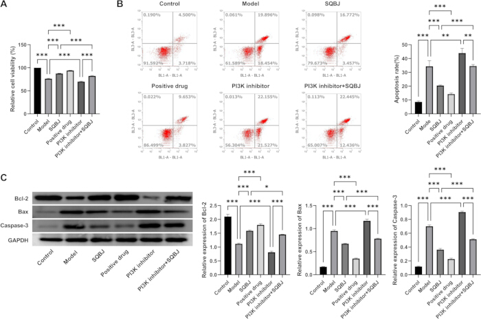
Figure 5. Regulation of GES-1 cell proliferation and apoptosis by SQBJ in association with PI3K/AKT-related signaling. (A) Relative cell viability measured by CCK-8 assay. (B) Cell apoptosis detected by Annexin V/PI double staining using flow cytometry. (C) Protein expression of Bcl-2, Bax, and Caspase-3 analyzed by western blot, with GAPDH as an internal control. Data are presented as mean ± SD from three independent experiments. n=3. * p < 0.05, *** p < 0.001. Please click here to view a larger version of this figure.
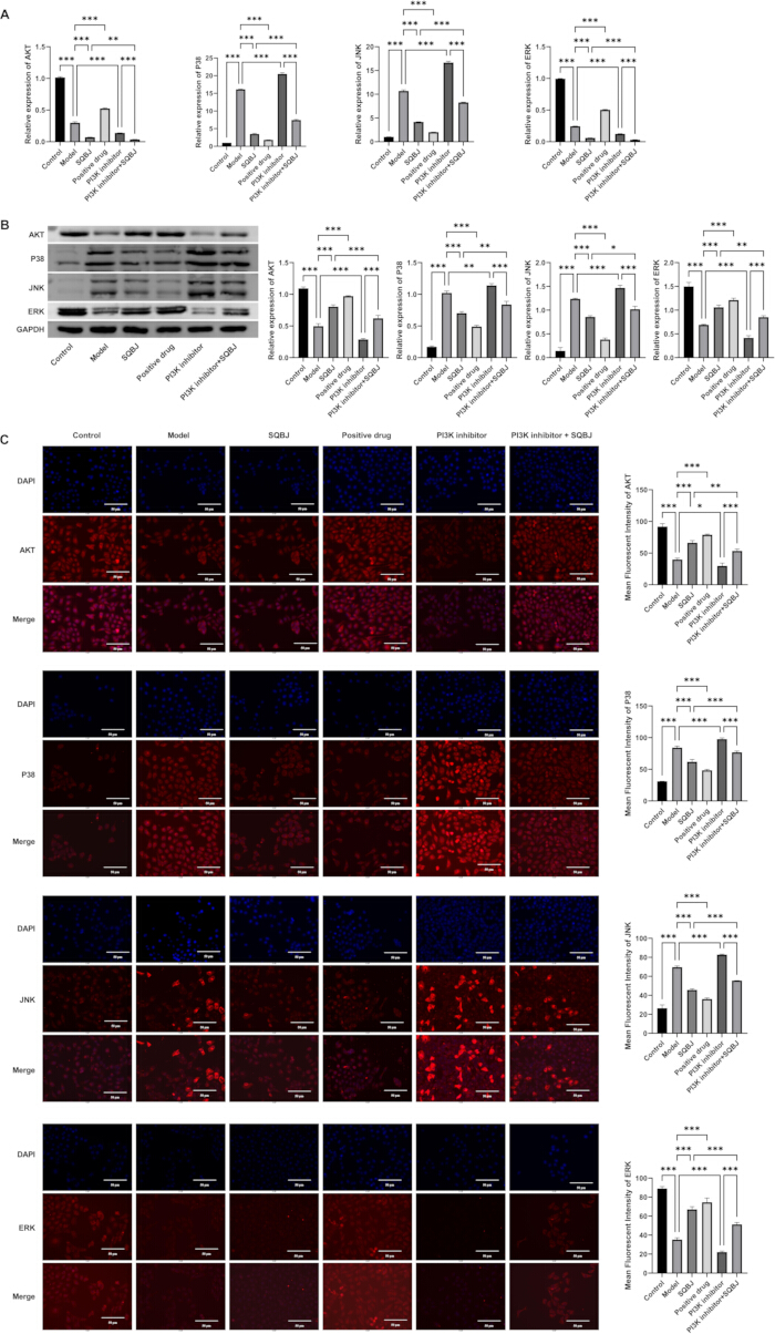
Figure 6. Regulation of PI3K/AKT- and MAPK-related signaling markers in ethanol-injured GES-1 cells. (A) mRNA expression of PI3K, AKT, p38, JNK, and ERK assessed by qPCR. (B) Protein levels of AKT, p38, JNK, and ERK detected by western blot, with GAPDH as an internal control. (C) Representative immunofluorescence staining images of AKT, p38, JNK, and ERK combined with DAPI-stained nuclei, together with quantification of mean fluorescence intensity. Scale bars = 50 µm. Data are presented as mean ± SD from three independent experiments. * p < 0.05, ** p < 0.01, *** p < 0.001. Please click here to view a larger version of this figure.
Supplemental Figure S1. Study workflow and experimental design. The schematic summarizes the overall experimental strategy of this study. SQBJ was prepared from Panax notoginseng and Bletilla striata at a 1:1 ratio, followed by aqueous extraction and HPLC-based quality control. Network pharmacology and molecular docking analyses were used to identify overlapping targets and prioritize the PI3K/AKT pathway for experimental validation. Ethanol-induced injury was established in GES-1 cells using 500 mmol/L ethanol for 4 h, and SQBJ concentrations of 5, 10, and 50 µg/mL were screened to determine the optimal intervention condition. Subsequent assays evaluated cell viability, inflammatory mediator levels, oxidative stress, mitochondrial function, apoptosis, and PI3K/AKT-MAPK-related signaling. The integrated analysis supports the protective effect of SQBJ against ethanol-induced GES-1 cell injury in vitro.Please click here to download this file.
Supplemental File 1. Network pharmacology target datasets. This file contains the supporting datasets used for the network pharmacology analysis, including gastric ulcer-associated genes/loci, disease-target relevance scores, and related target lists used to identify shared targets between SQBJ bioactive components and gastric ulcer-associated targets. These data support the analyses shown in Figure 1.Please click here to download this file.
Supplemental File 2: Raw numerical data supporting all figures in the manuscript. This Excel file contains separate sheets for each figure, including the original absorbance values, fluorescence intensities, and densitometry measurements from all three independent experiments.Please click here to download this file.
Discussion
Loading...
$$\rightleftharpoonup{xx}$$
$$\longleftharp{xx}$$,
$$\longrightharp{xx}$$,
The primary objective of this investigation was to evaluate whether SQBJ, a classical TCM compound formula composed of Panax notoginseng and Bletilla striata., possesses cytoprotective activity against ethanol-triggered gastric mucosal epithelial damage within a GES-1 cell model, with involvement of PI3K/AKT-related signaling and associated reductions in inflammatory mediators, oxidative stress indicators, mitochondrial dysfunction, and apoptotic events. The principal findings of the present work are as follows: network pharmacology analysis identified the PI3K/AKT–MAPK axis as a potential mechanistic hub of SQBJ in GU treatment; in ethanol-challenged GES-1 cells, a 10 µg/mL SQBJ extract dose-dependently and time-dependently restored cell viability, suppressed inflammatory and oxidative biomarkers, preserved mitochondrial membrane potential (ΔΨm) and ATP production, and inhibited apoptosis via Bcl-2/Bax/caspase-3 regulation; these effects were substantially weakened by the PI3K inhibitor LY294002, supporting a contributory role for PI3K/AKT-related signaling during SQBJ-mediated protection of ethanol-injured GES-1 cells.
The network pharmacology results revealed 126 overlapping targets between SQBJ and GU, with prominent enrichment in the PI3K/AKT and MAPK cascades. This multi-target prediction framework is consistent with the emerging paradigm of TCM network pharmacology16, in which a complex formula achieves synergistic efficacy by engaging numerous disease-relevant nodes simultaneously17. Notably, querying the PPI hub topology implicated PIK3CA as a central regulatory node—an observation corroborated by prior network pharmacological studies of TCM formulas in gastrointestinal disease, which consistently identify AKT1, TP53, and TNF as high-degree hubs17. The molecular docking results confirming the binding stability of core SQBJ constituents—principally quercetin and ginsenosides—to PI3K and AKT proteins are consistent with recent reports demonstrating nanomolar-range affinity of quercetin for AKT1 (PDB: 1UNQ) and the PI3K catalytic subunit18. The convergent identification of the PI3K/AKT pathway across PPI topology, KEGG enrichment, and molecular docking analyses provided the main rationale for selecting this pathway for experimental validation. Although several other pathways, including focal adhesion, cancer-related pathways, MAPK signaling, Nrf2/HO-1, and mTOR-related biological processes, were also enriched, the present study focused on PI3K/AKT because it is closely associated with gastric epithelial cell survival, mitochondrial function, oxidative stress, and apoptosis. MAPK signaling was selected as a downstream stress-related pathway for partial validation because of its close biological relationship with PI3K/AKT and ethanol-induced oxidative injury. Other enriched pathways were not experimentally examined in this study due to limitations in experimental scope and sample availability, and they should be further validated in future studies.
The potential synergistic effect of Panax notoginseng and Bletilla striata in SQBJ may be explained by the complementary target profiles suggested by the network pharmacology and molecular docking results in Figure 1, together with previously reported pharmacological evidence. According to the docking results shown in Figure 1F, the core active compounds of SQBJ exhibited stable binding conformations with key receptors associated with PI3K/AKT and MAPK signaling architecture, suggesting that different chemical constituents in SQBJ may converge on overlapping but functionally complementary signaling nodes. Ginsenosides from Panax notoginseng, such as Rg1 and Rb1, have been reported to regulate PI3K/AKT-related survival signaling and attenuate epithelial cell apoptosis and barrier dysfunction19, while Panax notoginseng saponins have demonstrated a regulatory role on autophagic flux and cell death cascades through the upstream PI3K/AKT/mTOR axis20. In contrast, Bletilla striata polysaccharides are mainly associated with mucosal protection, anti-inflammatory activity, antioxidant defense, and inhibition of MAPK/NF-κB signaling21. Therefore, Panax notoginseng may primarily activate cell-survival signaling, whereas Bletilla striata may primarily suppress inflammatory and oxidative stress-related injury. More recently, BSP-based in situ gel formulations have been shown to maintain mucosal barrier integrity and attenuate inflammation-associated apoptosis in peptic ulcer models22. In the present study, 10 µg/mL SQBJ aqueous extract showed a marked protective effect in ethanol-injured GES-1 cells. However, because SQBJ is a complex herbal extract and omeprazole is a single chemical drug, the comparison between SQBJ and omeprazole should be interpreted only as a positive-control reference rather than a strict dose-equivalence or potency comparison. Further pharmacological studies are required to establish dose conversion, active-compound equivalence, and comparative efficacy. Combined with the docking results in Figure 1F and previous studies on Panax notoginseng and Bletilla striata, these findings suggest a potential multi-component and multi-target cooperative mechanism. Nonetheless, since the current investigation did not perform a parallel evaluation of Panax notoginseng alone, Bletilla striata .alone, and their combined administration, the synergistic effect of the two herbs should be interpreted cautiously and requires further verification by component-splitting and compatibility experiments.
More specifically, the molecular docking results provide a possible mechanistic explanation for the compatibility of Panax notoginseng and Bletilla striata. Compounds derived from Panax notoginseng may interact with PI3K/AKT-related targets and thereby enhance cell survival signaling, whereas compounds derived from Bletilla striata may interact with targets involved in MAPK-mediated inflammatory and oxidative stress responses. This target complementarity is consistent with the experimental observation that SQBJ modulated PI3K/AKT- and MAPK-associated markers in ethanol-injured GES-1 cells. Nevertheless, docking analysis only predicts potential binding affinity and cannot independently prove direct compound-target binding or pharmacological synergy. Experimental binding validation, such as surface plasmon resonance, isothermal titration microcalorimetry, thermal shift testing, drug affinity-responsive target stability evaluation, or pull-down assays, is required to confirm the predicted interactions between SQBJ constituents and PI3K/AKT-MAPK pathway-related targets. Therefore, future studies should combine molecular docking, single-herb intervention, herb-pair compatibility analysis, and in vivo .experiments to fully elucidate how these components interact collaboratively.
The dual anti-inflammatory and antioxidant properties of SQBJ observed herein closely align with the established pathophysiological model of ethanol-induced gastric injury, in which direct mucosal contact with ethanol triggers a cascade of ROS overproduction, lipid peroxidation (reflected by elevated MDA), and depletion of endogenous antioxidant enzymes (SOD and GSH)13,23. Ethanol-induced oxidative stress subsequently activates NF-κB-driven expression profiles of downstream inflammatory mediators, notably TNF-α and IL-65, both of which exhibited a remarkable upregulation under ethanol challenge, whereas their levels were suppressed following administration of SQBJ. The reduction in CRP, TNF-α, and IL-6 suggests that SQBJ attenuated ethanol-induced inflammatory responses in GES-1 cells. Because NF-κB signaling was not directly examined in this study, the involvement of NF-κB can only be inferred from inflammatory cytokine changes and previous reports, rather than directly demonstrated9. Compared with sinapic acid24 and other natural compounds that attenuate ethanol-induced inflammation primarily through Nrf2/HO-1 activation, SQBJ may engage a broader signaling network that includes PI3K/AKT-related signaling.
Preservation of mitochondrial function represents an underappreciated but critical axis of gastric mucosal defense. Ethanol exposure is well documented to produce mitochondrial ROS, disrupt Δψm, and trigger mitochondrial permeability transition, with ROS predominantly co-localizing within mitochondria in gastric epithelial cells4. Our JC-1 staining and ATP quantification data demonstrated that SQBJ significantly restored Δψm and intracellular ATP levels in ethanol-challenged GES-1 cells, and these effects were attenuated by LY294002 pretreatment. A mechanistically similar observation was reported for astragaloside IV (AS-IV), which preserved ΔΨm and prevented mitochondria-dependent apoptosis in ethanol-injured rat gastric mucosa by modulating Bax/Bcl-2 expression and inhibiting caspase-9/3 activation25. Extensive literature documents that the PI3K/AKT cascade can phosphorylate and neutralize the pro-apoptotic factor BAD, thereby preventing mitochondrial cytochrome c release and preserving ΔΨm6. Our pharmacological inhibition data provide preliminary evidence linking PI3K/AKT-related signaling to the mitochondria-protective property of this TCM formula.
Regulation of the Bcl-2 family and caspase-3 cascade represents an important apoptotic mechanism by which SQBJ may reduce ethanol-induced apoptosis. In the Model group, immunoblotting demonstrated reduced Bcl-2 expression, accompanied by Bax activation and cleaved caspase-3, a pattern consistent with intrinsic mitochondrial apoptosis and supported by Annexin V/PI differential staining. SQBJ treatment reversed the observed imbalance, and this effect was weakened by PI3K inhibition. Our analytical observations run parallel with data documented in a previous flavonoid formulation report (DOFF) in GES-1 cells, where Bcl-2 upregulation and caspase pathway suppression via PI3K/AKT signaling correlated with recovery from ethanol-induced apoptosis. Prior studies on BSP have similarly demonstrated that polysaccharide-mediated gastroprotection involves enhancement of antioxidant capacity and inhibition of the apoptotic pathway in gastric tissue7. The present work suggests that the anti-apoptotic effect of complete SQBJ aqueous extract is associated with PI3K/AKT-related signaling and MAPK-related changes, extending prior single-component observations while requiring further confirmation by genetic or additional pharmacological approaches.
The crosstalk between PI3K/AKT and MAPK (p38/JNK/ERK) signaling pathways observed in the present study adds mechanistic context to the current understanding of ethanol-induced gastric epithelial injury. We found that ethanol challenge reduced AKT-related signaling markers while increasing p38, JNK, and ERK signals, indicating suppression of survival-associated signaling alongside concurrent initiation of stress-related MAPK signaling. SQBJ treatment reversed these changes; however, these effects were markedly weakened by LY294002. Notably, although the PI3K inhibitor + SQBJ cohort exhibited marginal recovery in comparison with the single inhibitor cohort, the degree of improvement was limited and did not demonstrate a clear protective effect, suggesting that SQBJ-mediated protection is closely linked to intact PI3K/AKT-related signaling. Oxidative stress is a well-established activator of JNK and p38 MAPK 26, and our data suggest that the antioxidant effects of SQBJ—through restoring SOD and GSH—may indirectly suppress MAPK activation by reducing the ROS burden that drives these stress kinases. This regulatory pattern is consistent with the multitarget therapeutic philosophy of TCM formulas. However, because direct pathway interaction experiments were not performed, the present data cannot prove that MAPK inhibition is strictly downstream of PI3K/AKT activation.
A few constraints regarding the current research warrant consideration. Primarily, our experimental observations were derived solely from a cell-based environment employing the GES-1 immortalized human gastric mucosal epithelial lineage. Therefore, although the present results demonstrate that SQBJ protects gastric epithelial cells from ethanol-induced injury, they cannot directly prove that SQBJ alleviates ethanol-induced gastric ulcer in vivo. Future studies should establish rodent models of ethanol-induced gastric ulcer to evaluate gastric lesion area, histopathological damage, mucosal inflammatory infiltration, reactive oxygen dynamics, and the engagement of the PI3K/AKT-MAPK pathway within target gastric tissues. Second, the SQBJ aqueous extract used in this study is a complex mixture of bioactive constituents. Although HPLC fingerprinting and quantitative analysis of four representative markers were performed for quality control, broader chemical profiling such as UPLC-QTOF-MS, LC-MS/MS, or metabolomic analysis was not conducted. Therefore, unidentified or low-abundance compounds may also contribute to the observed biological effects. Future studies should combine comprehensive phytochemical characterization with component-specific experiments to clarify the contribution of ginsenosides, Bletilla striata polysaccharides, militarine, and other constituents to the PI3K/AKT-related protective effects. Third, the mechanistic exploration was mainly based on pharmacological inhibition with LY294002. Although LY294002 is widely used as a PI3K inhibitor, it is not completely specific and may affect other kinases or signaling processes at certain concentrations. Therefore, off-target effects may have contributed to the observed results. Future studies should use complementary strategies, such as PI3K or AKT. siRNA knockdown, AKT activators, additional PI3K inhibitors, or genetic rescue experiments, to clarify the upstream regulatory function assigned to PI3K/AKT signaling. Fourth, the cell death profiling within our investigation was primarily restricted to the parameters of Bax, Bcl-2, and cleaved caspase-3. While these variables reflect mitochondrial apoptosis-related regulation, additional upstream mitochondrial apoptotic indicators, such as cytochrome c release, caspase-9 activation, mitochondrial Bax translocation, and apoptosome formation, were not examined. Therefore, the conclusion that SQBJ regulates mitochondrial apoptosis should be interpreted cautiously and requires further verification using a broader panel of mitochondrial apoptotic markers. Fifth, although this study measured downstream inflammatory cytokines (specifically CRP, TNF-α, and IL-6), direct documentation of NF-κB pathway activation was not evaluated. Detection of p-NF-κB p65, total NF-κB p65, IκBα degradation, or the kinetics of nuclear translocation of this transcription factor would strengthen our analytical insights into the anti-inflammatory effect of SQBJ. Notwithstanding these limitations, this investigation offers a preliminary laboratory framework characterizing the potential application of SQBJ in GU-related gastric mucosal injury and supports the broader integration of network pharmacology-guided experimental validation as a productive approach for decoding the pharmacological mechanisms of TCM compound formulas.
Disclosures
Loading...
$$\rightleftharpoonup{xx}$$
$$\longleftharp{xx}$$,
$$\longrightharp{xx}$$,
The authors have no conflicts of interest to declare.
Acknowledgements
Loading...
$$\rightleftharpoonup{xx}$$
$$\longleftharp{xx}$$,
$$\longrightharp{xx}$$,
This study was funded by Jiangsu Provincial Traditional Chinese Medicine Science and Technology Development Plan Project-General Project (2023), Project Number: MS2023118.
Materials
| Name | Company | Catalog Number | Comments |
|---|---|---|---|
| Apoptosis assay | Annexin V-FITC/PI Apoptosis Detection Kit | Beyotime | C1062L |
| ATP assay | Enhanced ATP Assay Kit | Beyotime | S0027 |
| Buffer | PBS | Solarbio | — |
| Cell digestion reagent | 0.25% trypsin-EDTA | Gibco | 25200072 |
| Cell line | GES-1 human gastric mucosal epithelial cells | Cell Bank, Chinese Academy of Sciences | SCSP-533 |
| Cell viability assay | CCK-8 reagent | — | — |
| Confocal microscope | LSM 880 confocal microscope | Zeiss | LSM 880 |
| Culture medium | RPMI-1640 | Gibco | — |
| ELISA kit | Human CRP ELISA Kit | R&D Systems | DCPR00 |
| ELISA kit | Human IL-6 ELISA Kit | R&D Systems | DTAM00D |
| ELISA kit | Human TNF-α ELISA Kit | R&D Systems | — |
| Flow cytometer | FACSCalibur / FACSCanto II | BD Biosciences | — |
| Herbal material | Bletilla striata (Thunb. ex A. Murray) Rchb. f. (tuber) | Anguo Traditional Chinese Medicine Market | Voucher No. BS-20240301 |
| Herbal material | Panax notoginseng (Burk.) F. H. Chen (root and rhizome) | Anguo Traditional Chinese Medicine Market | Voucher No. PN-20240301 |
| Herbal material | Sanqi Baiji San powder | In-house preparation | Voucher No. SQBJ-20240301 |
| HPLC column | ZORBAX SB-C18 column | Agilent Technologies | 4.6 mm × 250 mm, 5 μm |
| HPLC system | 1260 Infinity II HPLC system | Agilent Technologies | 1260 Infinity II |
| IF secondary antibody | Alexa Fluor 594–conjugated anti-rabbit IgG | Invitrogen | A11012 |
| Inverted microscope | CKX41 inverted phase-contrast microscope | Olympus | CKX41 |
| Luminescence reader | Infinite 200 PRO microplate reader | Tecan | Infinite 200 PRO |
| Membrane | PVDF membrane | Millipore | IPVH00010 |
| Microplate reader | iMark microplate reader | Bio-Rad | iMark |
| Mitochondrial membrane potential kit | JC-1 Assay Kit | Beyotime | C2006 |
| Nuclear stain | DAPI | Beyotime | C1002 |
| Online platform | Online bioinformatics plotting platform | Bioinformatics online analysis platform | Online platform |
| Online platform | Web-based enrichment analysis platform | Metascape | Online platform |
| Oxidative stress assay | GSH Assay Kit | Beyotime | S0052 |
| Oxidative stress assay | MDA Assay Kit | Beyotime | S0131 |
| Oxidative stress assay | SOD Assay Kit | Beyotime | S0109 |
| PI3K inhibitor | LY294002 | MCE | S1105 |
| Positive control drug | Omeprazole | MCE | HY-B0053 |
| Primary antibody | AKT | Cell Signaling Technology | 4691 |
| Primary antibody | Bax | Cell Signaling Technology | 5023 |
| Primary antibody | Bcl-2 | Cell Signaling Technology | 15071 |
| Primary antibody | Cleaved Caspase-3 Asp175 | Cell Signaling Technology | 9664 |
| Primary antibody | ERK1/2 | Cell Signaling Technology | 4695 |
| Primary antibody | GAPDH | Abcam | ab8245 |
| Primary antibody | JNK / SAPK-JNK | Cell Signaling Technology | 9252 |
| Primary antibody | p-AKT Ser473 | Cell Signaling Technology | 4060 |
| Primary antibody | p-ERK1/2 Thr202/Tyr204 | Cell Signaling Technology | 4370 |
| Primary antibody | p-JNK Thr183/Tyr185 | Cell Signaling Technology | 4668 |
| Primary antibody | p-p38 MAPK Thr180/Tyr182 | Cell Signaling Technology | 4511 |
| Primary antibody | p-PI3K p85 Tyr458 / p55 Tyr199 | Cell Signaling Technology | 4228 |
| Primary antibody | p38 MAPK | Cell Signaling Technology | 8690 |
| Primary antibody | PI3K p85 | Cell Signaling Technology | 4257 |
| Primer | AKT forward | — | — |
| Primer | AKT reverse | — | — |
| Primer | ERK forward | — | — |
| Primer | ERK reverse | — | — |
| Primer | GAPDH forward | — | — |
| Primer | GAPDH reverse | — | — |
| Primer | JNK forward | — | — |
| Primer | JNK reverse | — | — |
| Primer | p38 forward | — | — |
| Primer | p38 reverse | — | — |
| Primer | PI3K forward: | — | — |
| Primer | PI3K reverse | — | — |
| Protease inhibitor | PMSF | Beyotime | ST506 |
| Protein assay | BCA Protein Assay Kit | Beyotime | P0010 |
| Protein lysis buffer | RIPA buffer | Beyotime | P0013B |
| Public database | DrugBank | DrugBank Online | Online database |
| Public database | GeneCards | Weizmann Institute of Science | Online database |
| Public database | Online Mendelian Inheritance in Man | Johns Hopkins University | Online database |
| Public database | PubChem compound database | National Center for Biotechnology Information | Online database |
| Public database | RCSB Protein Data Bank | RCSB PDB | Online database |
| Public database | STRING protein interaction database | STRING Consortium | Online database |
| Public database | Traditional Chinese Medicine Systems Pharmacology Database and Analysis Platform | TCMSP | Online database |
| Public database | Universal Protein Resource | UniProt Consortium | Online database |
| Real-time PCR system | CFX96 Real-Time PCR System | Bio-Rad | CFX96 |
| Reverse transcription kit | PrimeScript RT Reagent Kit with gDNA Eraser | Takara Bio | RR047A |
| RNA extraction reagent | TRIzol reagent | — | — |
| ROS probe | DCFH-DA | Beyotime | S0033 |
| Secondary antibody | HRP-conjugated anti-rabbit IgG | Cell Signaling Technology | 7074 |
| Software | Cytoscape | Cytoscape Consortium | 3.9.1 |
| Software | GraphPad Prism | GraphPad Software | Prism 9.5.1 |
| Software | Image analysis software | National Institutes of Health | ImageJ |
| Software | ImageJ | NIH | ImageJ |
| Software | Molecular docking software | AutoDock Tools | Version not specified |
| Software | Molecular format conversion software | OpenBabel | Version 2.3.1 |
| Software | Molecular visualization software | PyMOL | Version 2.3.1 |
| Software | Network visualization software | Cytoscape Consortium | Version 3.9.1 |
| Software | R statistical software | R Foundation for Statistical Computing | Version not specified |
| Software | Statistical analysis software | GraphPad Software | Version 9.5.1 |
| Supplement | Fetal bovine serum | Gibco | — |
| Supplement | Penicillin–streptomycin | Gibco | 15140122 |
Reprints and Permissions
Request permission to reuse the text or figures of this JoVE article
Request Permission