This study evaluates miR-21-5p expression, its association with cell adhesion molecule 1, and its relationships with migration, proliferation, inflammatory-marker expression, and survival in non-small cell lung cancer tissues and cell lines.
Research Article
The miR-21-5p-Cell Adhesion Molecule 1 Axis in Non-Small Cell Lung Cancer Progression and Prognosis
83 views
⸱
September 11th, 2026
* These authors contributed equally
In This Article
Summary
Abstract
Evidence indicates that miR-21-5p is an important molecular regulator in non-small cell lung cancer (NSCLC). Cell adhesion molecule 1 (CADM1) is a potential tumor suppressor, but the regulatory relationship between miR-21-5p and CADM1 in the progression of NSCLC remains unclear. Quantitative reverse-transcription PCR (qRT-PCR) was used to detect the expression of miR-21-5p and CADM1 in NSCLC tissues and cells; target binding of miR-21-5p and CADM1 was verified by dual luciferase reporter gene assay; the cell migration ability was detected by the Transwell assay; the clinicopathological data and survival analysis were combined to explore the clinical significance of miR-21-5p; the function enrichment of miR-21-5p target genes was analyzed by bioinformatics. The expression of miR-21-5p in NSCLC tissues was significantly higher than in adjacent normal tissues, and patients with high miR-21-5p expression had a shorter overall survival. High expression of miR-21-5p is an independent risk factor for the survival of NSCLC patients; overexpression of miR-21-5p promoted cell migration in NSCLC cells and upregulated the expression of inflammatory factors; the dual luciferase reporter gene assay confirmed that miR-21-5p directly targeted CADM1; overexpression of CADM1 inhibited cell migration in NSCLC cells and downregulated the expression of inflammatory factors; bioinformatics analysis showed that miR-21-5p target genes were enriched in multiple signaling pathways related to tumor occurrence and development. miR-21-5p promotes NSCLC progression by targeting and inhibiting CADM1 and may serve as a prognostic biomarker and therapeutic target.
Introduction
Across the globe, lung cancer ranks as a leading cause of cancer-related mortality worldwide, with its incidence steadily rising1,2. Among East Asian populations, lung cancer incidence and mortality reach 222.1 and 155.5 per 100,000 men, versus 158.1 and 87.3 per 100,000 women, respectively. Non-small cell lung cancer (NSCLC) accounts for ~80% of all lung malignancies and encompasses three major histological subtypes3. Compared with highly aggressive small cell lung cancer (SCLC), NSCLC progresses and metastasizes more slowly; however, vague early clinical symptoms result in over 75% of patients receiving diagnoses at intermediate or advanced stages, contributing to persistently poor 5-year overall survival4. Lung tumorigenesis is driven by oncogenic activation and tumor suppressor gene silencing5, highlighting an urgent need to dissect the molecular underpinnings of NSCLC to advance novel therapeutics6.
MicroRNAs (miRNAs, 18–25 nucleotides in length) mediate post-transcriptional gene repression by binding to the 3′ untranslated region (3′-UTR) of target mRNAs, triggering translational suppression or mRNA degradation7. Biogenesis of miRNAs initiates with RNA polymerase II transcription to generate primary miRNA transcripts, which are then cleaved by RNase III enzymes to form hairpin-shaped precursor miRNAs (pre-miRNAs)8. While most miRNAs repress gene expression via partial or full sequence complementarity to mRNA 3′-UTRs, certain miRNAs can boost mRNA translation under specific cell cycle contexts9. To date, hundreds of human miRNAs have been identified, which govern core biological processes including cell proliferation, differentiation, and apoptosis10. Previously, detection of aberrantly expressed miRNAs remained technically challenging; the advent of high-throughput sequencing has enabled rapid, unbiased profiling of dysregulated miRNAs in disease tissues11. Here, we focus on miR-21-5p, one of the earliest-characterized RNA polymerase II-transcribed miRNAs, whose oncogenic regulatory functions are under intensive investigation12.
miR-21-5p is encoded within an intron of the TMEM49 gene and is consistently overexpressed across multiple human malignancies, including esophageal, colorectal, and lung cancers13. Existing preclinical NSCLC data confirm its oncogenic activity: elevated miR-21-5p accelerates tumor proliferation, epithelial-mesenchymal transition (EMT), and distant metastasis by silencing anti-oncogenic targets such as PTEN and PDCD414. Nevertheless, prior work largely focuses on miR-21-driven apoptotic and proliferative signaling, and few reports systematically interrogate the miR-21-5p/CADM1 regulatory axis in NSCLC15. Cell adhesion molecule 1 (CADM1) is a well-established tumor suppressor across epithelial cancers16; it preserves intercellular adhesion to block tumor cell detachment and metastatic dissemination, and its downregulation correlates with advanced tumor stage and worse survival in NSCLC patient cohorts. Mechanistically, CADM1 also restrains pro-tumor nuclear factor kappa B (NF-κB) inflammatory signaling, which fuels metastasis by inducing pro-inflammatory cytokine secretion and remodeling the tumor microenvironment17. Despite separate documentation of miR-21-5p oncogenicity and CADM1 tumor-suppressive effects, three critical knowledge gaps persist for the miR-21-5p/CADM1 axis in NSCLC: first, direct binding and repression of CADM1 by miR-21-5p lack experimental validation in lung cancer cells; second, whether this axis drives malignant phenotypes via downstream inflammatory pathways remains undefined; third, the prognostic predictive capacity of combined miR-21-5p/CADM1 expression signatures has not been assessed in clinical NSCLC specimens.
Based on these unresolved questions, we propose a central mechanistic hypothesis: upregulated miR-21-5p in NSCLC directly binds the CADM1 3′-UTR to suppress CADM1 expression, thereby derepressing CADM1-inhibited metastatic inflammatory signaling, thereby accelerating NSCLC migration and conferring an inferior clinical prognosis. This study pursues three predefined research aims: (1) Verify direct molecular targeting of CADM1 by miR-21-5p and evaluate migration in A549 and H1299 NSCLC cell lines; (2) Elucidate whether the miR-21-5p/CADM1 cascade modulates metastatic potential through inflammatory signal transduction; (3) Evaluate the clinical prognostic value of the miR-21-5p/CADM1 expression signature to identify new diagnostic biomarkers and therapeutic candidates for metastatic NSCLC.
Protocol
This retrospective study was reviewed and approved by the Ethics Committee of Quzhou Hospital of Traditional Chinese Medicine (approval no. 202402208). Written informed consent was obtained from every enrolled patient or a legal guardian before sample collection.
Sample collection
Primary NSCLC tumor tissues were surgically harvested from enrolled patients, while their matched adjacent normal lung tissues were retrospectively collected from the National Engineering Research Center for Clinical Biopsy (Quzhou City, Zhejiang Province, China). A total of 157 NSCLC patient specimens consecutively enrolled between January 2019 and December 2024 were included in this cohort study. The sample size of 157 paired specimens was statistically justified based on previously published NSCLC miRNA prognostic research: assuming a moderate effect size of 0.5, α = 0.05, statistical power of 80%, the minimum required sample size was calculated as 122 cases. Considering potential specimen loss, severe tissue necrosis, and patients lost to follow-up, we consecutively enrolled 157 eligible patients from January 2019 to December 2024 to guarantee sufficient statistical power. All tumor and matched adjacent normal lung tissues were harvested from the same patient during surgical resection, forming paired samples for subsequent comparative analyses.
Inclusion criteria were: (1) Patients pathologically confirmed as primary NSCLC via postoperative biopsy; (2) Complete clinical baseline, histopathological staging, and long-term follow-up survival data; (3) No prior radiotherapy, chemotherapy, or targeted therapy before tissue sampling; (4) Complete paired tumor and paracancerous tissue specimens available.
Exclusion criteria were: (1) Combined secondary malignant tumors of other organs; (2) Patients lost to follow-up within 3 months after surgery; (3) Specimens with severe necrosis or insufficient tissue volume for subsequent molecular detection.
All fresh tissue samples were placed in liquid nitrogen within 15 min after surgical resection, transferred to an ultra-low-temperature freezer, and stored at −80 °C for RNA and protein extraction. Clinical follow-up was performed regularly, and overall survival was measured in months from pathological diagnosis until death or the last follow-up cutoff.
Stratification of miR-21-5p expression level
miR-21-5p relative expression values of all 157 patients were sorted in ascending order, and the cohort was stratified by median expression cutoff. Owing to multiple tied values around the median, patients with expression below the median were assigned to the low-expression group (n = 61); patients with expression equal to or greater than the median, including all tied observations at the median, were classified into the high-expression group (n = 96). This tied-value allocation rule was pre-specified prior to grouping to resolve duplicate median readings, and the grouping result was cross-verified against the raw qRT-PCR quantification data.
Cell culture
Human NSCLC cell lines A549 and H1299 were obtained from a commercial supplier (see Table of Materials). A549 cells were maintained in RPMI-1640 medium, and H1299 cells were cultured in DMEM. Both cell culture media were enriched with 10% heat-inactivated fetal bovine serum (FBS), penicillin (100 U/mL), and streptomycin (100 µg/mL). Cells were cultured under standard conditions at 37 °C in a humidified incubator supplied with 5% CO₂.
Short tandem repeat (STR) profiling was performed to authenticate the identity of A549 and H1299 cell lines prior to all functional experiments. Mycoplasma contamination testing was conducted monthly using a commercial PCR-based mycoplasma detection kit, and only mycoplasma-negative cells were used for subsequent assays.
Cells in the logarithmic growth phase with intact morphology were harvested for transient transfection. A lipid-based transfection reagent (see Table of Materials) was used according to the manufacturer’s instructions. Downstream cellular assays were conducted 50 h after transfection.
Cell transfection
For transfection, cells in the logarithmic growth phase with intact morphology were seeded in culture plates 24 h in advance to reach 60%–70% confluence. Transfection was performed with a lipid-based transfection reagent (see Table of Materials) according to the manufacturer’s instructions. Four experimental groups were used: mimic negative control (mimic-NC), miR-21-5p mimic, inhibitor negative control (inhibitor-NC), and miR-21-5p inhibitor. The nucleotide sequences are provided in Supplemental File 1.
Total RNA was extracted 50 h after transfection, and quantitative reverse-transcription PCR (qRT-PCR) was performed to measure the relative expression of miR-21-5p and verify transfection efficiency. Cellular migration assays were performed 50 h after transfection. For transient transfection, cells were treated with the miR-21-5p mimic or mimic negative control (mimic-NC) at a final concentration of 50 nM, whereas the miR-21-5p inhibitor and inhibitor negative control (inhibitor-NC) were introduced at 100 nM. To avoid confounding effects attributable to differences in nucleic acid input, the total amount of transfected nucleic acid was kept identical across all four treatment groups.
Cell proliferation was evaluated using the Cell Counting Kit-8 (CCK-8) assay. Stably transfected cells were plated in 96-well plates at 3 × 10³ cells/well, with six technical replicates established for each group. At 24, 48, and 72 h after seeding, 10 µL of CCK-8 reagent was added to each well, followed by incubation at 37 °C for 2 h. Cell proliferation was quantified by measuring the absorbance at 450 nm using a microplate reader.
All transient transfection, CCK-8 and migration experiments were performed with no less than three independent biological replicates, and each biological replicate contained 3–6 technical replicates as described above.
Quantitative reverse-transcription PCR
Total RNA from NSCLC cells and paired clinical tumor and adjacent normal tissues was isolated with a phenol/guanidine-based RNA extraction reagent (see Table of Materials). RNA purity and concentration were determined by ultraviolet spectrophotometry; only samples with optical-density (OD) 260/280 and OD 260/230 ratios of 1.8–2.1 underwent reverse transcription. Primer sequences are provided in Supplemental File 1. The reverse transcription system contained 500 ng of total RNA, 2 µL of reverse transcription buffer, 1 µL of dNTP mixture, 0.5 µL of reverse transcriptase, 1 µL of specific stem-loop primer for miRNA or oligo(dT) primer for mRNA, and RNase-free water to a total volume of 20 µL. The qRT-PCR amplification system (20 µL total volume) consisted of 10 µL of SYBR Green qPCR master mix, 0.4 µL of forward primer (10 µM), 0.4 µL of reverse primer (10 µM), 2 µL of diluted cDNA template, and 7.2 µL of RNase-free water.
Messenger RNA for CADM1 and GAPDH was reverse-transcribed with a reverse-transcription kit, whereas miR-21-5p was reverse-transcribed with stem-loop primers and exogenous cel-miR-39 as a spike-in control. Exogenous cel-miR-39 spike-in was added to each RNA sample immediately after total RNA isolation and before reverse transcription. As a synthetic C. elegans microRNA with no homologous sequence in human transcripts, cel-miR-39 can correct the deviation caused by differential RNA extraction efficiency, RNA degradation loss, and reverse transcription efficiency variation across different clinical tissue samples, serving as an exogenous universal normalization control for miRNA quantification. qRT-PCR was performed with a fluorescent DNA-binding dye-based master mix on a real-time PCR system (see Table of Materials): 95 °C for 3 min, followed by 40 cycles at 95 °C for 10 s, 60 °C for 30 s, and 72 °C for 20 s, with melting-curve analysis to confirm specific amplification. Each sample had three biological replicates with three technical replicates each, and relative expression was calculated using the 2⁻ΔΔCt method. Full primer sequences for CRP, TNF-α, and IL-6 mRNA amplification are provided in Supplemental File 1 alongside CADM1, GAPDH, and miR-21-5p primer sequences.
Establishment of lentivirus-mediated stable miR-21-5p expression
Commercial lentiviral vectors LV-miR-21-5p and empty control LV-NC were packaged in 293T cells. The miR-21-5p precursor sequence was inserted into a lentiviral backbone carrying puromycin resistance and GFP reporter genes. Lentiviral supernatants were collected at 48 h and 72 h post-transfection, concentrated, and purified for cell infection.
A549 and H1299 cells were seeded 24 h before infection to reach 50–60% confluence. Cells were infected at MOI = 10 (A549) and MOI = 15 (H1299) with 5 µg/mL polybrene to improve transfection efficiency. Medium was replaced 24 h post-infection, and cells were cultured for another 48 h prior to puromycin screening.
Stable cell selection was conducted with 5 µg/mL puromycin for 10 consecutive days; uninfected blank cells were used to verify complete elimination of non-transduced cells within 7 days. Single GFP-positive clones were isolated and expanded to generate stable monoclonal cell lines LV-21 and LV-NC.
For CADM1 rescue assays, commercial LV-CADM1 overexpression and sh-CADM1 knockdown lentiviruses were prepared. Four experimental groups were established: LV-NC, LV-miR-21-5p, LV-CADM1, and LV-miR-21-5p + LV-CADM1 rescue group. CADM1 lentiviruses were infected at the same MOI and puromycin screening conditions as miR-21-5p vectors. All rescue proliferation and migration experiments were repeated with three independent biological replicates.
Validation of stable miR-21-5p overexpression
Total RNA was extracted from harvested LV-21 and LV-NC stable cell clones. qRT-PCR was conducted to detect the relative expression level of hsa-miR-21-5p using cel-miR-39 as an exogenous reference. Cell clones with more than 8-fold upregulation of miR-21-5p relative to the LV-NC group were retained for all subsequent functional assays, confirming the successful construction of miR-21-5p-stably overexpressing cell lines.
Dual-luciferase reporter assay
Wild-type CADM1 3'-UTR reporter plasmid (WT-CADM1) containing the predicted miR-21-5p binding sequence and mutant CADM1 3'-UTR reporter plasmid (MUT-CADM1) with a mutated binding seed region were constructed. A549 cells were seeded into 24-well plates 24 h before transfection to reach 60%–70% confluence. Each well was co-transfected with 500 ng of WT/MUT reporter plasmid, 50 nM miR-21-5p mimic or mimic-NC, and Renilla luciferase internal reference plasmid at a fixed mass ratio. After 48 h of post-transfection incubation, cells were fully lysed, and firefly and Renilla luciferase luminescence signals were sequentially detected using a dual-luciferase detection kit. The relative luciferase activity was calculated as the ratio of firefly luminescence value to Renilla luminescence value. Each experimental group contained three independent biological replicates, with three technical replicates per biological replicate, and a two-group independent sample t-test was used for statistical comparison of relative luciferase activity between mimic-NC and miR-21-5p mimic groups.
Transwell migration assay
For the Transwell migration assay, stably transfected cells were suspended in serum-free medium and plated into the upper inserts at 4 × 10⁴ cells per well. The lower chambers were filled with complete culture medium supplemented with 10% FBS to establish a chemotactic gradient. Following incubation for 48 h at 37 °C in a humidified atmosphere containing 5% CO₂, cells remaining on the upper surface of the membrane were carefully removed. Cells that had migrated to the underside of the membrane were fixed in methanol and stained with crystal violet. Migration was quantified by counting cells in five randomly selected microscopic fields per insert. Each experiment was performed using three independent biological replicates, with three technical replicates included for each biological replicate.
Statistical analyses
All in vitro experiments were conducted with a minimum of three independent biological replicates, each accompanied by corresponding technical replicates. Data distribution was first evaluated using the Shapiro-Wilk normality test to guide statistical analysis. For paired comparisons between tumor and adjacent normal tissues, either the paired t-test or the paired Wilcoxon signed-rank test was applied, depending on data distribution. Comparisons between two independent groups were analyzed using either the independent t-test or the Mann-Whitney U test. Differences among multiple groups, including rescue experiments, were assessed by one-way analysis of variance (ANOVA) followed by Bonferroni's post hoc correction, whereas repeated-measures ANOVA was used to analyze longitudinal datasets such as the CCK-8 proliferation and wound-healing assays. Associations between categorical clinical variables were examined using the χ2 test, and the relationship between miR-21-5p and CADM1 expression was evaluated by Pearson correlation analysis. Patient survival was analyzed using Kaplan-Meier curves with comparisons performed by the log-rank test. Independent prognostic factors were identified through univariate and multivariate Cox proportional hazards regression analyses. Statistical significance was defined as P < 0.05. Multivariate Cox regression adjusted for age, sex, histological type, miR-21-5p level, and lymph node status (reference: age ≤ 60, female, squamous carcinoma, low miR-21-5p, node-negative); Schoenfeld residuals checked the proportional hazards assumption, variables with univariate P < 0.10 were included via enter selection.
Results
Correlation between miR-21-5p expression and clinicopathological characteristics of NSCLC patients
A total of 157 NSCLC tumor samples were stratified into miR-21-5p low-expression (n = 61) and high-expression (n = 96) groups based on median miR-21-5p levels, and chi-square analyses revealed no significant correlations of miR-21-5p expression with patient age (χ2 = 1.36, P = 0.2435), sex (χ2 = 1.06, P = 0.3032), histological type (χ2 = 1.09, P = 0.2965) or tumor size (χ2 = 5.623, P = 0.0601), whereas high miR-21-5p expression was significantly associated with advanced tumor grade (χ2 = 16.25, P = 0.00006), positive lymph node metastasis (χ2 = 12.17, P = 0.00049) and higher T stage (χ2 = 4.30, P = 0.117), suggesting miR-21-5p exerts oncogenic effects facilitating NSCLC progression (Table 1).
Multivariable Cox proportional hazards regression analysis for independent prognostic factors of NSCLC
Multivariable Cox regression analysis incorporating age, sex, histological type, miR-21-5p expression and lymph node metastasis revealed that high miR-21-5p expression (hazard ratio [HR] = 3.066; 95% confidence interval [CI]: 1.412–6.659; P = 0.005) and positive lymph node metastasis (HR = 22.295; 95% CI: 3.039–163.588; P = 0.002) were independent unfavorable prognostic risk factors for poor overall survival of NSCLC patients, while patient age, sex, and histological subtype exerted no significant independent prognostic impacts (all P > 0.05) (Table 2).
Clinical expression characteristics of miR-21-5p and its correlation with CADM1 and inflammatory factors
qRT-PCR detection of paired NSCLC tumor and adjacent normal tissues showed significantly elevated relative miR-21-5p levels in cancer tissues compared with normal controls (Figure 1A, P < 0.001). Kaplan-Meier survival curves showed that patients with high miR-21-5p expression had significantly poorer overall survival than those with low miR-21-5p expression (Figure 1B, P = 0.0027). The mRNA abundances of inflammatory mediators C-reactive protein (CRP), tumor necrosis factor alpha (TNF-α), and interleukin-6 (IL-6) were all markedly upregulated in NSCLC specimens (Figure 1C, P < 0.01, P < 0.001). Pearson correlation analysis revealed a strongly significant negative correlation between miR-21-5p and CADM1 mRNA expression in clinical samples (r = -0.7773, 95% CI: -0.8325 to -0.7076, P < 0.0001, Figure 1D).
Cellular functional validation of miR-21-5p in NSCLC cells
qRT-PCR confirmed successful transfection efficiency of miR-21-5p mimic in A549 and H1299 cells (Figure 2A). Transwell assays showed that miR-21-5p overexpression significantly increased relative migration capacity and cell migration counts in both cell lines (Figure 2B,C). qRT-PCR detection of inflammatory cytokines revealed markedly increased mRNA levels of CRP, TNF-α, and IL-6 after miR-21-5p mimic transfection, whereas miR-21-5p inhibitor treatment reversed this upregulation (Figure 2D). Rescue experiments verified that miR-21-5p mimic drastically suppressed CADM1 transcription, and LV-CADM1 co-transfection fully restored CADM1 mRNA expression in A549 and H1299 cells (Figure 2E,F). Collectively, these cellular data demonstrated that miR-21-5p promotes NSCLC cell migration and inflammatory response via repressing CADM1.
Effects of miR-21-5p on NSCLC cell proliferation
CCK-8 proliferation curves at 24 h, 48 h, and 72 h showed that miR-21-5p mimic transfection markedly increased OD values at 450 nm in both A549 and H1299 cells, while miR-21-5p inhibitor significantly suppressed cell viability, with statistically significant differences observed at 72 h (Figure 3A,B; P < 0.001). These data demonstrated that miR-21-5p exerts a pro-proliferative effect on NSCLC cells.
Validation of CADM1 as the direct target of miR-21-5p via bioinformatics prediction and dual-luciferase reporter assay
Three online miRNA target prediction databases (miRWalk, StarBase, TargetScan) were intersected to screen candidate downstream genes of miR-21-5p, and 95 overlapping target genes were obtained (Figure 4A). Gene Ontology (GO) enrichment analysis indicated that these candidate genes were mainly enriched in biological processes, cellular components, and molecular functions related to cell metabolism, stress response, and transcription regulation (Figure 4B). Kyoto Encyclopedia of Genes and Genomes (KEGG) pathway enrichment further revealed prominent enrichment in tumor-associated signaling cascades, including mitogen-activated protein kinase (MAPK) and Ras pathways (Figure 4C). Dual-luciferase reporter assay demonstrated that miR-21-5p mimic significantly suppressed the relative luciferase activity of wild-type CADM1 3'-UTR reporter, while no inhibitory effect was observed in mutant CADM1 3'-UTR group (Figure 4D, P < 0.01, ns: not significant), confirming the direct binding between miR-21-5p and CADM1.
Promotion of NSCLC malignant phenotypes via suppression of CADM1 in rescue experiments
qRT-PCR verified the transfection efficiency of miR-21-5p mimic and inhibitor in A549 and H1299 cells (Figure 5A). CADM1 overexpression (LV-CADM1) reduced mRNA levels of pro-inflammatory factors CRP, TNF-α, and IL-6, whereas CADM1 knockdown with small interfering RNA (si-CADM1) markedly elevated their expression (Figure 5B). Transwell migration assays showed that LV-CADM1 transfection significantly suppressed the migratory capacity of both NSCLC cell lines (Figure 5C,D). CCK-8 proliferation detection further demonstrated that LV-21-mediated pro-proliferative effect was fully offset by LV-CADM1 co-transfection, whereas si-CADM1-induced cell growth acceleration could be rescued by miR-21-5p inhibitor (Figure 5E,F). Collectively, these rescue data validated that CADM1 acts as a functional downstream mediator responsible for miR-21-5p-triggered proliferation, migration, and inflammatory activation in NSCLC.
DATA AVAILABILITY:
The source data supporting the findings of this study are provided in Supplemental File 2 and are available with the published article.
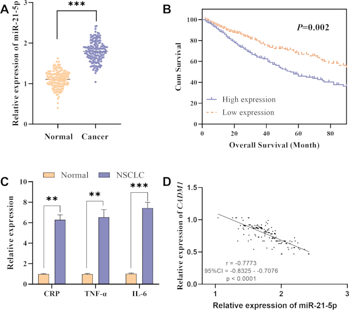
Figure 1: Expression profiles of miR-21-5p, inflammatory factors, and their correlation with CADM1 in clinical NSCLC samples. (A) Relative expression of miR-21-5p in normal lung tissues and NSCLC tumor tissues. (B) Kaplan-Meier overall survival curves of NSCLC patients stratified by miR-21-5p expression level. (C) Relative mRNA expression of inflammatory factors CRP, TNF-α, and IL-6 in normal and NSCLC tissues. (D) Pearson correlation scatter plot showing the negative linear correlation between miR-21-5p and CADM1 mRNA expression. **P < 0.01, ***P < 0.001. For Panel C: statistical comparisons were performed between paired tumor tissues and adjacent normal tissues for CRP, TNF-α, and IL-6, respectively; **P < 0.01, ***P < 0.001 denote significant differences vs matched normal lung tissues. All panels included ≥3 independent biological replicates; qRT-PCR data present mean ± standard deviation; paired t-test was adopted for inter-group comparison; scatter plot correlation analysis used Pearson test. Abbreviations: CADM1 = cell adhesion molecule 1; CI = confidence interval; CRP = C-reactive protein; IL-6 = interleukin-6; NSCLC = non-small cell lung cancer; TNF-α = tumor necrosis factor alpha. Please click here to view a larger version of this figure.
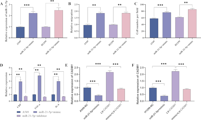
Figure 2: Cellular phenotypes and CADM1 expression regulated by miR-21-5p in A549 and H1299 NSCLC cells. (A) qRT-PCR detection of miR-21-5p expression after mimic transfection. (B) Statistical quantification of relative cell migration from Transwell assays. (C) Quantification of migrated cell number per microscopic field. (D) Relative mRNA expression of CRP, TNF-α, and IL-6 under miR-21-5p overexpression or inhibition. (E) Relative CADM1 mRNA expression in A549 rescue groups (NC, miR-21-5p mimic, LV-CADM1, mimic+LV-CADM1). (F) Relative CADM1 mRNA expression in H1299 rescue groups. **P < 0.01, ***P < 0.001. All cellular assays were repeated for three independent biological replicates with three technical replicates each; error bars represent mean ± SD; inter-group comparisons adopted one-way ANOVA with Bonferroni correction; group labels strictly match the transfection groups described in Methods. Abbreviations: CADM1 = cell adhesion molecule 1; CRP = C-reactive protein; IL-6 = interleukin-6; LV = lentiviral vector; NC = negative control; NSCLC = non-small cell lung cancer; qRT-PCR = quantitative reverse-transcription PCR; TNF-α = tumor necrosis factor alpha. Please click here to view a larger version of this figure.
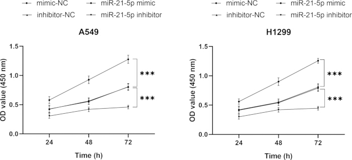
Figure 3: CCK-8 proliferation curves reflecting cell viability after miR-21-5p overexpression or knockdown. (A) Time-dependent OD₄₅₀ absorbance curves of A549 cells transfected with mimic-NC, miR-21-5p mimic, inhibitor-NC, or miR-21-5p inhibitor. (B) Time-dependent OD₄₅₀ absorbance curves of H1299 cells under the four transfection groups. ***P < 0.001. CCK-8 assays contained six technical replicates per group across 3 biological repeats; data are presented as mean ± SD; repeated-measures ANOVA was used for time-point comparisons. Abbreviations: CCK-8 = Cell Counting Kit-8; NC = negative control; OD = optical density. Please click here to view a larger version of this figure.
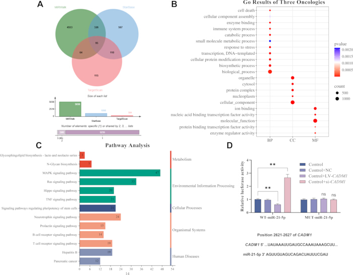
Figure 4: Bioinformatics screening and dual-luciferase assay for miR-21-5p target gene CADM1. (A) Venn diagram illustrating the common target genes of miR-21-5p identified by the miRWalk, StarBase, and TargetScan prediction databases. (B) Bubble plot of GO functional enrichment analysis for intersected target genes. (C) Bar plot of the top enriched KEGG signaling pathways. (D) Dual-luciferase reporter activity of wild-type and mutant CADM1 3'-UTR after miR-21-5p overexpression. **P < 0.01; ns, no significance. Panel D only adopts WT-CADM1 3'-UTR and MUT-CADM1 3'-UTR co-transfected with miR-21-5p mimic/mimic-NC for dual-luciferase detection; the labels Control/overexpression/interference shown in raw plotting drafts were adjusted to standard WT/MUT grouping in the final figures. Target prediction analysis used three online databases; enrichment P values were adjusted via Benjamini-Hochberg FDR correction; dual-luciferase experiments had three biological replicates (three technical replicates each); error bars represent mean ± SD; two-group t-test for statistical comparison. Abbreviations: CADM1 = cell adhesion molecule 1; GO = Gene Ontology; KEGG = Kyoto Encyclopedia of Genes and Genomes; MUT = mutant; NC = negative control; ns = not significant; UTR = untranslated region; WT = wild type. Please click here to view a larger version of this figure.
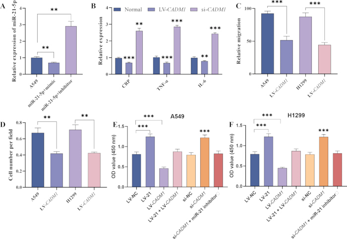
Figure 5: Functional rescue experiments verifying the miR-21-5p/CADM1 regulatory axis in A549 and H1299 cells. (A) Relative miR-21-5p expression after mimic or inhibitor transfection. (B) Relative mRNA expression of CRP, TNF-α, and IL-6 in LV-CADM1 and si-CADM1 groups. (C) Statistical quantification of relative migration ability under CADM1 overexpression. (D) Quantification of migrated cell number per microscopic field in LV-CADM1 groups. (E) CCK-8 OD₄₅₀ values of A549 rescue groups (LV-NC, LV-21, LV-CADM1, LV-21+LV-CADM1, si-NC, si-CADM1, si-CADM1+miR-21 inhibitor). (F) CCK-8 OD₄₅₀ values of H1299 rescue groups with identical transfection treatments. **P < 0.01, ***P < 0.001. All rescue assays included three independent biological replicates; error bars indicate mean ± SD, one-way ANOVA with Bonferroni post-hoc test for multi-group comparisons. Abbreviations: CADM1 = cell adhesion molecule 1; CRP = C-reactive protein; IL-6 = interleukin-6; LV = lentiviral vector; NC = negative control; OD = optical density; qRT-PCR = quantitative reverse-transcription PCR; si = small interfering RNA; TNF-α = tumor necrosis factor alpha. Please click here to view a larger version of this figure.
| miR-21-5p expression | Total | x2 | P value | |||
| Low | High | |||||
| Age (years) | ≤60 | 24 | 29 | 53 | 1.357 | 0.244 |
| ˃60 | 37 | 67 | 104 | |||
| Total | 61 | 96 | 157 | |||
| Sex | Male | 42 | 73 | 115 | 1.06 | 0.3032 |
| Female | 19 | 23 | 42 | |||
| Total | 61 | 96 | 157 | |||
| Histologic type | Squamous cell carcinoma | 31 | 41 | 72 | 1.09 | 0.2965 |
| Adenocarcinoma | 30 | 55 | 85 | |||
| Total | 61 | 96 | 157 | |||
| Tumor grade | Ⅰ-Ⅱ | 55 | 58 | 113 | 16.25 | 0.00006 |
| Ⅲ | 6 | 38 | 44 | |||
| Total | 61 | 96 | 157 | |||
| Lymph node metastasis | Negative | 41 | 37 | 78 | 12.17 | 0.00049 |
| Positive | 20 | 59 | 79 | |||
| Total | 61 | 96 | 157 | |||
| Tumor size(cm) | ≤3 | 20 | 23 | 43 | 5.623 | 0.0601 |
| 3-7 | 40 | 62 | 102 | |||
| ˃7 | 1 | 11 | 12 | |||
| Total | 61 | 96 | 157 | |||
| T stage | T1 | 15 | 17 | 32 | 4.295 | 0.117 |
| T2 | 36 | 52 | 88 | |||
| T3 | 10 | 27 | 37 | |||
| Total | 61 | 96 | 157 | |||
Table 1: Association between miR-21-5p expression and clinicopathological characteristics in patients with non-small cell lung cancer. Values are patient counts. Associations were assessed with the χ2 test. Abbreviation: NSCLC = non-small cell lung cancer. Each χ2 test was performed for categorical clinicopathological variables, all analyses adopted full 157 patient samples.
| n | P value | Relative risk | 95% confidence interval | |||
| Lower | Upper | |||||
| Age (years) | ≤60 | 53 | 0.503 | 1.282 | 0.62 | 2.652 |
| ˃60 | 104 | |||||
| Sex | Male | 115 | 0.339 | 1.418 | 0.693 | 2.901 |
| Female | 42 | |||||
| Histologic type | Squamous cell carcinoma | 72 | 0.515 | 1.261 | 0.627 | 2.537 |
| Adenocarcinoma | 85 | |||||
| miR-21-5p expression | High | 96 | 0.005 | 3.066 | 1.412 | 6.659 |
| Low | 61 | |||||
| Lymph node metastasis | Negative | 78 | 0.002 | 22.295 | 3.039 | 163.588 |
| Positive | 79 | |||||
Table 2: Multivariable Cox proportional-hazards analysis of overall survival in patients with non-small cell lung cancer. Values are hazard ratios with 95% confidence intervals. Abbreviations: CI = confidence interval; NSCLC = non-small cell lung cancer. Multivariable Cox regression was conducted on complete clinical cohort data, HR and 95% CI were calculated for independent prognostic screening.
Supplemental File 1: Oligonucleotide and primer sequences used for transfection and quantitative reverse-transcription PCR. The file lists the miR-21-5p mimic and inhibitor, their respective negative controls, and primers for CADM1, GAPDH, hsa-miR-21-5p, and cel-miR-39. All sequences are presented in the 5′–3′ direction. Abbreviations: CADM1 = cell adhesion molecule 1; cel = Caenorhabditis elegans; GAPDH = glyceraldehyde-3-phosphate dehydrogenase; hsa = Homo sapiens; miR = microRNA; PCR = polymerase chain reaction. Please click here to download this file.
Supplemental File 2. Source data supporting the figures. This file contains the underlying datasets and statistical analyses used to generate the figures presented in the manuscript, including the clinical characteristics, expression analyses, survival data, and associated statistical results. These source data support the findings reported in the main text. Please click here to download this file.
Discussion
This study systematically confirmed elevated miR-21-5p expression in NSCLC clinical tissues, which correlated with enhanced inflammatory factor transcription, stronger cell migration and proliferation capacity, while dual-luciferase, qRT-PCR and rescue experiments identified CADM1 as its direct functional target, bioinformatics prediction further indicated that miR-21-5p downstream targets were enriched in MAPK and Ras oncogenic cascades, providing preliminary evidence that the miR-21-5p/CADM1 axis may be involved in tumor malignant progression-related signaling pathways.
Clinical cohort analysis revealed that high miR-21-5p expression was closely associated with advanced tumor grade, lymph node metastasis, and shorter overall survival. Multivariate Cox regression confirmed miR-21-5p as an independent adverse prognostic factor, suggesting that miR-21-5p may serve as a potential biomarker to evaluate the progression risk and survival outcome of NSCLC patients.
In vitro functional assays illustrated that miR-21-5p overexpression facilitated NSCLC cell proliferation, migration, and inflammatory response, whereas CADM1 upregulation reversed all these oncogenic phenotypes; negative expression correlation between miR-21-5p and CADM1 in patient samples, together with rescue cellular data, fully clarified the regulatory mechanism that miR-21-5p drives NSCLC malignant behaviors via directly inhibiting CADM1.
Consistent with prior studies reporting the oncogenic role of miR-21-5p in lung carcinoma, this work further supplements intact clinical correlation data and complete rescue validation to solidify its tumor-promoting function; distinct from existing research, which only focuses on a single metastasis phenotype, the present study simultaneously links miR-21-5p to cell proliferation and tumor inflammatory microenvironment through the CADM1 target, expanding the regulatory network of miR-21-5p in NSCLC.
Based on the above preliminary evidence, detection of tissue miR-21-5p expression has potential auxiliary value for NSCLC risk stratification and prognostic assessment, and the miR-21-5p/CADM1 axis may provide novel candidate molecular targets for developing anti-progression intervention strategies for NSCLC, though its clinical therapeutic efficacy remains to be verified.
This study has several limitations. It used in vitro cell assays and a single retrospective clinical cohort without in vivo xenograft or independent external-cohort validation. Additional proliferation assays (e.g., EdU and colony-formation assays), invasion, apoptosis, and cell-cycle assays were not performed. Peripheral-blood testing for noninvasive diagnostic potential, CADM1 protein-level validation, the full overlapping target-gene list, and systematic protein-protein interaction (PPI) network analysis were also unavailable. Future work should address these limitations and further examine upstream regulators of miR-21-5p and downstream CADM1 signaling.
Collectively, this work offers preliminary evidence that miR-21-5p acts as an oncogenic microRNA, accelerating NSCLC proliferation, migration, and inflammation by directly targeting CADM1, and its high expression predicts unfavorable patient survival.
Disclosures
The authors have no relevant financial or non-financial interests to disclose.
Materials
| Name | Company | Catalog Number | Comments |
|---|---|---|---|
| 10% Neutral Buffered Formalin | Fisher Scientific (Fisherbrand™) | SF100-4 (typical) | Tissue fixative; tissues sliced to <5 mm thickness, fixed at room temperature for 14-26 hours |
| 24-Well Cell Culture Plate | Corning | 3526 | Plate for seeding transfected cells and performing wound healing (scratch) assay |
| A549 Cell Line | ATCC (or Shanghai Resource Center) | CCL-185 | Human non-small cell lung cancer (NSCLC) cell line (adenocarcinoma) |
| CCK-8 assay kit | Dojindo | CK04 | Proliferation detection |
| Cell Culture Medium (unspecified) | Gibco (Thermo Fisher Scientific) | Various (e.g., RPMI-1640: 11875093, F-12K: 21127022) | Basal medium for NSCLC cell lines; exact composition depends on cell line used |
| cel-miR-39 (Exogenous Spike-in Control) | GenePharma (Shanghai, China) | Custom (e.g., B010302) | External reference for miRNA expression normalization, added during RNA isolation |
| CO2 Incubator (Humidified) | Thermo Fisher Scientific (Heracell) | Heracell 150i (50116047) | Provides 37°C, 5% CO2, and saturated humidity for cell culture |
| CRP/TNF-α/IL-6 primer pairs | Sangon Biotech | custom synthesis | Full sequences in Supplemental File 1 |
| Crystal violet staining solution | Solarbio | G1062 | Migrated cell staining |
| DMEM medium | Gibco | 11965092 | For H1299/293T culture |
| Dual-luciferase reporter assay kit | Promega | E1910 | WT/MUT CADM1 3'UTR detection |
| Exogenous cel-miR-39 | Qiagen | custom spike-in | Added post-RNA isolation for normalization |
| Fetal Bovine Serum (FBS) | Gibco (Thermo Fisher Scientific) | 10437028 | Supplement added to culture medium at 10% final concentration to support cell growth |
| Fine Pipette Tip (e.g., 10 µL tip) | Axygen (Corning) | T-300 | Used to manually scrape a monolayer of cells to create a "wound" gap |
| GAPDH (Endogenous Control) | Multiple vendors (e.g., Sangon Biotech, RiboBio; custom primers) | Custom | mRNA normalization reference gene used in qRT-PCR |
| GO/KEGG (clusterProfiler annotation) | Bioconductor | 2026 build | |
| GraphPad Prism | GraphPad Software | v9.5.1 | Plotting & in vitro statistics |
| H1299 Cell Line | ATCC (or Shanghai Resource Center) | CRL-5803 | Human non-small cell lung cancer (NSCLC) cell line derived from lymph node metastasis |
| Heat-inactivated FBS | Gibco | 10099141 | 10% working concentration |
| HEK-293T cell line | ATCC | CRL-3216 | RRID:CVCL_0063; STR tested monthly, mycoplasma detection negative |
| Lentivirus Packaging Mix | Genecopoeia (or Takara Bio) | HPK-LvT-100 (Genecopoeia) | Provides packaging plasmids (gag/pol, rev, vsv-g) for lentivirus production |
| Lentivirus-21 Vector (LV-21) | GeneChem (Shanghai, China) or Genecopoeia | Custom | Lentiviral vector containing miR-21-5p for constructing stable expression cell lines |
| Lentivirus-NC Vector (LV-NC) | GeneChem (Shanghai, China) or Genecopoeia | Custom | Negative control lentiviral vector without miR-21-5p insert |
| Lipid transfection reagent | Thermo Fisher | L3000015 | For transient transfection |
| Lipofectamine 3000 Transfection Reagent | Invitrogen (Thermo Fisher Scientific) | L3000015 or L3000008 | Lipid-based transfection reagent; 50 hours post-transfection for subsequent experiments |
| Methanol | Sinopharm | 67-56-1 | Cell fixation |
| Methanol (Fixative/Staining Diluent) | Sigma-Aldrich | 34860 | Used as a fixative or diluent for crystal violet staining |
| miR-21-5p inhibitor/inhibitor-NC | GenePharma | custom synthesis | Sequences in Supplemental File 1 |
| miR-21-5p mimic/mimic-NC | GenePharma | custom synthesis | Sequences in Supplemental File 1 |
| miRDB | Washington University | v6.0 | |
| miRWalk | University of Heidelberg | v3.0 | |
| National Biopsy Engineering Center | National Biopsy Engineering Center (China) | N/A | Institutional provider of lung cancer tissue samples with recorded clinical data and histopathologic diagnosis |
| NSCLC Tissue (for primary culture) | Shanghai Rui Bao He Biotechnology Co. | Custom order | Tissue source; cultured in 10% FBS medium at 37°C; cells with good morphology in logarithmic growth phase selected for transfection |
| PCR Instrument | Applied Biosystems (Thermo Fisher Scientific) | 7500 Fast (4406984) | Obtain amplification products for qRT-PCR |
| Penicillin-Streptomycin | Solarbio | P1400 | 100 U/mL penicillin, 100 μg/mL streptomycin |
| Pipettes (Single & Multi-channel) | Eppendorf | Research Plus series | For precise liquid handling across all experiments |
| Polybrene | Sigma-Aldrich | H9268 | Final 5 μg/mL for lentivirus infection |
| Puromycin | InvivoGen | ant-pr-1 | 5 μg/mL for stable cell screening |
| Puromycin Dihydrochloride | Thermo Fisher Scientific (InvivoGen) | ant-pr-1 (InvivoGen); P7255 (Sigma-Aldrich) | Selection antibiotic, used at 5 μg/mL to establish stably transfected cell lines |
| R | R Foundation | v4.2.1 | Package: clusterProfiler, ggplot2 |
| Reverse transcription kit | Takara | RR047 | Stem-loop RT for miRNA, oligo(dT) for mRNA |
| RNase-free EP Tubes | Axygen (Corning) | MCT-150-C | 1.5 mL RNase-free tubes for RNA storage and processing |
| RPMI-1640 medium | Gibco | 11875093 | For A549 culture |
| si-CADM1 | GenePharma | custom synthesis | Sequences in Supplemental File 1 |
| si-NC | GenePharma | custom synthesis | Sequences in Supplemental File 1 |
| SPSS | IBM | v26.0 | Clinical/Cox regression analysis |
| SYBR Green qPCR Master Mix | Roche | 4913914001 | qRT-PCR amplification |
| TargetScan | MIT | v8.2 | |
| Total RNA extraction reagent | Takara | 9109 | Phenol/guanidine-based lysis |
| Trans-well Chamber | Corning (Costar) | 3422 (24-well, 8.0 µm) | For cell transfection in medium at room temperature; chemical primers added and incubated for 48 h |
| Transwell migration chambers | Corning | 3422 | 8 μm pore size |
| TRIzol Reagent | Invitrogen (Thermo Fisher Scientific) | 15596026 | Total RNA extraction from tissue samples; RNA quality evaluated by OD260/280 and OD260/230 ratios |
| Trypsin-EDTA (0.25%) | Gibco (Thermo Fisher Scientific) | 25200072 | For detaching cells from culture vessels before seeding into 24-well plates |
References
- Bautista-Sánchez D, et al. The promising role of miR-21 as a cancer biomarker and its importance in RNA-based therapeutics. Mol Ther Nucleic Acids. 2020;20:409-20.
- Zhou J, et al. Global burden of lung cancer in 2022 and projections to 2050: incidence and mortality estimates from GLOBOCAN. Cancer Epidemiol. 2024;93:102693.
- Nicholson AG, et al. The 2021 WHO classification of lung tumors: impact of advances since 2015. J Thorac Oncol. 2022;17(3):362-87.
- The Lancet Regional Health – Europe. NSCLC: bridging the gap between clinical progress and real-world application. Lancet Reg Health Eur. 2024;38:100878.
- Reyes J, et al. Oncogenic and tumor-suppressive forces converge on a progenitor niche at the benign-to-malignant transition. Cell. 2026;189(10):2875-97.e53.
- Yu L, Yang R, Long Z, Tao Q, Liu B. Targeted therapy of non-small cell lung cancer: mechanisms and clinical trials. Front Oncol. 2024;14:1451230.
- O'Brien J, Hayder H, Zayed Y, Peng C. Overview of microRNA biogenesis, mechanisms of actions, and circulation. Front Endocrinol (Lausanne). 2018;9:402.
- Treiber T, Treiber N, Meister G. Regulation of microRNA biogenesis and its crosstalk with other cellular pathways. Nat Rev Mol Cell Biol. 2019;20(1):5-20.
- Ohno S, Kuroda M. Exosome-mediated targeted delivery of miRNAs. Methods Mol Biol. 2016;1448:261-70.
- Ell B, Kang Y. MicroRNAs as regulators of bone homeostasis and bone metastasis. Bonekey Rep. 2014;3:549.
- Dedeoğlu BG. High-throughput approaches for microRNA expression analysis. Methods Mol Biol. 2014;1107:91-103.
- Qu K, et al. Circulating miRNA-21-5p as a diagnostic biomarker for pancreatic cancer: evidence from comprehensive miRNA expression profiling analysis and clinical validation. Sci Rep. 2017;7(1):1692.
- Xu H, et al. Engineered exosomes transporting the lncRNA, SVIL-AS1, inhibit the progression of lung cancer via targeting miR-21-5p. Am J Cancer Res. 2024;14(7):3335-47.
- Chawra HS, et al. MicroRNA-21's role in PTEN suppression and PI3K/AKT activation: implications for cancer biology. Pathol Res Pract. 2024;254:155091.
- Huang Y, Feng G. MiR-423-5p aggravates lung adenocarcinoma via targeting CADM1. Thorac Cancer. 2021;12(2):210-7.
- Hartsough EJ, et al. CADM1 is a TWIST1-regulated suppressor of invasion and survival. Cell Death Dis. 2019;10(4):281.
- Li H, Gao J, Zhang S. Functional and clinical characteristics of cell adhesion molecule CADM1 in cancer. Front Cell Dev Biol. 2021;9:714298.