This protocol describes a standardized mechanical acupuncture method using a vibratory stimulation device that delivers reproducible needle stimulation at the HT7 acupuncture point in rats. Validated in a cocaine-induced locomotor activity model, this method provides a quantitative and reproducible platform for investigating the peripheral and central neurophysiological mechanisms underlying acupuncture-mediated effects.
Method Article
A Mechanical Acupuncture Instrument for Reproducible Vibratory Stimulation of Acupuncture Points in Rats
* These authors contributed equally
In This Article
Summary
Abstract
Although manual acupuncture is widely used in experimental and clinical research, its application is limited by poor reproducibility resulting from practitioner-dependent variability in needle manipulation. To overcome this limitation, a mechanical acupuncture instrument (MAI) was developed to deliver controlled vibratory stimulation through an inserted acupuncture needle without the use of electrical current. This article presents a standardized protocol for applying MAI stimulation to the HT7 acupuncture point in rats, including animal preparation, localization of the acupuncture point, needle insertion, configuration of stimulation parameters, and behavioral assessment. Representative validation studies showed that MAI stimulation at HT7 reduced cocaine-induced locomotor activity and preferentially activated mechanoreceptor-associated A-fiber pathways. Further studies confirmed the involvement of peripheral ulnar afferents, the dorsal column pathway, and mesolimbic reward circuitry in mediating these effects. The representative results presented in this article are based on previously published validation studies using the MAI platform. This protocol provides a reproducible and quantitative method for investigating the neurophysiological mechanisms of acupuncture stimulation in experimental animal models.
Introduction
Manual acupuncture is a traditional needle-based stimulation method that delivers dynamic mechanical input to peripheral tissues through direct needle manipulation and has been widely used in both clinical and experimental settings1,2. Unlike electroacupuncture (EA), manual acupuncture generates complex mechanical stimulation, including tissue deformation and activation of peripheral mechanoreceptors, through physical manipulation of the inserted needle2. These mechanical components are considered important contributors to the characteristic physiological effects of acupuncture and are thought to engage somatosensory afferent pathways differently from electrical stimulation3,4.
Despite its physiological relevance and broad use, the experimental application of manual acupuncture remains limited by poor reproducibility2. The therapeutic effects of manual acupuncture largely depend on operator-dependent factors, including needle manipulation techniques, stimulation intensity, frequency, and duration, which can vary substantially among practitioners and experimental conditions1. This variability presents a major obstacle for mechanistic studies attempting to establish causal relationships between acupuncture stimulation and neurophysiological outcomes.
To overcome these limitations, EA has frequently been adopted as an alternative method because it enables precise control of stimulation parameters such as frequency, amplitude, and duration. However, EA introduces electrical input that may recruit neural pathways distinct from those activated by manual needling5. Consequently, it remains difficult to isolate and evaluate the specific contribution of mechanical stimulation, which represents a fundamental component of traditional manual acupuncture. Therefore, a reproducible method capable of delivering controlled mechanical stimulation without electrical confounds is needed for mechanistic acupuncture research.
To address this limitation, a mechanical acupuncture instrument (MAI) was developed in 2013 to reproduce vibration-based mechanical stimulation while enabling quantitative control of stimulation parameters3,5. This device delivers controlled vibratory stimulation directly to an inserted acupuncture needle, thereby standardizing the mechanical input applied to tissue. Using this approach, previous studies demonstrated that mechanical acupuncture modulates somatosensory, reward-related, and autonomic neural circuits across multiple experimental models4,6,7,8. For example, mechanical stimulation at HT7 acupoints attenuated cocaine-induced behavioral responses and alcohol dependence through modulation of mesolimbic dopamine circuitry, endogenous opioid pathways, and somatosensory relay circuits6,7. In addition, combined mechanical and electrical stimulation applied to neurogenic spots suppressed hypertension through opioid-mediated mechanisms in central autonomic regions5. Collectively, approximately 30 studies using this platform have contributed to the mechanistic understanding of acupuncture in addiction, autonomic regulation, and other experimental conditions.
Unlike general vibration-only stimulation systems, this protocol provides an integrated workflow for needle-coupled, non-electrical, vibration-based mechanical acupuncture in awake rats. It combines device assembly, needle-motor coupling, insertion-depth standardization, accelerometer-based calibration, acupuncture point localization, and representative behavioral validation using HT7 stimulation in a rat cocaine-induced locomotor activity model. Thus, this article provides a reproducible experimental protocol for controlled mechanical acupuncture stimulation rather than presenting a new therapeutic device.
The standardized and quantitatively controlled output of the MAI may also provide a useful platform for future integration with data-driven analytical approaches. Because stimulation parameters such as vibration frequency, acceleration, insertion depth, and stimulation duration can be precisely defined and reproduced, these variables may serve as structured inputs for computational modeling. Recent studies have shown that machine learning, deep learning, and transfer learning approaches can improve feature extraction, signal classification, pattern recognition, and optimization in complex biomedical images and vibration-based signal datasets9,10,11,12. Similar approaches could potentially be applied to MAI-based acupuncture research by integrating stimulation parameters with behavioral, neurophysiological, and vibration-output data to support signal analysis, behavioral classification, stimulation-parameter optimization, and prediction of acupuncture-related responses. Thus, future studies combining MAI-based stimulation with machine learning methodologies may further enhance the precision, reproducibility, and mechanistic interpretation of acupuncture research.
Importantly, the representative validation data presented in this article are reproduced, adapted, or summarized from previously published studies using the MAI platform. The primary objective of this article is not to report new experimental findings, but rather to provide a detailed, step-by-step protocol for the reproducible application of standardized mechanical acupuncture stimulation in experimental animal models.
Protocol
All animal protocols described in this article were approved by the Institutional Animal Care and Use Committees (IACUCs) of Daegu Haany University (DHU2012-008, DHU2022-12) and Yonsei University College of Medicine (# 2023-0006) and were conducted in accordance with the National Institutes of Health Guide for the Care and Use of Laboratory Animals.
1. Preparation of the mechanical acupuncture instrument
- Assemble the mechanical acupuncture instrument (MAI) (Figure 1).
- Prepare a mechanical acupuncture instrument (MAI) consisting of a custom-made control unit and a mechanical vibrator connected to an acupuncture needle (Figure 1D). Use the control unit to adjust stimulation intensity, vibration frequency, and stimulation duration3,4,13.
- Use a sterile acupuncture needle with a diameter of 0.10 mm, a 10-mm needle shaft, and a 10-mm handle. Fix a rubber stopper to the needle shaft at a distance of 3 mm from the needle tip to standardize insertion depth.
- Attach the vibration motor to the acupuncture needle using a stable clip-type connector. Confirm that the needle does not slip during vibration and that the stopper remains fixed during stimulation.
- Configure stimulation parameters.
- Set the stimulation condition to continuous vibration at 85 Hz and 1.3 m/s2 for 20 s, which generates stable acceleration waveforms during stimulation (Figure 1E).
NOTE: Previous validation experiments13 showed that manual acupuncture generated vibration frequencies of approximately 70–90 Hz and acceleration values averaging approximately 1.3 m/s2 (Figure 1A,B). Based on these measurements, the MAI was configured to deliver vibratory stimulation at 85 Hz and 1.3 m/s2, which closely approximates the mechanical characteristics of manual acupuncture while providing stable and reproducible output. The 20 s stimulation duration was adopted from previously published parameter-optimization studies13 in a rat cocaine-induced locomotor activity model, in which different stimulation durations and acceleration intensities were compared to identify an effective and well-tolerated stimulation condition. In this protocol, “frequency” refers to the dominant frequency component of needle-tip vibration measured from the accelerometer signal. This value reflects the vibration transmitted to the needle tip during manual handle-flicking manipulation rather than the slower rhythm of repeated hand movements by the operator. - Apply the 85 Hz, 1.3 m/s2 condition to generate a stable and reproducible vibration pattern comparable to manual needle manipulation (Figure 1E).
- Set the stimulation condition to continuous vibration at 85 Hz and 1.3 m/s2 for 20 s, which generates stable acceleration waveforms during stimulation (Figure 1E).
- Validate vibration output.
- To validate stable vibration output, firmly attach the needle tip to the accelerometer so that mechanical vibration from the motor is directly transmitted from the needle to the sensor.
- Record the acceleration signal during the stimulation period using a data acquisition system and analyze the signal with LabVIEW or equivalent software.
- Define the acceleration value as the stable needle-tip acceleration measured during the plateau phase of stimulation after the initial transient response, and calculate the dominant vibration frequency from the acceleration signal.
- Perform calibration before each experimental session and whenever the motor, connector, needle, or needle-motor coupling is replaced or adjusted.
- Proceed with animal experiments only when the measured dominant frequency and acceleration remain stable during the plateau phase and are within an acceptable range of the target stimulation parameters, such as approximately ±10% of 85 Hz and 1.3 m/s2.
NOTE: Representative acceleration traces generated during manual acupuncture and MAI stimulation are shown in Figure 1B,E.
2. Animal preparation and cocaine-induced locomotor activity model
- Prepare experimental animals.
- Use male Sprague-Dawley rats weighing 270–320 g for cocaine-induced locomotor activity experiments3,4,5. House animals under a 12-h light/dark cycle with free access to food and water.
- Assign 6–8 animals to each experimental group unless otherwise specified.
- Habituate animals.
- Habituate each rat to the experimental procedures 1 day before testing. Perform gentle handling and sham acupuncture manipulation without needle insertion for 2–3 min.
- Place the animals in the locomotor activity chamber for 90 min to reduce stress-related behavioral responses during awake stimulation procedures.
- Before the acupuncture experiment, habituate rats to grab-handling and light manual restraint daily for the same duration as the planned acupuncture procedure.
- During stimulation, gently hold the animal by hand to stabilize the body and forelimb without excessive pressure. Lightly restrain non-acupuncture control animals in the same manner and for the same duration, but without needle insertion, to control for generalized effects of immobilization stress.
NOTE: Operators should be trained to perform restraint, needle insertion, and stimulation consistently. - If persistent struggling, vocalization, escape attempts, or other excessive stress responses are observed, stop the procedure and provide additional habituation before testing.
NOTE: Perform all acupuncture stimulation procedures in awake animals under light manual restraint.
3. Acupuncture point localization
- Locate HT7 and LI5 acupuncture points.
- Locate acupoints using the transpositional method, in which the human acupoint location is mapped onto the corresponding anatomical site in the animal.
- Identify HT7 (Shenmen) at the transverse crease of the forepaw wrist, radial to the tendon of the flexor carpi ulnaris muscle3,4,5,8.
NOTE: This location corresponds anatomically to the ulnar tunnel/Guyon’s canal region near the ulnar nerve. In contrast, LI5 is located at the radial end of the dorsal side of the wrist, between the tendons of the extensor pollicis muscle and the extensor pollicis brevis muscle. LI5 is positioned on the opposite side of HT7 and is approximately 5 mm apart (Figure 1F). These anatomical landmarks were used to ensure reproducible localization of the acupuncture points14.
- Locate control acupuncture points.
- Use LI5 as a nearby wrist control point. Locate LI5 approximately 5 mm from HT7 near the distal end of the radius between the tendons of the palmaris longus and flexor carpi radialis muscles.
4. Mechanical stimulation procedure
- Perform locomotor activity experiments.
- Place the rat in the open-field chamber and allow 60 min of habituation. Record baseline locomotor activity for 30 min.
- Inject cocaine intraperitoneally at 15 mg/kg to induce locomotor activation3,4,5. Lightly restrain the animal 1 min after cocaine injection.
- To standardize insertion depth, mount a rubber stopper on the needle shaft at a point 3 mm from the needle tip (Figure 1D). Ensure the stopper is securely fastened so it remains stable during insertion and vibration.
- During needle insertion, advance the needle until the stopper gently contacts the skin surface, while avoiding excessive skin compression that could alter the effective insertion depth.
- Apply mechanical stimulation.
- Apply continuous vibratory stimulation at 85 Hz and 1.3 m/s2 for 20 s, the optimized stimulation condition identified in parameter validation experiments (Figure 2A–C). Maintain the needle in place for 1 min after insertion, then withdraw it.
- Continue recording locomotor activity for up to 60 min after cocaine injection. Analyze locomotor activity in 10-min intervals or as total distance traveled over 60 min (Figure 1C).
- Apply 20-s stimulation as the standard condition because this duration produced robust inhibition of cocaine-induced locomotor activity in previous validation experiments (Figure 2A–C).
NOTE: Continuous 40-s stimulation induced stress-like behaviors, including vocalization and avoidance responses, and was therefore excluded from standard stimulation conditions.
- Perform stimulus-parameter validation experiments.
- Compare stimulation durations of 0 s, 10 s, 20 s, and 40 s or compare different acceleration settings, such as 0.7 and 1.3 m/s2, to validate stimulation parameters (Figure 2).
- Apply 50-Hz stimulation as a Meissner-corpuscle-biased condition and 200-Hz stimulation as a Pacinian-corpuscle-biased condition for mechanoreceptor-related experiments, as previously described4,8.
5. Data acquisition and statistical analysis
- Measure locomotor activity using an overhead video-tracking system. Place animals in a square open-field chamber (40 cm × 40 cm × 45 cm) and quantify total distance traveled using video-tracking software.
- Record acceleration-time traces from accelerometer output during vibration calibration experiments. Report dominant frequency, acceleration amplitude, and stable vibration duration (Figure 1D,E).
- Assess data distribution for normality before parametric statistical analysis. When data satisfied the assumptions of normality, analyze group differences using one-way or two-way analysis of variance, followed by appropriate post hoc comparisons.
- Perform behavioral tracking and analysis by investigators blinded to experimental group allocation whenever possible.
- Express behavioral and physiological data as mean ± SEM. Analyze locomotor activity using one-way or two-way repeated-measures analysis of variance (ANOVA) followed by Tukey post hoc testing.
NOTE: Sample sizes were selected based on previous studies using the same rat cocaine-induced locomotor activity model and mechanical acupuncture stimulation protocol. To ensure sufficient statistical power, 6–8 rats were used per group. Animals were excluded from analysis only when predefined technical or procedural issues occurred, including incorrect acupuncture point localization, device malfunction, unstable vibration output during calibration, or failure of video tracking.
Results
Validation of reproducible mechanical stimulation
The representative device configuration and vibration-output validation shown in Figure 1 were generated for the present manuscript, whereas the quantitative reference ranges for manual acupuncture-derived vibration parameters were summarized from prior validation studies.
The MAI was validated by comparing the mechanical output generated during manual acupuncture manipulation with that generated by device-driven stimulation (Figure 1). Manual manipulation of an acupuncture needle produced abrupt and irregular acceleration peaks at the needle tip, with substantial variability across operators13. In the original validation study, acceleration values ranged from 0.1 m/s2 to 2.9 m/s2, with an average value of approximately 1.3 m/s2, and dominant vibration frequencies of approximately 70–90 Hz were detected during manual needle manipulation (Figure 1A,B)3,13.
Based on these measurements, the MAI was configured to deliver reproducible vibratory stimulation at 85 Hz and 1.3 m/s2. Under these conditions, the device generated stable and continuous acceleration waveforms during stimulation, demonstrating that manual-like needle vibration can be converted into quantifiable and reproducible mechanical input (Figure 1D,E).
This observation represents a direct mechanical validation of device output rather than a behavioral or mechanistic interpretation. These findings validate the MAI platform's ability to standardize mechanical acupuncture stimulation while preserving vibration characteristics comparable to those of manual acupuncture manipulation.
Behavioral validation in a cocaine-induced locomotor activity model
The behavioral data shown in Figure 2 are presented as representative validation data from prior MAI studies using a rat cocaine-induced locomotor activity model, unless otherwise indicated in the figure legend.
The biological effectiveness of the protocol was evaluated using a cocaine-induced locomotor activity model (Figure 2). Cocaine administration markedly increased locomotor activity, whereas MAI stimulation at HT7 significantly attenuated cocaine-induced hyperlocomotion4,5,7,8,13. These experiments were generally performed with 6–8 rats per group, and statistical significance was determined using ANOVA-based comparisons as reported in the original studies. The inhibitory effect was stimulation-duration dependent and was most robust following 20 s of stimulation at 85 Hz and 1.3 m/s2 (Figure 2A–C).
Stimulation parameter optimization experiments further demonstrated that the inhibitory effect depended on stimulation intensity and acupuncture point location. Mechanical stimulation delivered at HT7 significantly reduced cocaine-induced locomotor responses, whereas stimulation of the nearby control point LI5 under identical conditions failed to suppress locomotor activity4,13. Thus, the direct behavioral observation was that HT7 stimulation reduced cocaine-induced locomotor activity, whereas LI5 stimulation did not produce the same inhibitory effect under matched stimulation conditions (Figure 2D,E). These findings indicate that the behavioral effect was acupuncture point-specific rather than a nonspecific consequence of restraint or wrist stimulation.
Prior validation evidence for the peripheral and central pathways involved in MAI stimulation
The results in this section are summarized from prior mechanistic validation studies. Peripheral and central validation experiments further supported the specificity of the MAI protocol (Figure 3). The inhibitory effect of HT7 stimulation was significantly reduced by local anesthetic injection and abolished by ulnar nerve transection, indicating that intact peripheral ulnar afferent signaling is required for the behavioral effects of mechanical acupuncture stimulation3,13. In contrast, radial or median nerve transection did not abolish the inhibitory effect of HT7 stimulation, supporting the anatomical specificity of the ulnar afferent pathway. These experiments were performed with group sizes reported in the original studies, typically 6–8 rats per group unless otherwise specified.
Representative recordings demonstrated that MAI stimulation preferentially activated large myelinated A-fiber pathways associated with mechanoreceptors, including Pacinian and Meissner corpuscles, whereas relatively weaker responses were observed in C/Aδ-fiber nociceptive pathway5,13. This statement refers to direct electrophysiological observations of afferent responses. The interpretation that these responses reflect mechanoreceptor-associated signaling is presented as a pathway-level inference based on prior validation studies. In addition, ulnar mechanical stimulation activated the dorsal column–cuneate nucleus pathway, and disruption of this pathway prevented the inhibitory effect on cocaine-induced locomotor responses3. Subsequent studies further demonstrated involvement of the lateral hypothalamus, medial prefrontal cortex-lateral habenula pathway, rostromedial tegmental nucleus, and mesolimbic dopaminergic circuitry in MAI-mediated suppression of cocaine-related behavioral responses (Figure 4)3,4,7,8,13,15. These central pathway studies included pathway disruption or inhibition approaches, such as electrolytic lesion, ibotenic acid-induced chemical lesion, and halorhodopsin-mediated optogenetic inhibition, to test whether these circuits were required for the behavioral effects of HT7 stimulation. Because these experiments involve specialized circuit-level manipulations, they are summarized here as prior validation evidence rather than presented as step-by-step experimental procedures.
Collectively, these representative results demonstrate that MAI-based stimulation provides a reproducible and quantifiable mechanical acupuncture protocol capable of engaging defined peripheral mechanoreceptor pathways and modulating reward-related neural circuits and behavioral responses.
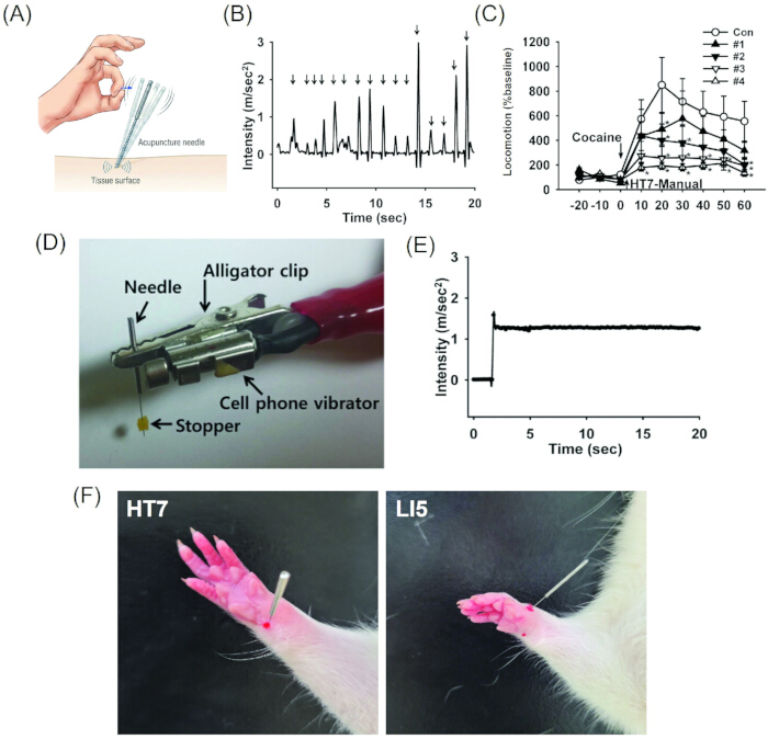
Figure 1: Development and validation of the mechanical acupuncture instrument (MAI). (A) Schematic illustration showing variability in manual acupuncture manipulation across operators. (B) Representative acceleration traces generated during manual acupuncture manipulation. (C) Effect of manual acupuncture at HT7 performed by four different operators on cocaine-induced locomotor activity, illustrating inter-operator variability in the behavioral effects of manual stimulation. (D) Annotated image of the assembled MAI showing the needle, alligator clip, vibration motor, and rubber stopper used to standardize the 3-mm insertion depth. (E) Representative acceleration waveform generated by the MAI at 85 Hz and 1.3 m/s2, demonstrating stable and reproducible mechanical stimulation. (F) Representative images showing the anatomical locations of HT7 and LI5 in the rat forepaw. Red dots indicate the acupuncture point locations used for needle insertion. HT7 was identified at the transverse wrist crease near the flexor carpi ulnaris tendon, whereas LI5 was identified on the radial side of the dorsal wrist between the extensor pollicis and extensor pollicis brevis tendons, providing reproducible anatomical landmarks for acupoint localization. This figure has been adapted with permission from Kim et al.13. Please click here to view a larger version of this figure.
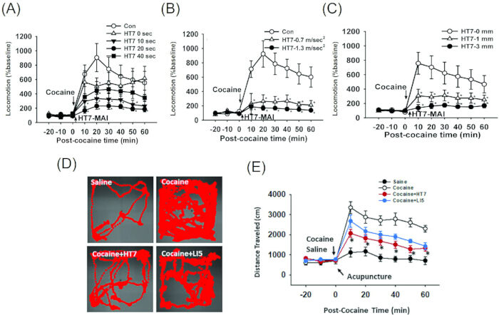
Figure 2: Optimization of mechanical stimulation parameters in the cocaine-induced locomotor activity model. (A–C) Effects of stimulation duration, stimulation intensity, and needle insertion depth on cocaine-induced locomotor activity. (A) HT7-MAI stimulation was applied for 0, 10, 20, or 40 s at 85 Hz and 1.3 m/s2. (B) HT7-MAI stimulation was applied for 20 s at different acceleration intensities. (C) HT7-MAI stimulation was applied at different needle insertion depths. (D) Representative movement traces during locomotor activity testing after saline, cocaine, cocaine plus HT7 stimulation, or cocaine plus LI5 stimulation. (E) Mechanical acupuncture stimulation at HT7, but not LI5, delivered at 85 Hz and 1.3 m/s2 for 20 s, significantly attenuated cocaine-induced enhancement of locomotor activity. Data are presented as mean ± SEM. *p < 0.05 versus cocaine-treated control. This figure has been adapted with permission from Kim et al.13. Please click here to view a larger version of this figure.
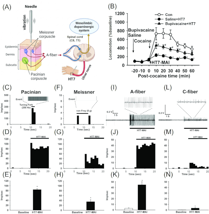
Figure 3: Peripheral afferent and mechanoreceptor validation of MAI-mediated acupuncture effects. (A) Schematic diagram of mechanical acupuncture-induced peripheral-to-central signaling. Vibratory stimulation at HT7 activates mechanoreceptors, including Meissner and Pacinian corpuscles, in cutaneous and subcutaneous tissues. These signals are transmitted via large myelinated A-fiber afferents of the ulnar nerve to C8–T1 spinal segments and subsequently influence the mesolimbic dopaminergic system. (B) Effect of local anesthetic injection on the inhibitory effect of HT7-MAI stimulation. Bupivacaine injection around HT7 reduced the suppressive effect of HT7 stimulation on cocaine-induced locomotor activity, indicating the involvement of local peripheral afferent signaling. (C–H) Somatic afferent responses of Pacinian corpuscle-associated fibers and Meissner corpuscle-associated fibers during MAI stimulation at HT7. (I–N) Responses of A-fibers and C-fibers during MAI stimulation at HT7. MAI stimulation robustly activated A-fiber afferents and produced weaker C-fiber responses. Data are presented as mean ± SEM. *p < 0.05 versus baseline or cocaine-treated control, as appropriate. This figure has been adapted with permission from Kim et al.13. Please click here to view a larger version of this figure.
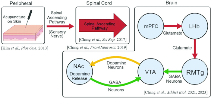
Figure 4: Proposed peripheral-to-central neural pathway underlying MAI-mediated modulation of cocaine-induced responses and mesolimbic dopaminergic system. MAI stimulation on the skin activates peripheral sensory nerves and transmits somatosensory signals through spinal ascending pathways. These inputs are relayed to brain circuits involving the mPFC, LHb, RMTg, VTA, and NAc. Activation of the mPFC–LHb–RMTg pathway may enhance GABAergic inhibition of VTA dopamine neurons, thereby reducing dopamine release in the NAc and suppressing cocaine-induced psychomotor activation. Please click here to view a larger version of this figure.
Discussion
This study presents a standardized protocol for applying mechanical acupuncture using a vibratory stimulation device that enables reproducible delivery of vibration-based mechanical stimulation. By providing quantitative control of stimulation frequency, acceleration intensity, and duration, this approach addresses a major limitation of traditional manual acupuncture, which is inherently dependent on practitioner technique and difficult to standardize.
One of the major strengths of this protocol is its ability to isolate and standardize the mechanical component of acupuncture stimulation. Unlike manual acupuncture, which varies substantially among practitioners, the MAI generates stable and reproducible vibratory stimulation directly through the inserted acupuncture needle. This reproducibility is particularly important for mechanistic studies investigating the neural and physiological basis of acupuncture effects.
Compared with electroacupuncture (EA), the present protocol allows mechanical stimulation to be investigated without introducing external electrical current. This distinction is important because electrical stimulation may directly depolarize peripheral nerve fibers according to electrical excitability, electrode configuration, current intensity, and tissue impedance16,17. In contrast, the MAI primarily generates mechanical deformation of local cutaneous and subcutaneous tissues surrounding the inserted needle. Therefore, this protocol more closely reproduces the physical component of manual acupuncture while enabling stimulation parameters to be quantitatively controlled.
Previous validation studies demonstrated that MAI stimulation at HT7 suppresses cocaine-induced locomotor activity through peripheral afferent mechanisms13. The inhibitory effect was blocked by local anesthesia at HT7 and abolished by ulnar nerve transection, indicating that intact ulnar afferent signaling is required for the behavioral effects of mechanical acupuncture stimulation. Importantly, the inhibitory effect was not prevented by blockade of C/Aδ-fiber pathways and was not reproduced by selective nociceptive stimulation, suggesting that MAI stimulation primarily activates large myelinated A-fiber pathways rather than nociceptive afferents.
The mechanoreceptor-based nature of this protocol further distinguishes MAI stimulation from EA. The stimulation frequency described here represents the dominant frequency of oscillatory vibration transmitted to the needle tip, not the rhythm of repeated manual hand movement. Previous studies showed that vibration frequencies associated with Meissner and Pacinian corpuscle activation effectively suppressed cocaine-induced locomotor responses5,18. These findings support the interpretation that vibration-based mechanical acupuncture engages low-threshold mechanoreceptor-associated afferents in superficial and deep tissues and transmits signals through large A-fiber pathways of the ulnar nerve3,13. Thus, the MAI represents an intermediate experimental model between manual acupuncture and EA by preserving the needle-based mechanical component of manual acupuncture while enabling stimulation to be standardized and quantified.
The biological validity of this protocol is further supported by pathway-level evidence. Ulnar mechanical stimulation activated the dorsal column–cuneate nucleus pathway, and disruption of this pathway abolished the inhibitory effect on cocaine-induced locomotor activity3. In contrast, lesions of the spinothalamic tract did not prevent the behavioral effect, suggesting that innocuous tactile and vibration-related somatosensory pathways, rather than nociceptive signaling, are involved. Mechanistically, these prior studies indicate that MAI-based HT7 stimulation engages a peripheral-to-central pathway linking large myelinated A-fiber afferents in the ulnar nerve to mesolimbic dopamine regulation. These somatosensory signals are transmitted through the dorsal column-cuneate nucleus pathway and subsequently recruit downstream mesolimbic modulatory circuits, including the mPFC–LHb–RMTg–VTA–NAc pathway. This pathway may contribute to the suppression of cocaine-induced locomotor activity and dopamine-related behavioral responses4,6,7,8. Collectively, these findings indicate that the MAI protocol provides a reproducible experimental platform for investigating how defined peripheral mechanical stimulation modulates central neural circuits and reward-related behaviors.
Several critical steps should be considered for the successful application of this protocol. First, acupuncture point localization must be anatomically precise. For HT7 stimulation in rodents, the transverse wrist crease and flexor carpi ulnaris tendon serve as essential anatomical landmarks13,19. Second, insertion depth should be standardized using the stopper because uncontrolled variation in insertion depth may increase experimental variability20. Third, stable needle-motor coupling is required to ensure accurate transmission of vibration to the needle tip. Finally, excessive stimulation should be avoided. Previous validation studies showed that prolonged continuous stimulation, such as 40-s vibration, induced stress-like behavioral responses, whereas 20-s stimulation produced robust behavioral effects with improved tolerability13.
Despite these advantages, several limitations should be acknowledged. The stimulation generated by the MAI represents only a subset of manual acupuncture techniques and does not fully reproduce the multidirectional manipulations performed by experienced practitioners. In addition, most validations of this protocol have been performed in animal models, and its direct applicability to human clinical settings remains to be further investigated.
The MAI does not reproduce the full multidimensional complexity of traditional manual acupuncture, including lifting-thrusting, rotation, bidirectional tissue deformation, and practitioner-dependent tactile feedback. Therefore, vibratory stimulation delivered by the MAI should not be interpreted as a complete representation of manual acupuncture. Instead, this protocol standardizes the vibration-based mechanical component transmitted to the needle tip during manual handle-flicking manipulation.
Although this protocol was validated primarily using HT7 stimulation in male Sprague-Dawley rats in a cocaine-induced locomotor activity model, the MAI can be adapted to other acupuncture points and experimental models. However, parameter re-optimization may be required because tissue depth, local anatomy, mechanoreceptor distribution, and afferent nerve composition may differ across acupuncture points, species, strains, sexes, and disease conditions.
Future applications of this approach may include its integration into standardized neuromodulation research platforms and its adaptation for translational and clinical acupuncture studies. Further refinement of the device may enable more precise control of stimulation patterns and facilitate broader application across multiple disease models. Ultimately, this protocol may help bridge traditional acupuncture practice with modern neuroscience by enabling reproducible and quantifiable investigation of acupuncture-mediated physiological mechanisms.
Disclosures
The authors declare that they have no competing financial interests or conflicts of interest related to this study.
Acknowledgements
This work was supported by the Korea Institute of Oriental Medicine (KIOM) under Grant No. KSN2512011, the Korea Health Technology R&D Project through the Korea Health Industry Development Institute (KHIDI), funded by the Ministry of Health & Welfare, Republic of Korea (RS-2024-00443893), and the National Research Foundation of Korea (NRF) grant funded by the Korea government (RS-2024-00349070). We would like to express our sincere gratitude to the Acupuncture Point Research Team at the Korea Institute of Oriental Medicine, the Department of Meridian and Acupuncture Point Studies at Daegu Haany University, and the Department of Physiology at Yonsei University College of Medicine for their invaluable support and insightful discussions related to the development and validation of the mechanical acupuncture instrument.
Materials
| Name | Company | Catalog Number | Comments |
|---|---|---|---|
| Accelerometer | PCB Piezotronics | PO-AXA-12-01 | Used for vibration measurement |
| Acupuncture needle (0.10 mm × 10 mm) | Dongbang Acupuncture Inc. | DB102 | Sterile disposable acupuncture needle |
| Alligator clip connector | N/A | N/A | Used to connect vibrator to acupuncture needle |
| Bupivacaine hydrochloride (0.5%) | Huons Pharm, Korea | HIRA Code (199502333) | Used for local anesthesia experiments |
| Cocaine hydrochloride | Macfarlan Smith Ltd | 53-21-4 | Used for cocaine-induced locomotor experiments |
| Control unit | Custom-made | N/A | Used to adjust stimulation frequency, acceleration intensity, and duration |
| Data acquisition software | LabVIEW | LabVIEW 7 Express (7.0) | Used for vibration signal analysis |
| Data acquisition system | National Instruments | DAQ-NI USB-6200 | Used for signal digitization |
| Mechanical acupuncture instrument (MAI) | Custom-made | N/A | Custom-built device consisting of a control unit, vibration motor, clip-type connector, acupuncture needle, and rubber stopper |
| Open-field locomotor chamber | Custom-made | square open field box (40 cm × 40 cm× 45cm) made of black acrylic. | Used for locomotor activity analysis |
| Rubber stopper | N/A | N/A | Mounted 3 mm from the needle tip to standardize insertion depth |
| Sprague-Dawley rats | Orient Bio | Crl:CD(SD)IGS | Male, 270–320 g |
| Vibration motor | Motor Bank | MB-0412V | Used for mechanical vibratory stimulation |
| Video tracking software | EthoVision XT | Ethovision 3.1 | Used for locomotor tracking |
References
- Langevin HM, et al. Biomechanical response to acupuncture needling in humans. J Appl Physiol (1985). 2001;91(6):2471-2478.
- Langevin HM, et al. Manual and electrical needle stimulation in acupuncture research: Pitfalls and challenges of heterogeneity. J Altern Complement Med. 2015;21(3):113-128.
- Chang S, et al. Involvement of the cuneate nucleus in the acupuncture inhibition of drug-seeking behaviors. Front Neurosci. 2019;13:928.
- Chang S, et al. Acupuncture reduces cocaine psychomotor responses by activating the rostromedial tegmental nucleus. Addict Biol. 2021;26(6):e13052.
- Shin JH, et al. Paired mechanical and electrical acupuncture of neurogenic spots induces opioid-mediated suppression of hypertension in rats. J Physiol Sci. 2020;70(1):14.
- Chang S, et al. Acupuncture attenuates alcohol dependence through activation of endorphinergic input to the nucleus accumbens from the arcuate nucleus. Sci Adv. 2019;5(9):eaax1342.
- Chang S, et al. Mediation of mPFC-LHb pathway in acupuncture inhibition of cocaine psychomotor activity. Addict Biol. 2023;28(10):e13321.
- Ahn D, et al. Role of lateral hypothalamus in acupuncture inhibition of cocaine psychomotor activity. Int J Mol Sci. 2021;22(11):5994.
- He J, et al. Cancer detection for small-size and ambiguous tumors based on semantic FPN and transformer. PLoS One. 2023;18(2):e0275194.
- Zhang J, Yu H, Ning X. Variational mode decomposition based on refined composite multiscale dispersion entropy and its application in sEMG denoising. J Electromyogr Kinesiol. 2026;89:103176.
- Zhu H, Rong C, Liu H. Deep deterministic policy gradient algorithm based on dung beetle optimization and priority experience replay mechanism. Sci Rep. 2025;15(1):13978.
- Su H, et al. A novel transfer learning framework based on deep contrastive convolution for wind turbine bearing fault localization. IEEE Trans Reliab. 2025;74(4):5579-5591.
- Kim SA, et al. Peripheral afferent mechanisms underlying acupuncture inhibition of cocaine behavioral effects in rats. PLoS One. 2013;8(11):e81018.
- Jin W, et al. Acupuncture reduces relapse to cocaine-seeking behavior via activation of GABA neurons in the ventral tegmental area. Addiction Biology. 2018;23(1):165-181.
- Kim, DH, et al. Acupuncture points can be identified as cutaneous neurogenic inflammatory spots. Sci Rep. 2017;7:15214.
- Merrill DR, Bikson M, Jefferys JG. Electrical stimulation of excitable tissue: Design of efficacious and safe protocols. J Neurosci Methods. 2005;141(2):171-198.
- Rattay F. The basic mechanism for the electrical stimulation of the nervous system. Neuroscience. 1999;89(2):335-346.
- Abraira VE, Ginty DD. The sensory neurons of touch. Neuron. 2013;79(4):618-639.
- Yin CS, et al. A proposed transpositional acupoint system in a mouse and rat model. Res Vet Sci. 2008;84(2):159-165.
- Macpherson H, et al. Standards for reporting interventions in controlled trials of acupuncture: The STRICTA recommendations. Acupunct Med. 2002;20(1):22-25.
Reprints and Permissions
This article has been published
Video Coming Soon