This study demonstrates that hesperidin pretreatment protects rat skeletal muscle against tourniquet-induced ischemia–reperfusion injury by enhancing Nrf2-mediated antioxidant defenses while attenuating NF-κB-associated inflammation and NLRP3/caspase-1-associated pyroptotic and apoptotic responses.
Research Article
Hesperidin Mitigates Skeletal Muscle Ischemia–Reperfusion Injury via Nrf2, NF-κB, and NLRP3 Pathways
In This Article
Summary
Abstract
The application of tourniquets during limb surgery can lead to ischemia–reperfusion (IR) injury, a pathological process characterized by excessive oxidative stress, increased inflammatory signaling, and progressive muscle fiber damage. Hesperidin, a bioactive flavonoid derived from citrus fruits, is known for its antioxidant and anti-inflammatory properties. This study evaluated the protective effects of hesperidin against lower-limb skeletal muscle IR injury and investigated the molecular pathways potentially associated with these effects. Male Wistar rats were allocated to four groups: control, hesperidin alone, IR (2 h of ischemia followed by 2 h of reperfusion), and IR + hesperidin (100 mg/kg administered 30 min before ischemia). Histological examination of the gastrocnemius muscle was performed, and plasma markers of muscle injury, including creatine kinase (CK), lactate dehydrogenase (LDH), and alkaline phosphatase (ALP), were measured. Oxidative stress and antioxidant parameters (MDA, SOD, CAT, and GPx), components of the Nrf2/HO-1/NQO1 pathway, inflammatory mediators (NF-κB, TNF-α, IL-6, and IL-1β), pyroptosis-associated markers (NLRP3 and caspase-1), and apoptosis-associated proteins (Bax and caspase-3) were also assessed. IR resulted in pronounced muscle fiber disruption, elevated plasma injury markers, increased lipid peroxidation, and reduced antioxidant capacity. IR also decreased Nrf2, HO-1, and NQO1 levels while increasing NF-κB, NLRP3, caspase-1, pro-inflammatory cytokines, and apoptosis-associated proteins. Hesperidin pretreatment markedly attenuated these changes by preserving muscle structural integrity, reducing plasma injury markers and lipid peroxidation, restoring antioxidant enzyme activity, increasing Nrf2/HO-1/NQO1-associated antioxidant responses, and reducing NF-κB-associated inflammatory, NLRP3/caspase-1-associated pyroptotic, and apoptotic responses. Overall, hesperidin pretreatment protected skeletal muscle against tourniquet-induced IR injury and was associated with enhanced Nrf2-mediated antioxidant defenses and attenuation of NF-κB-associated inflammation and NLRP3/caspase-1-associated responses. These findings support further investigation of hesperidin as a potential prophylactic intervention for reducing tourniquet-associated lower-limb IR injury.
Introduction
In orthopedic and other extremity surgeries, tourniquets are commonly applied to create a bloodless operative field, typically for 1–2 h and occasionally for up to 3 h, thereby improving surgical accuracy. Although effective, tourniquet application inevitably induces localized ischemia followed by reperfusion, a sequence that can result in ischemia–reperfusion injury (IRI). Studies across multiple organ systems have demonstrated that IRI is characterized by an inflammatory response initiated upon restoration of blood flow to previously oxygen-deprived tissue. Notably, tissue damage occurring during reperfusion may exceed that sustained during the ischemic period itself1,2. Clinically, tourniquet use has been associated with persistent postoperative pain lasting 2–6 weeks3,4, increased limb edema at follow-up intervals of 2 and 6 weeks5,6, and delayed functional recovery, including reduced joint mobility and quadriceps weakness that may persist for several weeks6.
During the ischemic phase, mitochondrial oxidative phosphorylation is suppressed, forcing cells to shift toward anaerobic metabolism and disrupting electron transport. As ATP stores decline, Na⁺/K⁺-ATPase activity becomes insufficient, resulting in the intracellular accumulation of Ca2⁺, H⁺, and Na⁺. Excess intracellular calcium destabilizes cellular membranes, impairs mitochondrial integrity, and activates inflammatory and apoptotic signaling cascades7. Upon restoration of blood flow, a surge in reactive oxygen species (ROS), complement activation, and leukocyte recruitment further exacerbates tissue injury8,9. ROS promote leukocyte activation, chemotaxis, cytokine gene transcription, and NF-κB-associated signaling, thereby contributing to cellular damage. Complement activation and the release of pro-inflammatory mediators, including TNF-α, IL-1, and IL-6, further impair microvascular perfusion10. Infiltrating leukocytes subsequently release ROS, proteases, and elastases into the interstitial space, increasing vascular permeability and promoting edema, thrombosis, and parenchymal cell death11.
Hesperidin, a flavanone glycoside abundant in citrus fruits, is widely recognized for its antioxidant, anti-inflammatory, and anticancer properties. Recent studies have shown that hesperidin can attenuate oxidative stress and inflammatory responses associated with IRI12,13. Hesperidin has also demonstrated protective effects against chemically induced liver injury and has been reported to inhibit hepatocarcinogenesis by modulating oxidative stress, inflammation, and abnormal cell proliferation14. Based on this evidence, the hypothesis of the present study was that hesperidin pretreatment would attenuate skeletal muscle ischemia–reperfusion injury in a rat tourniquet model by enhancing Nrf2/HO-1/NQO1-associated antioxidant defenses while attenuating NF-κB-associated inflammation, NLRP3/caspase-1-associated pyroptotic responses, and apoptotic signaling. Accordingly, the present study evaluated the protective effects of hesperidin against lower-limb skeletal muscle ischemia–reperfusion injury and investigated the molecular pathways potentially associated with its antioxidant and anti-inflammatory effects, with particular emphasis on the Nrf2/HO-1/NQO1, NF-κB, and NLRP3/caspase-1 pathways.
Protocol
All experimental procedures involving animals were approved by the Ethics Committee of Umm Al-Qura University, Saudi Arabia (approval code: HAPO-02-K-012-2026-05-3459). All procedures were conducted in accordance with the institutional guidelines for the care and use of laboratory animals.
Animals
Adult male Wistar albino rats, 3–4 months of age and weighing an average of 322 ± 12 g, were used in the study. The animals were housed 10 per type IV cage on wood-shaving bedding and maintained at 24°C under a 12 h light/dark cycle, with unrestricted access to standard laboratory chow and water. Before the experimental procedures, all animals were fasted overnight but were allowed unrestricted access to water.
Experimental groups and induction of ischemia–reperfusion
Animals were randomly assigned to four experimental groups, with 10 rats in each group. Randomization was performed using a computer-generated random-number sequence. Anesthesia was induced by intraperitoneal administration of xylazine (8 mg/kg) and ketamine (60 mg/kg). Except for the control and hesperidin-only groups, ischemia was induced by placing a tightly secured rubber tourniquet around the proximal region of the left hind limb to occlude the common femoral artery and its collateral branches. A rubber tourniquet (width 5 mm) was applied around the proximal left hind limb at the level of the greater trochanter. Occlusion was standardized by applying a consistent tension using a spring tension gauge set at 2.5 N, ensuring complete occlusion of the common femoral artery and its collateral branches. Successful induction of ischemia was confirmed by limb cyanosis and a noticeable decrease in surface temperature. After 2 h of ischemia, the tourniquet was removed, and reperfusion was allowed to proceed for an additional 2 h. Reperfusion was confirmed by restoration of limb warmth and normal pink coloration, the presence of a palpable pulse, and a reduction in edema, consistent with previously described criteria15,16. Subsequent experimental analyses were performed by evaluators blinded to the treatment conditions.
The control group (C; n = 10) received 0.50 mL of 0.5% carboxymethylcellulose (CMC) orally by gavage, 30 min before the sham procedure. These animals were anesthetized and handled in the same manner as the other groups, but were not subjected to tourniquet application, ischemia, or reperfusion. The hesperidin group (Hes; n = 10) received a single oral dose of hesperidin (100 mg/kg) dissolved in 0.5% carboxymethylcellulose (CMC) in distilled water by oral gavage 30 min before the sham procedure, following the regimen reported by Park et al.17 in a liver ischemia–reperfusion model. These animals underwent anesthesia and identical handling procedures but were not subjected to ischemia or reperfusion. This group was included to assess the baseline physiological effects of hesperidin alone.
The ischemia–reperfusion group (IR; n = 10) underwent 2 h of tourniquet-induced ischemia followed by 2 h of reperfusion according to the protocol described by Yildirim and Albayrak18. The IR + hesperidin group (IR + Hes; n = 10) received a single oral dose of hesperidin (100 mg/kg) by oral gavage 30 min before tourniquet application and was subsequently subjected to the same 2 h ischemia and 2 h reperfusion protocol used for the IR group.
At the end of the reperfusion period, the animals were euthanized, and whole blood was collected into lithium-heparin tubes. Plasma was separated by centrifugation at 1,400 × g for 10 min and stored at −25 °C until biochemical analysis. The left gastrocnemius muscle was excised and divided into three portions. One portion was fixed in 10% neutral-buffered formalin for histological examination; a second portion was snap-frozen and stored at −80 °C for RNA extraction; and a third portion was homogenized and stored at −25 °C for biochemical analyses.
Biochemical analyses
Blood samples were centrifuged at 1,400 × g for 10 min, and the resulting aliquots were stored at −25°C until analysis. Alkaline phosphatase (ALP), creatine kinase (CK), and lactate dehydrogenase (LDH) concentrations were quantified using commercially available enzyme-linked immunosorbent assay (ELISA) kits with detection ranges of 1.25–80 ng/mL, 31.25–2,000 pg/mL, and 1.563–100 ng/mL, respectively. All assays were performed according to the manufacturer's instructions.
Histological examination
Following euthanasia, the left gastrocnemius muscle was dissected and fixed in 10% neutral buffered formalin for a minimum of 24 h. The tissues were processed using standard histological procedures, embedded in paraffin, sectioned at 3 µm, and stained with hematoxylin and eosin for microscopic examination. For semiquantitative assessment of muscle injury, five randomly selected fields per section were examined at 400× magnification by two independent pathologists blinded to the experimental groups. Muscle injury was graded using a 5-point scoring system based on the degree of muscle fiber degeneration19, as follows: grade 0 (normal architecture), grade 1 (mild: isolated fibers with loss of striations), grade 2 (moderate: multiple degenerating fibers with interstitial widening), grade 3 (severe: widespread necrosis with marked edema and inflammation), and grade 4 (extremely severe: extensive necrosis with massive interstitial hemorrhage).
ELISA analysis of muscle homogenates
Gastrocnemius muscle samples were homogenized in ice-cold radioimmunoprecipitation assay (RIPA) lysis buffer containing 50 mM Tris-HCl (pH 7.4), 150 mM NaCl, 1% NP-40, 0.5% sodium deoxycholate, and 0.1% sodium dodecyl sulfate (SDS). The lysis buffer was supplemented with a protease inhibitor cocktail containing 1 mM phenylmethylsulfonyl fluoride, 1 µg/mL aprotinin, 1 µg/mL leupeptin, and 1 µg/mL pepstatin A. The homogenates were centrifuged at 12,000 × g for 15 min at 4 °C, and the resulting supernatants were collected and stored at −80 °C until analysis.
Commercially available ELISA kits were used to quantify markers of oxidative stress, antioxidant capacity, inflammatory responses, and apoptosis. The oxidative stress and antioxidant parameters assessed were superoxide dismutase (SOD; 0.16–10 ng/mL), catalase (CAT; 0.79–50 ng/mL), malondialdehyde (MDA; 31.25–2,000 ng/mL), and glutathione peroxidase (GPx; 15.63–1,000 pg/mL). Components associated with antioxidant signaling included nuclear factor erythroid 2-related factor 2 (Nrf2; 0.16–10 ng/mL), heme oxygenase-1 (HO-1; 0.16–10 ng/mL), and NAD(P)H quinone dehydrogenase 1 (NQO1; 46.875–3,000 pg/mL).
The inflammatory mediators assessed were nuclear factor kappa B (NF-κB; 0.16–10 ng/mL), tumor necrosis factor alpha (TNF-α; 15.63–1,000 pg/mL), interleukin-6 (IL-6; 7.82–500 pg/mL), and interleukin-1 beta (IL-1β; 15.63–1,000 pg/mL). Apoptosis- and pyroptosis-associated proteins included caspase-3 (0.313–20 ng/mL), Bax (0.16–10 ng/mL), and NLR family pyrin domain containing 3 (NLRP3; 0.312–20 ng/mL). All measurements were performed according to the manufacturer's instructions.
Quantitative PCR for caspase-1
Total RNA was isolated from frozen gastrocnemius muscle homogenates using a phenol-guanidine-based RNA isolation reagent followed by purification with a silica membrane-based RNA purification method. Complementary DNA (cDNA) was synthesized from 8 µL of RNA using a reverse transcription system. Quantitative polymerase chain reaction (qPCR) was subsequently performed using a real-time PCR system, with β-actin serving as the internal reference gene. The reaction mixture (20 µL total volume) consisted of 10 µL of 2× SYBR Green PCR Master Mix, 2 µL of cDNA template, 1 µL each of forward and reverse primers (10 µM), and 6 µL of RNase-free water. The cycling protocol included an initial denaturation at 95 °C for 10 min, followed by 40 cycles of denaturation at 95 °C for 15 s, annealing at 60 °C for 30 s, and extension at 72 °C for 30 s. A melt curve analysis was performed from 65 °C to 95 °C in 0.5 °C increments to verify amplification specificity. Each sample was run in duplicate, and no-template controls (NTCs) and no-reverse-transcription controls (NRTs) were included in each run. Amplification efficiency for each primer pair was determined from a standard curve (fourfold dilution series of pooled cDNA) and ranged from 90–105%. Relative caspase-1 expression was calculated using the 2−ΔΔCT method described by Livak and Schmittgen20. The primer sequences used for qPCR were as follows: caspase-1 forward, 5′-GACCGAGTGGTTCCCTCAAG-3′; caspase-1 reverse, 5′-GACGTGTACGAGTGGGTGTT-3′; β-actin forward, 5′-CCGAGATCTCACCGACTACCTCA-3′; and β-actin reverse, 5′-TCAAAGTCCAGAGCGACATAGCA-3′.
Statistical analysis
All statistical analyses were performed using statistical analysis software. The group size of 10 animals per experimental condition was determined prospectively using dedicated power-analysis software, with an effect size of 0.8, a significance level of α = 0.05, and a desired statistical power of 0.80.
Before parametric analyses were performed, normality and homogeneity of variance were evaluated using the Shapiro–Wilk and Levene's tests, respectively. Both tests yielded p > 0.05, supporting the use of parametric statistical procedures. Comparisons among the four experimental groups were therefore performed using one-way analysis of variance (ANOVA), followed by Tukey's multiple-comparison test for post hoc pairwise comparisons. A p-value < 0.05 was considered statistically significant. All quantitative data were expressed as mean ± standard deviation (SD).
Results
Improvement of ischemia–reperfusion-induced histological alterations by hesperidin
Histological examination of the gastrocnemius muscle revealed regularly arranged longitudinal muscle fibers in the control and hesperidin groups (Figure 1A–D). In contrast, the ischemia–reperfusion (IR) group exhibited marked muscle fiber degeneration and increased intercellular spacing (Figure 1E,F). Consistent with these morphological alterations, the muscle damage score (MDS) was approximately 24-fold higher in the IR group than in the control group. Hesperidin pretreatment markedly attenuated these histological alterations, with greater preservation of muscle fiber organization and reduced intercellular spacing in the IR + Hes group (Figure 1G,H). The MDS in the IR + Hes group was reduced to approximately 0.46-fold that of the IR group (Figure 1I). The complete group means ± SD, overall ANOVA results, and Tukey post hoc pairwise comparisons for MDS are provided in Supplementary Table 1 and Supplementary Table 2. These findings indicate that hesperidin pretreatment attenuated the histological manifestations of tourniquet-induced skeletal muscle IR injury.
Improvement of ischemia–reperfusion-induced biochemical alterations by hesperidin
As shown in Figure 2A–C, IR resulted in significant increases in plasma alkaline phosphatase (ALP), creatine kinase (CK), and lactate dehydrogenase (LDH) levels compared with the control group (p < 0.05). Relative to the control group, ALP, CK, and LDH levels increased approximately 2.5-, 7.1-, and 19-fold, respectively, indicating substantial biochemical evidence of muscle injury. Pretreatment with a single dose of hesperidin (100 mg/kg) 30 min before ischemia attenuated these increases, reducing ALP, CK, and LDH levels to approximately 0.71-, 0.30-, and 0.32-fold, respectively, of the corresponding IR values (Figure 2A–C). These biochemical changes were consistent with the histological preservation observed in the hesperidin-pretreated animals.
IR also markedly altered oxidative stress and antioxidant parameters in gastrocnemius muscle homogenates (Figure 2D–G). Malondialdehyde (MDA) levels increased approximately 2.7-fold relative to the control group, whereas superoxide dismutase (SOD), catalase (CAT), and glutathione peroxidase (GPx) levels decreased to approximately 0.28-, 0.12-, and 0.17-fold of control values, respectively. Hesperidin pretreatment attenuated these alterations. Relative to the IR group, SOD, CAT, and GPx levels increased approximately 1.95-, 4.79-, and 3.38-fold, respectively, whereas MDA levels decreased to approximately 0.54-fold of the IR value (Figure 2D–G).
Components associated with the Nrf2/HO-1/NQO1 antioxidant pathway were also reduced following IR (Figure 2H–J). Relative to the control group, Nrf2, HO-1, and NQO1 levels decreased to approximately 0.25-, 0.22-, and 0.26-fold, respectively. Hesperidin pretreatment partially restored these markers, increasing Nrf2, HO-1, and NQO1 levels approximately 2.28-, 3.13-, and 2.09-fold, respectively, relative to the IR group (Figure 2H–J). Collectively, these findings indicate that hesperidin pretreatment attenuated IR-induced oxidative stress and was associated with preservation of endogenous antioxidant defenses and Nrf2/HO-1/NQO1-associated responses.
Effects of hesperidin on ischemia–reperfusion-induced inflammatory, pyroptosis-associated, and apoptotic markers
IR markedly increased inflammatory markers in the gastrocnemius muscle (Figure 3A–D). Relative to the control group, NF-κB, TNF-α, IL-6, and IL-1β levels increased approximately 17.1-, 8.46-, 8.88-, and 9.64-fold, respectively. Hesperidin pretreatment attenuated these increases, reducing NF-κB, TNF-α, IL-6, and IL-1β levels to approximately 0.33-, 0.48-, 0.52-, and 0.54-fold of the corresponding IR values, respectively (Figure 3A–D). These findings indicate that hesperidin pretreatment was associated with attenuation of the inflammatory response induced by skeletal muscle IR.
IR also increased markers associated with pyroptotic and apoptotic responses (Figure 3E–H). Relative to the control group, NLRP3 increased approximately 4.96-fold, whereas relative caspase-1 gene expression increased approximately 3.44-fold. Caspase-3 and Bax levels increased approximately 5.10- and 14.8-fold, respectively. Hesperidin pretreatment reduced these IR-associated changes, with NLRP3 and caspase-1 decreasing to approximately 0.57- and 0.49-fold of the corresponding IR values, respectively. Caspase-3 and Bax were similarly reduced to approximately 0.33- and 0.29-fold of their respective IR values (Figure 3E–H).
Taken together, the results demonstrate that hesperidin pretreatment attenuated histological and biochemical evidence of skeletal muscle injury following tourniquet-induced IR. This protective effect was accompanied by reduced oxidative stress, preservation of endogenous antioxidant defenses, increased Nrf2/HO-1/NQO1-associated markers, and attenuation of NF-κB-associated inflammatory responses, NLRP3/caspase-1-associated responses, and apoptotic responses.
DATA AVAILABILITY:
The raw data supporting the findings of this study are provided as Supplementary File 1.
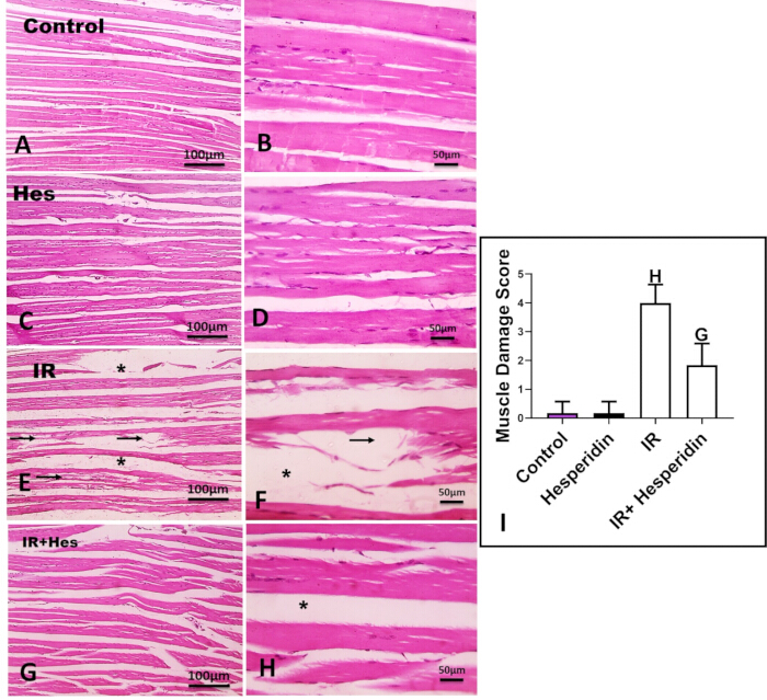
Figure 1: Histological alterations and muscle damage scores in gastrocnemius muscle following ischemia–reperfusion and hesperidin pretreatment. (A,B) Hematoxylin and eosin (H&E)-stained sections from the control group showing regularly arranged longitudinal gastrocnemius muscle fibers. (C,D) H&E-stained sections from the hesperidin (Hes) group showing preserved muscle morphology. (E,F) Sections from the ischemia–reperfusion (IR) group showing marked destruction and degeneration of longitudinal skeletal muscle fibers and increased intercellular spacing. (G,H) Sections from the IR + Hes group show greater preservation of longitudinal muscle fibers and reduced intercellular spacing compared with the IR group. Black arrows indicate muscle fiber destruction and degeneration, and asterisks indicate increased intercellular spacing. Scale bars = 100 µm in panels A, C, E, and G and 50 µm in panels B, D, F, and H. (I) Semiquantitative muscle damage score for the experimental groups (n = 10 per group). Data are expressed as mean ± SD. Letter "H" indicates a significant difference compared with the Control and Hesperidin groups (p < 0.05), while letter "G" indicates a significant difference compared with the IR group (p < 0.05). Please click here to view a larger version of this figure.
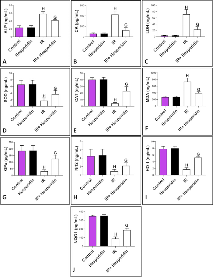
Figure 2: Effects of hesperidin pretreatment on biochemical markers of muscle injury, oxidative stress, and antioxidant responses following ischemia–reperfusion. (A–C) Plasma levels of alkaline phosphatase (ALP), creatine kinase (CK), and lactate dehydrogenase (LDH), respectively. (D–J) Gastrocnemius muscle levels of superoxide dismutase (SOD), catalase (CAT), malondialdehyde (MDA), glutathione peroxidase (GPx), nuclear factor erythroid 2-related factor 2 (Nrf2), heme oxygenase-1 (HO-1), and NAD(P)H quinone dehydrogenase 1 (NQO1), respectively (n = 10 per group). Data are expressed as mean ± SD. H indicates a significant difference compared with the control and hesperidin groups, whereas G indicates a significant difference compared with the IR group (p < 0.05). Please click here to view a larger version of this figure.
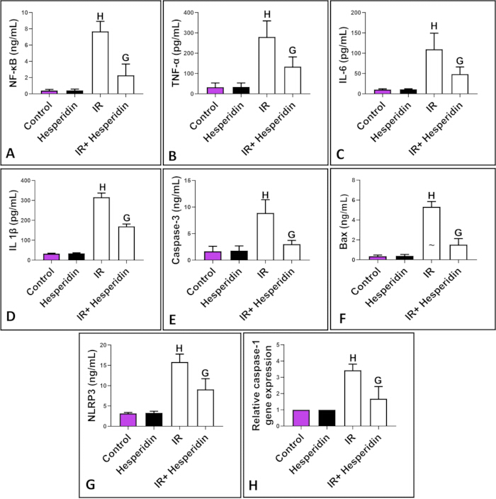
Figure 3: Effects of hesperidin pretreatment on inflammatory, pyroptosis-associated, and apoptotic markers following ischemia–reperfusion. (A–G) Levels of NF-κB, TNF-α, IL-6, IL-1β, caspase-3, Bax, and NLRP3, respectively, in gastrocnemius muscle homogenates. (H) Relative caspase-1 mRNA expression in gastrocnemius muscle tissue (n = 10 per group). Data are expressed as mean ± SD. H indicates a significant difference compared with the control and hesperidin groups, whereas G indicates a significant difference compared with the IR group (p < 0.05). Please click here to view a larger version of this figure.
Supplementary Table 1: Mean ± SD values for histological, biochemical, oxidative stress, antioxidant, inflammatory, pyroptosis-associated, and apoptosis-associated parameters across the four experimental groups, with corresponding one-way ANOVA results.Please click here to download this file.
Supplementary Table 2: Tukey post hoc pairwise comparison p values for all measured outcomes among the control, hesperidin, IR, and IR + hesperidin groups.Please click here to download this file.
Supplementary File 1: Raw data supporting the findings of this study. Please click here to download this file.
Discussion
Tourniquet-induced ischemia remains a standard technique in many surgical specialties, particularly orthopedics, to obtain a bloodless operative field and improve visualization. Despite its utility, this approach inevitably produces localized ischemic stress followed by reperfusion injury, which can adversely affect several postoperative outcomes21. Although advances in tourniquet design and evidence-based application strategies have reduced certain complications22, no clinically established intervention is currently available to prevent lower-limb ischemia–reperfusion injury (IRI). Accumulating evidence indicates that although the full spectrum of mechanisms underlying IR injury remains incompletely understood, reactive oxygen species (ROS) play a central pathogenic role, whereas endogenous antioxidant enzymes provide critical protection22. Supporting this concept, Park et al.23 reported that skeletal muscle IR markedly suppresses the transcription of major antioxidant enzymes, including SOD1, SOD2, CAT, and GPx. Consistent with these findings, the present results demonstrated a substantial reduction in gastrocnemius antioxidant activity accompanied by a marked increase in oxidative products following IR.
Histopathological analysis further demonstrated extensive degenerative changes in the gastrocnemius muscle, together with significant elevations in plasma CK and LDH, consistent with the findings of Herrero de la Parte et al.24. Hesperidin pretreatment preserved muscle architecture and reduced circulating markers of muscle injury. This structural preservation was accompanied by increased antioxidant enzyme levels and reduced oxidative stress. Similar protective effects of hesperidin have been documented in other experimental models. Abdel Raheem and Abdel Ghany25 showed that hesperidin reduced CK and LDH levels in doxorubicin-induced cardiotoxicity, whereas Park et al.17 demonstrated that hesperidin mitigated hepatic IR injury by enhancing antioxidant defenses, including CAT, GPx, and SOD. The preservation of muscle morphology and reduction in biochemical injury markers observed following hesperidin pretreatment may be associated with its antioxidant properties. These effects have been attributed to its ability to directly neutralize ROS and enhance endogenous antioxidant defenses26, as well as its potential to modulate the Nrf2 signaling pathway27.
Although previous studies have suggested crosstalk between Nrf2 and NF-κB, the precise molecular basis of this interaction remains incompletely defined28. One proposed mechanism is that increased Nrf2 activity may attenuate NF-κB-associated signaling by limiting ROS accumulation. Supporting this model, Jiang et al.29 demonstrated that nephritogenic injury increased phospho-p65 levels and NF-κB luciferase activity in parallel with elevated ROS; these effects were intensified by Nrf2 knockdown and attenuated by Nrf2 overexpression.
The present findings are consistent with this mechanistic framework. In the IR group, marked reductions in Nrf2, HO-1, and NQO1, key components associated with the antioxidant response, were accompanied by increased NF-κB levels and elevated pro-inflammatory cytokines. These results are consistent with the observations of Song et al.30, who reported Nrf2 downregulation in a lower-limb ischemia model, and Andrade Silva et al.31, who documented increased NF-κB-associated signaling in skeletal muscle following IR.
Hesperidin pretreatment attenuated these IR-induced alterations. Increased Nrf2, HO-1, and NQO1 levels were observed concurrently with reductions in NF-κB and associated inflammatory mediators. These findings are consistent with those of Liu et al.32, who demonstrated that hesperitin protects myocardial tissue against IR injury through mechanisms involving Nrf2, and Meng et al.33, who reported that hesperidin downregulates NF-κB-associated signaling in renal IR injury.
Mechanistically, the effects of hesperidin may be explained, at least in part, by modulation of the Keap1–Nrf2 axis, consistent with previous reports demonstrating that flavonoids can disrupt Keap1–Nrf2 binding, thereby facilitating Nrf2 translocation and subsequent expression of cytoprotective genes34. However, because Keap1 was not measured directly, this interpretation remains correlational. Nevertheless, the increased levels of Nrf2, HO-1, and NQO1 observed in hesperidin-treated animals, together with concurrent reductions in NF-κB-associated inflammatory and NLRP3-associated markers, provide a coherent correlative framework for the observed protective effects. Future studies employing pathway-specific inhibitors or genetic knockdown models are required to establish causal relationships.
Inflammation is a major contributor to IR pathology and is closely associated with inflammasome activation. Among the inflammasome complexes, NLRP3, comprising NLRP3, ASC, and caspase-1, is one of the most extensively characterized35. Following IR, NLRP3 activation increases rapidly and typically peaks within 24 h36. Excessive ROS production is recognized as an important upstream trigger of NLRP3 assembly37. Once activated, NLRP3 recruits ASC, promoting the cleavage of procaspase-1 into active caspase-1, which subsequently processes IL-1β and IL-18 into their mature forms and amplifies the inflammatory cascade38.
The present findings were consistent with involvement of this pathway, as IR markedly increased NLRP3 protein levels and caspase-1 mRNA expression in gastrocnemius muscle. Hesperidin pretreatment significantly attenuated these increases, consistent with the findings of Xie et al.39. These changes may be associated with hesperidin's antioxidant properties and the consequent reduction in ROS, an important upstream signal involved in NLRP3 activation. However, because cleaved caspase-1 and other direct markers of inflammasome activation were not measured, these findings should be interpreted as evidence of changes in NLRP3/caspase-1-associated responses rather than direct demonstration of inflammasome activation or pyroptosis.
Lower-limb IR injury also induces apoptotic responses associated with excessive ROS generation40. ROS can damage membrane lipids, proteins, and nucleic acids, ultimately contributing to cellular dysfunction and death41. In the present study, this response was reflected by significant increases in the apoptosis-associated markers Bax and caspase-3 in the IR group. Hesperidin pretreatment markedly attenuated these increases, consistent with the findings of Li et al.42, who demonstrated anti-apoptotic effects of hesperidin in hepatic IR injury.
The findings may have potential clinical relevance for orthopedic and vascular procedures requiring tourniquet application. The observed effects of hesperidin on antioxidant, inflammatory, NLRP3/caspase-1-associated, and apoptosis-associated markers support further investigation of this flavonoid as a potential prophylactic intervention. Administration 30 min before tourniquet inflation could represent a clinically feasible strategy in elective procedures if efficacy, pharmacokinetics, and safety are subsequently established in appropriate translational studies. Potential clinical outcomes of interest include postoperative pain, edema, functional recovery, length of hospitalization, and long-term limb function. However, these outcomes were not assessed in the present experimental model and therefore require direct evaluation in future studies.
Several limitations warrant consideration. First, the reperfusion period was limited to 2 h, allowing assessment of early molecular and histological changes but not chronic inflammation or functional recovery over subsequent days or weeks. Second, only a single dose of hesperidin (100 mg/kg) administered at a single time point (30 min before ischemia) was evaluated; dose–response relationships and alternative administration schedules therefore remain to be determined. Third, functional outcomes, such as muscle strength and ambulation, were not assessed. Fourth, the use of healthy young male rats may limit generalizability to older patients and individuals with comorbidities. Fifth, findings obtained from a single species require validation in larger animal models before clinical translation.
Sixth, assessment of Nrf2 and NF-κB-associated responses relied on total protein levels measured by ELISA in whole-tissue homogenates. This approach did not distinguish between cytoplasmic and nuclear protein pools or assess phosphorylation status, both of which are important determinants of transcriptional activity. Future studies incorporating nuclear/cytoplasmic fractionation, phospho-specific antibody detection (e.g., phospho-p65 measurement), or immunohistochemical assessment of nuclear translocation would provide more definitive evidence of pathway activation. Seventh, assessment of pyroptosis-associated signaling was limited to caspase-1 mRNA expression and NLRP3 protein levels. Caspase-1 mRNA does not undergo post-translational activation via proteolytic cleavage, a critical step in inflammasome activation and pyroptosis. Although reduced caspase-1 mRNA expression following hesperidin pretreatment suggests transcriptional regulation, direct measurement of cleaved caspase-1, ASC oligomerization, gasdermin D (GSDMD) cleavage, and mature IL-1β and IL-18 would provide stronger evidence of modulation of the pyroptotic pathway.
Eighth, apoptosis was assessed by measuring total Bax and caspase-3 protein levels by ELISA. These measurements did not establish the balance between pro- and anti-apoptotic proteins, such as the Bax/Bcl-2 ratio, or distinguish activated caspase-3 from its pro-form. Measurement of cleaved caspase-3 would provide more direct evidence of apoptotic execution. Furthermore, the absence of orthogonal validation by Western blotting or immunohistochemistry for key pathway components, including Nrf2, NF-κB, NLRP3, and apoptosis-associated proteins, limits the strength of the mechanistic conclusions. Future studies incorporating multiple complementary techniques for each pathway would strengthen validation of the observed molecular changes.
Ninth, although the control group received the same oral vehicle (0.5% carboxymethylcellulose) used for hesperidin administration, the study did not include a separate vehicle-treated ischemia–reperfusion group. Therefore, a potential interaction between the vehicle and ischemia–reperfusion-related outcomes cannot be completely excluded. Future studies may include an additional IR + vehicle group to further strengthen control of treatment-related effects. Finally, the precise molecular interactions underlying the observed effects, as well as the contributions of additional biological processes, require further investigation. Despite these limitations, the findings provide preclinical evidence that hesperidin pretreatment attenuates skeletal muscle injury following tourniquet-induced IR.
The findings indicate that pretreatment with hesperidin attenuated skeletal muscle injury following tourniquet-induced ischemia–reperfusion in rats. The protective effects were associated with increased levels of Nrf2/HO-1/NQO1 antioxidant pathway components and reduced levels of NF-κB-associated inflammatory markers, NLRP3/caspase-1-associated markers, and apoptosis-associated markers. Hesperidin pretreatment reduced oxidative stress, decreased pro-inflammatory cytokines, including TNF-α, IL-6, and IL-1β, and lowered Bax and caspase-3 levels. These molecular changes were accompanied by improved preservation of muscle fiber architecture and lower plasma concentrations of muscle injury markers, including CK, LDH, and ALP. Collectively, these preclinical findings support further investigation of hesperidin as a potential intervention for limiting lower-limb ischemia–reperfusion injury associated with tourniquet application. Validation in larger animal models and subsequent clinical studies will be required to determine efficacy, optimal dosing, pharmacokinetics, and safety before clinical application can be considered.
Disclosures
The author declares that the research was conducted in the absence of any commercial or financial relationships that could be construed as a potential conflict of interest. Generative artificial intelligence (AI) technologies, including ChatGPT (OpenAI), Claude (Anthropic), and SciSpace, were used solely for language refinement, literature organization, and editorial assistance during manuscript preparation. All scientific content, interpretations, citations, references, and conclusions were independently reviewed, verified, and approved by the author, who assumes full responsibility for the accuracy and integrity of the manuscript.
Acknowledgements
The authors extend their appreciation to Umm Al-Qura University, Saudi Arabia for funding this research work through grant number 26UQU4400419GSSR01.
Funding Statement
This research work was funded by Umm Al-Qura University, Saudi Arabia under grant number 26UQU4400419GSSR01.
Materials
| Name | Company | Catalog Number | Comments |
|---|---|---|---|
| ALP ELISA Kit (1.25–80 ng/mL) | ELK Biotechnology, China | ELK5657 | https://www.elkbiotech.com/product/ELK5657 |
| Aprotinin | Sigma-Aldrich, USA | A6279 | https://www.sigmaaldrich.com/US/en/product/sigma/a6279 |
| Bax ELISA Kit (0.16–10 ng/mL) | Novus Biologicals, USA | NBP2-69938 | https://www.novusbio.com/products/nbp2-69938 |
| β-actin Forward Primer (5′-CCGAGATCTCACCGACTACCTCA-3′) | Integrated DNA Technologies, USA | Custom | https://www.idtdna.com |
| β-actin Reverse Primer (5′-TCAAAGTCCAGAGCGACATAGCA-3′) | Integrated DNA Technologies, USA | Custom | https://www.idtdna.com |
| Carboxymethylcellulose (CMC), 0.5% | Sigma-Aldrich, USA | C5678 | https://www.sigmaaldrich.com/US/en/product/sigma/c5678 |
| Caspase-1 Forward Primer (5′-GACCGAGTGGTTCCCTCAAG-3′) | Integrated DNA Technologies, USA | Custom | https://www.idtdna.com |
| Caspase-1 Reverse Primer (5′-GACGTGTACGAGTGGGTGTT-3′) | Integrated DNA Technologies, USA | Custom | https://www.idtdna.com |
| Caspase-3 ELISA Kit (0.313–20 ng/mL) | Wuhan Fine Biotech Co., Ltd., China | ER0143 | https://www.finebiotech.com/product/ER0143 |
| CAT ELISA Kit (0.79–50 ng/mL) | ELK Biotechnology, China | ELK5986 | https://www.elkbiotech.com/product/ELK5986 |
| Centrifuge | Eppendorf, Germany | 5810 R | https://www.eppendorf.com/us-en/lab-equipment/centrifuges/5810-5810-r-centrifuges |
| CK ELISA Kit (31.25–2000 pg/mL) | Abcam, UK | ab285275 | https://www.abcam.com/products/ab285275 |
| Dimethyl sulfoxide (DMSO), 5% | Sigma-Aldrich, USA | D8418 | https://www.sigmaaldrich.com/US/en/product/sigma/d8418 |
| Eosin Y | Sigma-Aldrich, USA | 318906 | https://www.sigmaaldrich.com/US/en/product/aldrich/318906 |
| Ethanol (100%, 96%, 70%) | Sigma-Aldrich, USA | 32205 | https://www.sigmaaldrich.com/US/en/product/aldrich/32205 |
| Formalin, 10% neutral buffered | Sigma-Aldrich, USA | HT501128 | https://www.sigmaaldrich.com/US/en/product/sigma/ht501128 |
| G*Power 3.1 | Heinrich Heine University Düsseldorf, Germany | N/A | https://www.psychologie.hhu.de/arbeitsgruppen/allgemeine-psychologie-und-arbeitspsychologie/gpower |
| GPx ELISA Kit (15.63–1000 pg/mL) | ELK Biotechnology, China | ELK2222 | https://www.elkbiotech.com/product/ELK2222 |
| GraphPad Prism 8.0 | GraphPad Software, USA | N/A | https://www.graphpad.com |
| Hematoxylin | Sigma-Aldrich, USA | HHS128 | https://www.sigmaaldrich.com/US/en/product/sigma/hhs128 |
| Hesperidin | Sigma-Aldrich, USA | H5254 | https://www.sigmaaldrich.com/US/en/product/sigma/h5254 |
| HO-1 ELISA Kit (0.16–10 ng/mL) | ELK Biotechnology, China | ELK1477 | https://www.elkbiotech.com/product/ELK1477 |
| IL-1β ELISA Kit (15.63–1000 pg/mL) | ELK Biotechnology, China | ELK1272 | https://www.elkbiotech.com/product/ELK1272 |
| IL-6 ELISA Kit (7.82–500 pg/mL) | ELK Biotechnology, China | ELK1158 | https://www.elkbiotech.com/product/ELK1158 |
| Ketamine | Sigma-Aldrich, USA | K2753 | https://www.sigmaaldrich.com/US/en/product/sigma/k2753 |
| LDH ELISA Kit (1.563–100 ng/mL) | Wuhan Fine Biotech Co., Ltd., China | ER0647 | https://www.finebiotech.com/product/ER0647 |
| Leupeptin | Sigma-Aldrich, USA | L2884 | https://www.sigmaaldrich.com/US/en/product/sigma/l2884 |
| Light microscope | Olympus, Japan | BX53 | https://www.olympus-lifescience.com/en/microscopes/bx53/ |
| Lithium heparin tubes | BD Vacutainer, USA | 367886 | https://www.bd.com/en-us/products/blood-collection |
| MDA ELISA Kit (31.25–2000 ng/mL) | ELK Biotechnology, China | ELK10920 | https://www.elkbiotech.com/product/ELK10920 |
| Microtome | Leica Biosystems, Germany | RM2255 | https://www.leicabiosystems.com/us/histology-equipment/microtomes/ |
| Microsoft Excel | Microsoft Corporation, USA | N/A | https://www.microsoft.com/en-us/microsoft-365/excel |
| NF-κB ELISA Kit (0.16–10 ng/mL) | ELK Biotechnology, China | ELK1693 | https://www.elkbiotech.com/product/ELK1693 |
| NLRP3 ELISA Kit (0.312–20 ng/mL) | Novus Biologicals, USA | NBP3-42294 | https://www.novusbio.com/products/nbp3-42294 |
| NQO1 ELISA Kit (46.875–3000 pg/mL) | MyBioSource, USA | MBS7606601 | https://www.mybiosource.com/product/MBS7606601 |
| Nrf2 ELISA Kit (0.16–10 ng/mL) | ELK Biotechnology, China | ELK8043 | https://www.elkbiotech.com/product/ELK8043 |
| Paraffin wax | Sigma-Aldrich, USA | 327204 | https://www.sigmaaldrich.com/US/en/product/aldrich/327204 |
| Pepstatin A | Sigma-Aldrich, USA | P5318 | https://www.sigmaaldrich.com/US/en/product/sigma/p5318 |
| Protease inhibitor cocktail (PMSF) | Sigma-Aldrich, USA | P7626 | https://www.sigmaaldrich.com/US/en/product/sigma/p7626 |
| QIAzol Lysis Reagent | Qiagen, Germany | 79306 | https://www.qiagen.com/us/products/discovery-and-translational-research/dna-rna-purification/rna-purification/total-rna/qiazol-lysis-reagent |
| QuantiTect Reverse Transcription Kit | Qiagen, Germany | 205311 | https://www.qiagen.com |
| RIPA lysis buffer | Thermo Fisher Scientific, USA | 89900 | https://www.thermofisher.com/order/catalog/product/89900 |
| RNeasy Mini Kit | Qiagen, Germany | 74104 | https://www.qiagen.com/us/products/discovery-and-translational-research/dna-rna-purification/rna-purification/total-rna/rneasy-mini-kit |
| Rotor-Gene Q | Qiagen, Germany | 9001571 | https://www.qiagen.com |
| Sodium chloride, 0.9% | Sigma-Aldrich, USA | S7653 | https://www.sigmaaldrich.com/US/en/product/sigma/s7653 |
| SOD ELISA Kit (0.16–10 ng/mL) | ELK Biotechnology, China | ELK8178 | https://www.elkbiotech.com/product/ELK8178 |
| TissueLyser II | Qiagen, Germany | 85300 | https://www.qiagen.com |
| TNF-α ELISA Kit (15.63–1000 pg/mL) | ELK Biotechnology, China | ELK1396 | https://www.elkbiotech.com/product/ELK1396 |
| Type IV cage | Bioscape, Germany | N/A | https://www.bioscape.de |
| Xylene | Sigma-Aldrich, USA | 214736 | https://www.sigmaaldrich.com/US/en/product/aldrich/214736 |
| Xylazine | Sigma-Aldrich, USA | X1251 | https://www.sigmaaldrich.com/US/en/product/sigma/x1251 |
References
- Haglund U, Lundgren O. Intestinal ischemia and shock factors. Federation Proceedings. 1978;37(13):2729-2733.
- Blaisdell FW. The pathophysiology of skeletal muscle ischemia and the reperfusion syndrome: a review. Cardiovascular Surgery. 2002;10(6):620-630.
- Saied A, Zyaei A. Tourniquet use during plating of acute extra-articular tibial fractures: effects on final results of the operation. The Journal of Trauma. 2010;69(6):E94–E97.
- Konrad G, Markmiller M, Lenich A, Mayr E, Rüter A. Tourniquets may increase postoperative swelling and pain after internal fixation of ankle fractures. Clinical Orthopaedics and Related Research. 2005;(433):189-194.
- Kirkley A, et al. Tourniquet versus no tourniquet use in routine knee arthroscopy: a prospective, double-blind, randomized clinical trial. Arthroscopy. 2000;16(2):121-126.
- Ejaz A, et al. Faster recovery without the use of a tourniquet in total knee arthroplasty. Acta Orthopaedica. 2014;85(4):422-426.
- Anzell AR, Maizy R, Przyklenk K, Sanderson TH. Mitochondrial quality control and disease: insights into ischemia-reperfusion injury. Molecular Neurobiology. 2018;55(3):2547-2564.
- Toyokuni S. Reactive oxygen species-induced molecular damage and its application in pathology. Pathology International. 1999;49(2):91-102.
- Carden DL, Granger DN. Pathophysiology of ischaemia-reperfusion injury. The Journal of Pathology. 2000;190(3):255-266.
- Collard CD, et al. Complement activation following oxidative stress. Molecular Immunology. 1999;36(13-14):941-948.
- Panés J, Perry M, Granger DN. Leukocyte-endothelial cell adhesion: avenues for therapeutic intervention. British Journal of Pharmacology. 1999;126(3):537-550.
- Rong Z, et al. Hesperidin pretreatment protects hypoxia-ischemic brain injury in neonatal rat. Neuroscience. 2013;255:292-299.
- Wei D, et al. Hesperidin suppresses ovalbumin-induced airway inflammation in a mouse allergic asthma model. Inflammation. 2012;35(1):114-121.
- Kaur G, Tirkey N, Chopra K. Beneficial effect of hesperidin on lipopolysaccharide-induced hepatotoxicity. Toxicology. 2006;226(2-3):152-160.
- Apaydin Yildirim B, Albayrak S. Protective effects of crocin on experimental gastrocnemius muscle ischemia/reperfusion model in rat. The Eurasia Proceedings of Science, Technology, Engineering & Mathematics. 2018;3:116-120. Available from: https://epstem.net/index.php/epstem/article/view/129
- Pottecher J, et al. Skeletal muscle ischemia-reperfusion injury and cyclosporine A in the aging rat. Fundamental & Clinical Pharmacology. 2016;30(3):216-225.
- Park HK, Kang SW, Park MS. Hesperidin ameliorates hepatic ischemia-reperfusion injury in Sprague-Dawley rats. Transplantation Proceedings. 2019;51(8):2828-2832.
- Yildirim BA, Albayrak S. Effect of crocin on experimental gastrocnemius muscle ischemia/reperfusion injury in rat. Veterinary Research Forum. 2023;14(4):201-206.
- Jaćević V, Nepovimova E, Kuča K. Toxic injury to muscle tissue of rats following acute oximes exposure. Scientific Reports. 2019;9(1):1457.
- Livak KJ, Schmittgen TD. Analysis of relative gene expression data using real-time quantitative PCR and the 2(-Delta Delta C(T)) method. Methods. 2001;25(4):402-408.
- Liu D, Graham D, Gillies K, Gillies RM. Effects of tourniquet use on quadriceps function and pain in total knee arthroplasty. Knee Surgery & Related Research. 2014;26(4):207-213.
- Estebe JP, Davies JM, Richebe P. The pneumatic tourniquet: mechanical, ischaemia-reperfusion and systemic effects. European Journal of Anaesthesiology. 2011;28(6):404-411.
- Park UJ, et al. Remote ischemic preconditioning enhances the expression of genes encoding antioxidant enzymes and endoplasmic reticulum stress-related proteins in rat skeletal muscle. Vascular Specialist International. 2016;32(4):141-149.
- Herrero de la Parte B, et al. The prevention of ischemia-reperfusion injury in elderly rats after lower limb tourniquet use. Antioxidants. 2022;11(10):1936.
- Abdel-Raheem IT, Abdel-Ghany AA. Hesperidin alleviates doxorubicin-induced cardiotoxicity in rats. Journal of the Egyptian National Cancer Institute. 2009;21(2):175-184.
- Suarez J, Herrera MD, Marhuenda E. In vitro scavenger and antioxidant properties of hesperidin and neohesperidin dihydrochalcone. Phytomedicine. 1998;5(6):469-473.
- Xin S, et al. Therapeutic potential of hesperidin in diabetes mellitus-induced erectile dysfunction through Nrf2-mediated ferroptosis and oxidative stress. Andrology. 2025;13(7):1702-1714.
- Kim J, Cha YN, Surh YJ. A protective role of nuclear factor-erythroid 2-related factor-2 (Nrf2) in inflammatory disorders. Mutation Research. 2010;690(1-2):12-23.
- Jiang T, et al. Nrf2 suppresses lupus nephritis through inhibition of oxidative injury and the NF-κB-mediated inflammatory response. Kidney International. 2014;85(2):333-343.
- Song X, et al. Resveratrol alleviates vascular endothelial damage caused by lower-extremity ischemia reperfusion (I/R) through regulating Keap1/Nrf2 signaling-mediated oxidative stress. Evidence-Based Complementary and Alternative Medicine. 2021;2021:5556603.
- Andrade-Silva AR, et al. Effect of NFkappaB inhibition by CAPE on skeletal muscle ischemia-reperfusion injury. Journal of Surgical Research. 2009;153:254-262.
- Liu P, et al. Hesperetin modulates the Sirt1/Nrf2 signaling pathway in counteracting myocardial ischemia through suppression of oxidative stress, inflammation, and apoptosis. Biomedicine & Pharmacotherapy. 2021;139:111552.
- Meng X, et al. The protective effect of hesperidin against renal ischemia-reperfusion injury involves the TLR-4/NF-κB/iNOS pathway in rats. Physiology International. 2020;107(1):82-91.
- Huang Q, et al. Hesperidin ameliorates H2O2-induced bovine mammary epithelial cell oxidative stress via the Nrf2 signaling pathway. Journal of Animal Science and Biotechnology. 2024;15(1):57.
- Latz E. The inflammasomes: mechanisms of activation and function. Current Opinion in Immunology. 2010;22(1):28-33.
- Sandanger Ø, et al. The NLRP3 inflammasome is up-regulated in cardiac fibroblasts and mediates myocardial ischaemia-reperfusion injury. Cardiovascular Research. 2013;99(1):164-174.
- Sun HJ, et al. NLRP3 inflammasome activation contributes to VSMC phenotypic transformation and proliferation in hypertension. Cell Death & Disease. 2017;8(10).
- Artlett CM, Thacker JD. Molecular activation of the NLRP3 inflammasome in fibrosis: common threads linking divergent fibrogenic diseases. Antioxidants & Redox Signaling. 2015;22(13):1162-1175.
- Xie L, et al. The anti-depressive effects of hesperidin and the relative mechanisms based on the NLRP3 inflammatory signaling pathway. Frontiers in Pharmacology. 2020;11:1251.
- Gao S, et al. Remote limb ischaemic postconditioning protects against myocardial ischaemia/reperfusion injury in mice: activation of JAK/STAT3-mediated Nrf2-antioxidant signalling. Cellular Physiology and Biochemistry. 2017;43(3):1140-1151.
- Zuo L, et al. Biological and physiological role of reactive oxygen species—the good, the bad and the ugly. Acta Physiologica. 2015;214(3):329-348.
- Li S, et al. Hesperidin ameliorates liver ischemia/reperfusion injury via activation of the Akt pathway. Molecular Medicine Reports. 2020;22(6):4519-4530.