Mouse models are instrumental in advancing our understanding of embryonic development. Here, we describe a protocol to stain proteins and transcripts in whole-fixed mouse embryos, and how to quantitatively analyze the resulting staining in three dimensions. These techniques allow visualization and analysis of key developmental cell types and processes.
A subscription to JoVE is required to view this content. Sign in or start your free trial.
Method Article
Optimized Methods of Investigating Developmental Protein and Transcript Expression in Whole-mount Mouse Embryos
167 views
⸱
September 11th, 2026
In This Article
Summary
Abstract
Mouse models have been instrumental in advancing our understanding of embryonic development. Analyzing the expression and localization of proteins and transcripts within embryos is critical for elucidating cellular and molecular mechanisms underlying development and disease. However, traditional techniques used to analyze transcripts and proteins in mouse embryos are limited. RNA in situ hybridization permits whole-mount staining of transcripts but lacks multiplexing capability. By contrast, sectioning of embryos allows for multiplexed target staining, but only in two-dimensions (2D). More recently, techniques such as RNAscope and immunofluorescence staining have been applied to whole-mount embryos, allowing multiplexing of targets in three-dimensions (3D). These methods enable mapping of key interactions, pathways, and cell types necessary for embryonic development without the limitations of traditional techniques.
Here we describe optimized methods for whole-mount staining of fixed embryonic day (E) 8.5 and E9.5 mouse embryos using either RNAscope or immunofluorescence. Both enable multiplexed detection of up to 4 targets simultaneously in a single embryo. We also provide a method to combine immunofluorescence staining with RNAscope in the same embryo to visualize the interplay between proteins and transcripts during development. Furthermore, we describe a pipeline of confocal imaging and 3D analysis of target expression across the whole embryo and within defined developmental cell types using Zeiss Arivis Pro software. Together, these methods allow comprehensive analysis of multiplexed protein and transcript targets in 3D, providing a powerful system for dissecting cellular and molecular interactions in early to mid-embryogenesis.
Introduction
Mouse models have been instrumental for advancing progress in developmental biology, by allowing the understanding of human-relevant developmental processes in vivo due to the homology between mouse and human development and the ability to modulate gene expression by use of key techniques such as Cre-LoxP or CRISPR/Cas91,2. Around 90% of genomic regions are conserved between mice and humans3, and mice and humans share many developmental processes. For example, both mice and humans follow a similar timeline of embryogenesis, including the formation of a zygote that progresses to a blastocyst, followed by implantation and gastrulation4. Furthermore, the development of most organs is similar between mice and humans. For example, both the human and mouse heart follow a similar sequence of cardiac looping, septation of the atria and ventricles, valve formation, and outflow tract formation to form a four-chambered heart5. Thus, both developing and fully developed mouse and human hearts have significant structural similarity6,7.
In order to investigate the processes that occur in normal and pathological development, multiple techniques have been developed, allowing for the visualization of key proteins and transcripts spatially. Such techniques include immunofluorescence staining and RNA in situ hybridization.
RNA in situ hybridization was first described by Gall and Pardue8 and John, Brinstall, and Jones, in 19699, and Buongiorno-Nardelli and Amaldi10 in 1970. It is used to look at transcript expression in tissue8. Briefly, probes containing a complementary sequence of RNA to the desired target are selected and labeled with markers for detection. Such labels include radioactive labels, biotin, or digoxigenin. Fixed samples are permeabilized to allow probe penetration, and then the probe is hybridized to the tissue. Tissues can then either be directly imaged if radioactively labeled, or labels visualized using antibodies conjugated to alkaline phosphatase or a fluorescent dye. RNA in situ hybridization has the benefit of being able to stain whole-mount samples for 3D localization11. However, it is usually limited by its lack of multiplexing capacity of greater than 2 targets, unless optimized techniques with very specific applications are used, or DNA hybridization is used, such as M-FISH, which is used for fluorescent labeling of chromosomes12,13.
Immunofluorescence, first developed in 1941 by Coons et al.14,15, is a method of target protein detection in fixed or frozen samples. Briefly, samples are blocked to reduce non-specific binding to non-target areas of tissue. Then, a primary antibody specific to a target antigen is incubated with the samples. Primary antibodies can be directly conjugated to a fluorescent dye or protein or can be themselves targeted using a fluorescently conjugated secondary antibody targeting the species in which the primary antibody was raised. The benefit of using secondary antibodies over directly conjugated primary antibodies is that the signal is amplified, leading to greater sensitivity. Multiple primary antibodies raised in different species can be used to multiplex target detection, using directly conjugated or secondary antibodies conjugated to fluorophores with different excitation and emission spectra.
Traditional techniques such as RNA in situ hybridization and immunofluorescence are limited. Typically, RNA in situ hybridization only allows for the detection of a single target in a sample, so interaction between different cell types and pathways is extremely limited. Furthermore, immunofluorescence is traditionally performed in tissue sections rather than in whole-mount, limiting spatial resolution to 2D. More recent techniques have focused on multiplexing target detection in 3D systems. Optimization of immunofluorescence protocols has allowed for the use of the technique in whole-mount16. Furthermore, techniques have been developed to be able to multiplex transcript detection by using fluorescently labeled probes, in a similar manner to immunofluorescence.
RNAscope was first described in 2012 by Wang et al. It is based on RNA in situ hybridization technology17, which allows for spatial identification of transcripts in fixed samples. Briefly, fixed samples are permeabilized, allowing for infiltration of target probes that bind to complementary transcript sequences. RNAscope probes have a ‘double-Z design’ consisting of a region that is specific to an 18–25 base pair region of target RNA, a tail region containing 14 base pairs, and a spacer region that links two ‘Z probes’ together. The two tail regions together form a 28bp region that is bound by a pre-amplifier. Bound Z probe pairs are then amplified through the binding of a sequence of amplifiers that bind to sites on the pre-amplifier. Amplifiers can be specifically targeted using labelled probes tagged with fluorescent markers or horseradish peroxidase (HRP). The idea of the double Z design of the probes is to prevent amplification of unbound probes, reducing non-specific amplification, whereas the use of multiple amplifiers is to enhance sensitivity. The benefit of RNAscope over RNA in situ hybridization is the ability to multiplex transcript detection in a single sample by the use of distinct fluorescent markers for each target. RNAscope has been widely used in formalin-fixed paraffin-embedded sections of samples, allowing spatial transcript detection in 2D18. More recent studies have described whole-mount RNAscope staining in mouse embryos, allowing 3D analysis of key developmental processes19.
Here, we describe optimized methods of staining fixed E8.5 and E9.5 mouse embryos in whole-mount using both RNAscope in situ hybridization and immunofluorescence. This protocol describes a revised method of combining immunofluorescence and RNAscope that is successful for whole-mount E8.5/E9.5 embryos, allows for adding a further channel to whole-mount RNAscope staining by incorporating staining using a fourth RNAscope probe combined with the Opal 780 fluorophore, and details a widely applicable method for analysis of stained embryos. These methods can be used to further advance knowledge of key developmental processes such as cell migration and signaling pathways by allowing multiplexed visualization of up to 4 protein and transcript targets by immunofluorescence staining and RNAscope, and quantitative 3D analysis in wholemount E8.5/E9.5 mouse embryos during development.
Access restricted. Please log in or start a trial to view this content.
Protocol
All animal experiments were performed in compliance with the UK Animals (Scientific Procedures) Act 1986 and approved by the University of Oxford animal welfare review board and the Home Office (Project license PP7967396).
1. Dissection, dehydration, and storage of E8.5 and E9.5 mouse embryos
- Cull pregnant females using an approved method at E8.5 or E9.5 gestation.
NOTE: Mice in this study were culled using cervical dislocation followed by confirmation of death by exsanguination. Exsanguination is performed by making an incision in the ventricles, facilitating bleeding. Other methods, such as CO2 inhalation, may be used, but were not tested in this protocol. - Open the abdomen first by cutting through the skin, then the abdominal wall.
NOTE: Swab mice with 70% ethanol to reduce dander and bacterial contamination. When opening the abdominal wall, it is important to pull the wall away from the abdomen, then make a small incision. Cut the abdominal wall in a V-shape, avoiding inadvertently cutting the uterus. - Remove the uterus by cutting where the uterus meets the vagina. For both uterine horns, cut the uterus away from the abdominal fat, making small excisions until you reach the ovary.
NOTE: It is worthwhile to make a note of the position of each embryo within the uterus. Dissect out the ovary with the uterus to help maintain orientation. - Individually cut a single embryo from the uterine horn, within the surrounding uterus, and place it in a dish containing 1× Hanks’ Basic Salt Solution with 5mM EDTA.
- Using fine forceps, gently peel away the uterus from the embryo, placenta, and the decidua surrounding the two.
- Identify where the placenta meets the part of the maternal decidua surrounding the embryo; there should be a slight difference in color between these two sections. Carefully separate the embryo by cutting through the maternal decidua to disconnect the placenta and reveal the embryo.
- Gently tease the embryo within its yolk sac from the remaining tissue. Using fine forceps, remove the yolk sac surrounding the embryo. Remove the amnion. Count somites to determine the exact embryonic stage.
NOTE: The yolk sac surrounds the embryo and is thicker and more opaque. The amnion is thin, transparent, and clings tightly to the embryo. This needs to be carefully removed to ensure downstream probe/antibody penetration. For older embryos, a hole can be poked in the head with fine forceps to prevent probe trapping in the developing brain ventricles. It is important at this point to remove as much of the yolk sac as possible. Leftover yolk sac fluoresces strongly when anti-mouse secondary antibodies are used. - Optional: Store the yolk sac for genotyping by washing in 500 µL of PBS or 70% ethanol, depending on downstream genotyping, in a 1.5 mL Eppendorf tube to remove contaminating blood. Hold the yolk sac in the PBS using fine forceps and manually agitate. Take the washed yolk sac and place it in a clean 1.5 mL Eppendorf tube or genotyping plate. Store for up to 4 weeks at -20 °C or proceed to the preferred DNA extraction technique.
- Using a 2 mL low-density polyethylene (LDPE) Pasteur pipette, gently pipette the embryo and move to a 2 mL round-bottomed Eppendorf containing 1 mL of freshly made or frozen stock 4% paraformaldehyde (PFA), taking care to add minimal dissection media to the PFA.
NOTE: When dissecting E9.5 embryos, cut the end of the Pasteur pipette at an angle to increase the size of the opening. This is important to avoid damage to the embryo when pipetting. Use of round-bottomed 2 mL Eppendorf tubes is important to prevent the embryo from getting stuck in the bottom of the tube and allowing easier recovery. Avoid repeated freeze-thaw cycles of PFA.
CAUTION: PFA is toxic and carcinogenic. Use appropriate personal protective equipment (PPE) and do not inhale or ingest. Powdered PFA is dangerous if inhaled; therefore, it must be prepared in a fume hood. - Incubate the embryo in 4% PFA for 16–24 h at 4 °C with gentle rocking, with tubes at right angles to the direction of rocking.
- Dehydrate the embryo using washes in a methanol series. Using LDPE Pasteur pipettes and taking care not to pipette up the embryo, take off PFA and add 1 mL PBS. Wash for 10 min at room temperature (RT) with gentle rocking, with tubes at 90° to the plane of rocking. Repeat for washes in 25%, 50% 75% and 2× 100% methanol in PBS.
- Replace the last 100% methanol wash with 1.5 mL of fresh 100% methanol and store at -20 °C.
NOTE: Samples can be stored for years at -20 °C. Take care to avoid evaporation.
CAUTION: Methanol is flammable and toxic. Wear appropriate PPE and store in a flammables cabinet. Do not inhale or ingest.
2. Staining whole-mount E8.5 and E9.5 embryos for proteins using immunofluorescence
NOTE: All wash steps are 1 mL volume performed in a 2 mL round-bottomed Eppendorf tube. Perform 1mL wash steps with gentle rocking and tubes at 90° to the plane of rocking. Remove and replace liquid using a 2 mL LDPE Pasteur pipette, taking care not to pipette up the embryo. When using E9.5 embryos, cut the end of the Pasteur pipette to prevent damage to the embryo in the case of inadvertent pipetting. Dilute methanol in PBS. Embryos can be stained individually or in a tube with up to 12 pooled embryos. For E9.5 embryos: up to 2 conditions can be stained in a single tube. If using multiple conditions, cut the tail of one of the groups so they can be distinguished when imaging.
- Retrieve embryos from -20 °C storage and allow them to come to room temperature (RT) at 20–26 °C for 10–20 min. Make sure embryos have similar somite numbers to ensure comparison of similar developmental stages.
- Bleach and permeabilize the embryos in a solution of 100% methanol: Dimethyl sulfoxide (DMSO):30% hydrogen peroxide in a 4:1:1 ratio for 2 h.
CAUTION: Hydrogen peroxide is corrosive and irritant. It can cause serious eye damage and be harmful if swallowed. If hydrogen peroxide is in the eye, rinse with water for several minutes. Hydrogen peroxide is an aquatic hazard; dispose of it at an approved waste disposal facility. Wear appropriate PPE, including eye protection. Do not ingest. - Pre-heat the heat block to 98 °C, prepare the citrate unmasking buffer by diluting 100 µL of citrate unmasking buffer in 10.7 mL of distilled water.
- Rehydrate the samples by rinsing in 70% methanol, then washing in 50% methanol, 25% methanol, then PBS for 10 min each at RT with rocking.
- Preheat citrate unmasking buffer to 98 °C using the pre-heated heat block for at least 20 minutes. Wash the embryos in citrate unmasking buffer for 3 x 10 min at RT.
- After RT washes, add pre-heated citrate unmasking buffer to the embryos and heat in the heat block at 98 °C for 10 min. Remove from heat and allow to cool for at least 40 min.
NOTE: Do not use citrate unmasking buffer for antigen retrieval in E8.5 embryos, as it can lead to embryo dissociation. Embryos can become sticky when in citrate unmasking buffer, so keep the washes stationary to avoid embryo damage due to clumping. - Wash the embryos for 3 × 10 min in PBS + 0.1% Triton-X at RT with rocking. Block embryos for 16–24 h in a minimum of 150 µL/tube PBS + 0.1% Triton-X + 10% normal donkey serum at 4 °C with rocking.
- Prepare the primary antibodies by diluting them in PBS + 0.1% Triton-X + 10% normal donkey serum in recommended concentrations 1 h before placing on embryos and keep at 4 °C.
- Remove the blocking solution from the embryos and add a minimum of 150 µL/tube of the diluted antibody solution. Incubate for 1–3 days at 4 °C with rocking.
NOTE: Primary antibodies can be multiplexed as long as each antibody is raised in a different species. Use of antibodies raised in mice, and subsequent anti-mouse secondary antibodies, can lead to bright non-specific staining. Alternatively, in some instances, primary antibodies of a single species can be multiplexed, where primary antibodies are of a different isotype and combined with isotype-specific secondary antibodies. - Rinse embryos in PBS + 0.1% Triton-X, wash embryos for 6 × 30 min in PBS + 0.1% Triton-X at RT.
- Block secondary antibody by diluting appropriate fluorescently conjugated secondary antibodies and DAPI (or alternative nuclei marker) in 0.1% Triton-X + 10% normal donkey serum in recommended concentrations 1 h before placing on embryos at 4 °C in the dark.
- Remove wash and add diluted secondary antibody solution to the embryos and incubate for 16–24 h at 4 °C with rocking in the dark.
NOTE: We recommend using F(ab) fragment fluorescently conjugated secondary antibodies rather than whole antibodies. F(ab) fragments are smaller, therefore have better tissue penetration, and the lack of the Fc region helps to reduce non-specific background. - Rinse embryos in PBS + 0.1% Triton-X, then wash embryos for 6 × 30 min in PBS + 0.1% Triton-X at RT with rocking in the dark. Store for up to 4 weeks in PBS + 0.1% Triton-X at 4 °C in the dark.
3. Staining whole-mount E8.5 and E9.5 embryos for transcripts using RNAscope
NOTE: All wash steps are 1 mL in volume, performed in a 2 mL Eppendorf tube. Perform 1 mL wash steps with gentle rocking and tubes at 90° to the plane of rocking. Remove and replace liquid using a 2 mL LDPE Pasteur pipette, taking care not to pipette up the embryo. When using E9.5 embryos, cut the end of the Pasteur pipette to prevent damage to the embryo in the case of inadvertent pipetting. Methanol should be diluted with PBS. Switch on the hybridization oven prior to starting at 40 °C with some damp tissue at the bottom. Embryos can be stained individually or in a tube with up to 12 pooled embryos. For E9.5 embryos: up to 2 conditions can be stained in a single tube. If using multiple conditions, cut the tail of one of the groups so they can be distinguished when imaging.
- Prepare the bench, pipettes, and rack by cleaning with 70% ethanol followed by an RNase decontamination solution.
- Rehydrate embryos through a graded methanol series diluted in PBS of 75%, 50%, 25%, PBS, and PBS + 0.1% Tween 20, for 5 min per step with gentle rocking.
- Permeabilize embryos by incubating in ~75 µL (3 drops) of protease III for 20 min at RT with upright rocking.
NOTE: Permeabilization time can be optimized by embryonic stage/size. 20 min works well for E8.5/E9.5 embryos, but longer incubations might be needed to see deeper structures in E9.5 embryos. Too long permeabilization steps can lead to embryo dissociation. - Wash embryos in PBS + 0.01% Tween 20 for 3 × 5 min.
- Prepare RNAscope probes by putting approximately 150 µL (around 6 drops) per tube of C1 probes into an Eppendorf tube. Dilute any C2–C4 probes in the C1 probe at a concentration of 1:50 and incubate upright in a hybridization oven at 40 °C for 10 min prior to placing on embryos.
NOTE: For each set of RNAscope probes in an experiment, ‘C’ indicates the channel designation. A C1 probe is needed, as all other probes are diluted in the C1 probe. Make sure only one probe of each channel designation is used in each mix. Probe mixes can be reused at least 1× if stored at 4 °C for up to 6 weeks. - Remove the last wash and add 150 µL of probe mix to each tube of embryos. Incubate in a hybridization oven upright for 16–24 h at 40 °C.
- Remove probe mix and wash in 0.2× Saline-Sodium Citrate (SSC) buffer + 0.1% Tween 20 for 3 × 8 min at RT.
- Post-fix embryos in 4% PFA at RT with gentle rocking, with tubes at 90° to the plane of rocking.
- Wash in 0.2× SSC buffer + 0.1% Tween 20 for 3 × 8 min at RT.
- Incubate samples in approximately 150 µL (6 drops)/tube of Amp1 reagent for 30 min at 40 °C in a hybridization oven, keeping tubes upright.
- Wash in 0.2× SSC buffer + 0.1% Tween 20 for 3 × 8 min at RT.
- Incubate samples in approximately 150 µL (6 drops)/tube of Amp2 reagent for 30 min at 40 °C in a hybridization oven, keeping tubes upright.
- Wash in 0.2× SSC buffer + 0.1% Tween 20 for 3 × 8 min at RT.
- Incubate samples in approximately 150 µL (6 drops)/tube of Amp3 reagent for 15 min at 40 °C in a hybridization oven, keeping tubes upright.
- Wash in 0.2× SSC buffer + 0.1% Tween 20 for 3 × 8 min at RT.
- Incubate in 150 µL (6 drops) HRP-C1 for 15 min at 40 °C upright in a hybridization oven. Wash in 0.2× SSC buffer + 0.1% Tween 20 for 3 × 8 min at RT.
- Incubate samples with a fluorophore diluted in TSA buffer for 30 min at 40 °C in a hybridization oven, keeping tubes upright.
NOTE: RNAscope fluorophores can be used on different probes. TSA-vivid 570 is the brightest, then TSA-vivid 650, then TSA-vivid 520. Select fluorophores based on expected probe abundance. - Wash in 0.2× SSC buffer + 0.1% Tween 20 for 3 × 8 min at RT.
- In the dark, incubate samples in approximately 150 µL (6 drops)/tube of HRP-blocker reagent for 15 min at 40 °C in a hybridization oven, keeping tubes upright.
- Wash in 0.2× SSC buffer + 0.1% Tween 20 for 3 × 8 min at RT.
NOTE: The protocol can be paused here for 16–24 h by leaving the embryos in the last wash at RT. - Repeat steps 3.16–3.20 for each of probes C2–C4 if being used, swapping HRP-C1 for HRP C2/C3/C4 as required. Use a different fluorophore for each separate probe.
NOTE: If using C4 probes, the RNAscope 4-plex ancillary kit and the Opal 780 fluorophore are required. Opal 780 should only be used when multiplexing of 4-probes is needed and should be the last fluorophore used, as it is sensitive to degradation. When selecting fluorophores, it is important to consider the capabilities of the microscopy platform you will be using. Always check that the imaging system has appropriate excitation wavelengths, emission filtering, and detector sensitivity for all probes used. When staining with Opal 780, replace steps 3.17–3.19 with the following:- Incubate samples in 150 µL/tube TSA-DIG (diluted 1:100 in TSA-buffer) for 30 min at RT with gentle rocking.
- Wash in 0.2× SSC buffer + 0.1% Tween 20 for 3 × 8 min at RT.
- In the dark, incubate samples in approximately 150 µL (6 drops)/tube of HRP-blocker reagent for 15 min at 40° in a hybridization oven, keeping tubes upright.
- Wash in 0.2× SSC buffer + 0.1% Tween 20 for 3 × 8 min at RT.
- Incubate samples in 150 µL/tube of Opal Polaris 780 diluted in Antibody Diluent/Block for 45 min at 40 °C in a hybridization oven, keeping tubes upright
- In the dark, incubate samples in approximately 150 µL of DAPI diluted in 0.2 x SSC buffer + 0.1% Tween 20 (working concentration 1µg/mL) for 16–24 h at 4 °C with gentle rocking, keeping tubes upright
- Wash in 0.2× SSC buffer + 0.1% Tween 20 for 3 × 8 min at RT. Store samples short-term in PBS at 4 °C.
4. Combined RNAscope and immunofluorescence staining in whole-mount E8.5 embryos
- Follow steps 3.1–3.21 of the RNAscope protocol.
NOTE: Reduction of the time of the RNAscope permeabilization step might help with improved antibody performance on immunofluorescence. - Wash samples for 16–24 h in 1 mL of 0.2× SSC buffer + 0.1% Tween 20 at RT with rocking.
- Follow steps 2.7–2.12 of the immunofluorescence protocol.
5. Preparing whole-mount embryos for imaging using point scanning or spinning disk confocal microscopy
- After staining, using an LDPE Pasteur pipette, gently pipette up the embryo and pipette the embryo into either a multi-well imaging slide or a cavity slide, taking care to make sure the embryo does not dry out. If needed, orient the embryo as desired, taking care not to damage the sample.
NOTE: Cavity slides are beneficial for ensuring the orientation of embryos but can lead to embryo compression and tissue damage. Multi-well imaging slides are gentle on embryos but can lead to variation in imaging orientation and may introduce edge-of-well artifacts. - Optional – Tissue clearing: clear tissues by gently removing the last wash from the embryo and adding up to 200 µL of CUBIC-R solution within the imaging dish at RT. Keep the embryo in a single incubation CUBIC-R for a minimum of 40 min before imaging. Cleared samples should be imaged in the same CUBIC-R solution used for the incubation.
CAUTION: CUBIC-R is toxic when inhaled and can lead to eye and skin irritation. Avoid inhalation and use appropriate PPE when handling.
NOTE: Embryos become completely transparent shortly after exposure to CUBIC-R. Only add CUBIC-R directly to the imaging dish after orienting and take care not to lose the embryo. Partial reversal of clearing occurs if CUBIC-R is removed from the embryo and replaced with PBS. - If not performing tissue clearing, top up wells with PBS to ensure embryos do not become too dry. If using a cavity slide, cover the slide using a #1.5 coverslip. Add a lid to the multi-chamber imaging slide.
NOTE: Embryos can be imaged with a wide range of light microscopes. Due to sample thickness, it is pertinent to select a microscope that can image at an appropriate depth. Choose an objective appropriate for the desired structure. A 10x, low numerical aperture objective gives a good overview of whole embryo staining, but a higher magnification and numerical aperture objective might be necessary to resolve smaller or deeper structures in high resolution. Embryos can sometimes move in the imaging dish, introducing errors, so reducing imaging time where possible is important.
6. Analyzing nuclei for counts and signal intensity
NOTE: This protocol covers analysis of nuclei using Zeiss Arivis Pro; similar analyses may be possible using alternative image analysis programs. It works best with samples that are nuclear, e.g., transcripts or nuclear proteins. If analyzing non-nuclear proteins, it may be worth using a stain to visualize cell structure, e.g., CellMask, which stains cell membranes, and examining the expression of markers within the entire cell.
- Convert imaging files into SIS format using Zeiss Arivis SIS converter. Open the converter and select ‘Add files,’ then ‘Browse folder’. Select the source image folder. Select 1-to-1 conversion. Choose your output directory and select ‘start conversion’.
- Import SIS files into Zeiss Arivis Pro by selecting ‘file,’ then ‘open,’ then select the desired SIS file.
- Prepare the data for pipeline analysis by rotating images. To do so, select ‘Data’ then ‘Transformation gallery’. Select the ‘rotation’ function and rotate the embryo so the region of interest can align into a rigid rectangular box.
- Crop regions of interest by selecting ‘Data,’ then ‘Transformation gallery,’ selecting the ‘Crop’ option. Drag edges to crop to a region of interest and select ‘Ok’.
NOTE: For the head region in E8.5 embryos, crop region to head, cutting off the image at the notch close to the otic vesicle. For the heart region of E8.5 embryos, crop from below the notch close to the otic vesicle, to the bottom of the heart tube. When cropping rotated images, crop out the black regions created as an artifact of the rotation. - Start a new pipeline by selecting ‘Analysis’ then ‘Analysis Panel’. Select ‘new pipeline’.
- Input ROI: Set ROI to ‘current image set’, set channels to ‘All channels,’ and scaling to ‘100%’. Ensure ‘include Z’ is selected.
NOTE: Images can be downscaled (e.g., to 50%) if they are too large or processing is too slow; this will reduce the resolution of the images. - Find nuclei (marker #1) using the blob finder tool: Select ‘Add Operation,’ then find ‘Blob finder’. In the channel, select the channel containing the nuclei marker. Set the diameter to 6 µm and optimize the probability threshold based on staining intensity. Nuclei in the plane should be detected with appropriate separation between the nuclei. Set the split sensitivity. 55% is a reasonable value for most samples. Set ‘Normalization’ to the first time-point. Set output name, e.g., Blob finder (nuclei). Detected objects can be visualized by selecting the ‘eye’ icon in the top bar of the operation.
- Find the expression of marker #2 using the blob finder tool: Select ‘Add Operation’, then find ‘Blob finder’. In the channel, select the channel containing the nuclei marker. Set the diameter to 6 µm and optimize the probability threshold based on staining intensity. ‘Blobs’ positive for marker #2 in the plane should be detected with appropriate separation. Set the split sensitivity. 55% is a reasonable value for most samples. Set ‘Normalization’ to the first time-point. Set output name, e.g., Blob finder (channel 2). Detected objects can be visualized by selecting the ‘eye’ icon in the top bar of the operation.
- Find the expression of marker #3 using the blob finder tool: Select ‘Add Operation,’ then find ‘Blob finder’. In the channel, select the channel containing the nuclei marker. Set the diameter to 6 µm and optimize the probability threshold based on staining intensity. ‘Blobs’ positive for marker #3 in the plane should be detected with appropriate separation. Set the split sensitivity. 55% is a reasonable value for most samples. Set ‘Normalization’ to µ first time-point. Set output name, e.g., Blob finder (channel 3). Detected objects can be visualized by selecting the ‘eye’ icon in the top bar of the operation.
- Optional: volume filter – If there is a lot of debris in imaging, a volume filter can be used to remove artifacts. Select ‘Add Operation,’ then find ‘Object Feature Filter’. Set input to Blob finder (nuclei) (or other set name from step 6.7). Set ‘is type’ to ‘any’. In the dropdown menu, select Volume (volume) and set the value to > 6 µm3. Select ‘Add filter’. In the dropdown menu, select Volume (volume) and set the value to <3000 µm3. Name the output, e.g., ‘nuclei’. Run the pipeline to here by selecting the 3 lines button in the module and clicking ‘run pipeline to here’.
NOTE: Volume filters can also be used for the other channels if necessary. Optimize the greater than and less than values to ensure all positive objects are being selected, but omitting debris. - To identify nuclei that are positive for one other marker, use the intersect tool. Select ‘Add operation’ and find the ‘compartments’ tool. In the first dropdown box, add the nuclei output (e.g., blob finder (nuclei) or the volume filtered ‘nuclei’) and ensure the button to the left of the box is selected. In the inset dropdown box, select the desired channel (e.g., blob finder (channel 2)). Name the output, e.g., ‘nuclei with channel 2’. Repeat for all desired markers.
- To find nuclei that are positive for 2 or more other markers, select ‘Add operation’ and find the ‘Object Feature Filter’ tool. Set the input to the nuclei marker (e.g., ‘nuclei’, type= any and add filters from the previous step after ‘Has tag,’ e.g. Nuclei with channel 2’ and ‘Nuclei with channel 3’. Name output, e.g., ‘Nuclei with channel 2 and channel 3’. Run the pipeline to here
- Create custom features to count nuclei by selecting the data table icon in the top bar, selecting ‘create,’ and selecting ‘object count’. Give the feature an appropriate label (e.g., nuclei count) and select the desired output to quantify (e.g., nuclei) in the tag menu. Repeat this step for each group to be quantified.
- Create object groups by selecting ‘Add operation’ then ‘Create object groups’. Select each of the groups to be used in the analyses in the inputs. Label outputs as ‘Create object groups’. Run pipeline to here.
- Export summary counts by selecting ‘Add operation’ then ‘Export object features’. In inputs, select ‘create object groups’. Select a directory to save data. Mode is ‘single table’. Click features, and select the custom features created in step 6.13.
- Export nuclei detail by selecting ‘Add operation’ then ‘Export object features’. In inputs, select the nuclei detection created in step 6.7 (or 6.10 if using a volume filter). Select a directory to save data. Mode is ‘single table’. Click features, and select ‘intensities’ for each channel (#1, #2, #3, etc.) and other desired features to export, e.g., volume (volume).
- Store objects by selecting the objects needed for visualization/analysis in the store objects box.
- Save pipeline by reverting all steps, selecting the 3 lines in the top right, and clicking ‘Export’. Select ‘folder’ to save ‘pipeline’ in and save.
- To batch run a pipeline, reload Arivis. In the top bar, select ‘Analysis’ then ‘Batch analysis’. In the window, select the pipeline saved in step 6.18. In ‘files’ select the files for analysis and select ok. Once loaded in, any unwanted views can be deleted. Select ‘Ok’. In the next screen, select the directory to save data. Click ‘Run’. Data will automatically save in the selected output folder.
An overview of the workflow is shown in Figure 1.
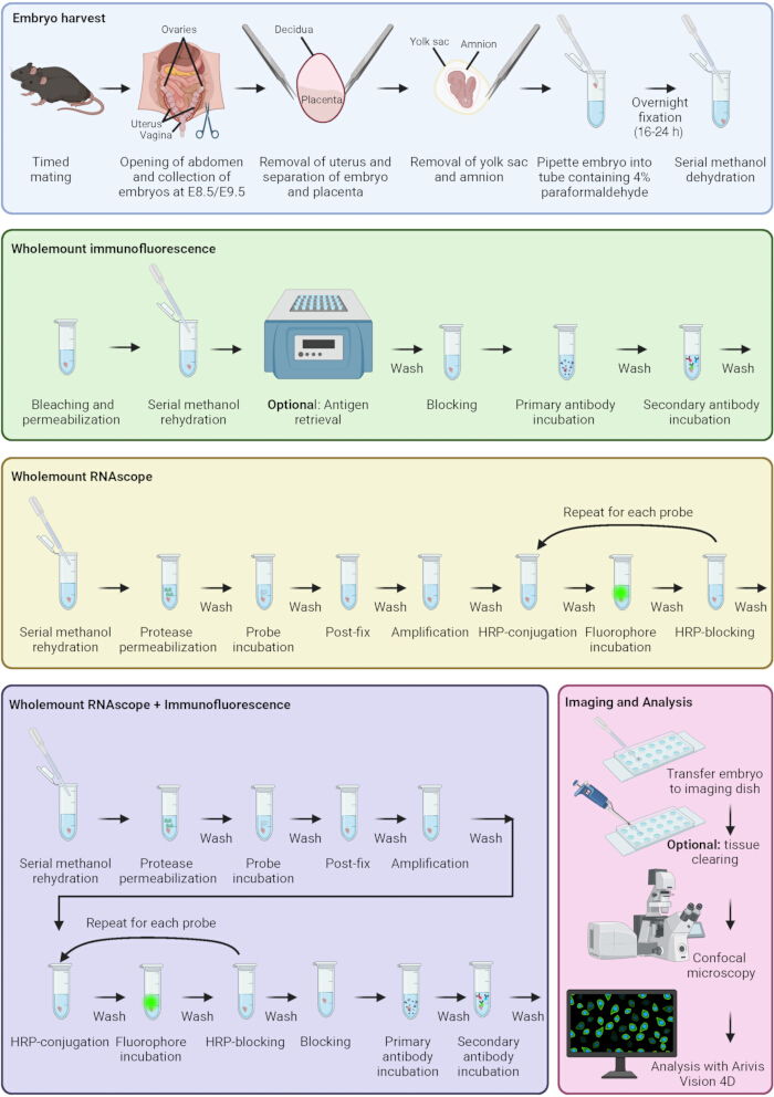
Figure 1: Immunofluorescence and RNAscope staining protocol. Schematic representing key stages of embryo harvest, immunofluorescence, RNAscope, combined immunofluorescence and RNAscope, sample imaging, and analysis. Colored boxes correspond to different protocol stages. Created in BioRender. Sparrow, D. (2026) https://BioRender.com/q6rmax8 Please click here to view a larger version of this figure.
Access restricted. Please log in or start a trial to view this content.
Results
E8.5 embryos were stained by immunofluorescence following this protocol to analyze the number of migrating neural crest cells that were in the G2/M phase of the cell cycle. Samples were stained using antibodies targeting PHH3 to visualize the G2/M phase of the cell cycle and SOX10 to visualize migrating neural crest cells. Embryos were further stained with appropriate secondary antibodies. Nuclei were stained with DAPI. Samples were imaged using the Olympus SpinSR spinning disk confocal microscope with a 10x objective (N...
Access restricted. Please log in or start a trial to view this content.
Discussion
Here, we describe an optimized method of analyzing gene and protein expression spatially within whole-mount E8.5 and E9.5 embryos. These methods have improved spatial resolution and multiplexing ability compared to traditional methods of RNA in situ hybridization and immunofluorescence of sections. This protocol builds upon existing recent protocols to examine developmental pathways in mouse embryos. One recent protocol used RNAscope and immunofluorescence staining to look at Wnt signaling in embryos, but staine...
Access restricted. Please log in or start a trial to view this content.
Disclosures
The authors declare no competing financial interest.
Acknowledgements
The authors thank the team at the Cellular Imaging Core facility, Centre for Human Genetics, University of Oxford, for providing technical support with the microscopy and image analysis included in this manuscript. The authors thank the staff in Biomedical Services, University of Oxford, for support in animal maintenance and timed matings to generate embryos.
Funding for this research was provided by British Heart Foundation Senior Fellowship FS/SBSRF/22/31022 (DBS), Additional Ventures Single Ventricle Fund (DBS), the John Fell Fund, University of Oxford 0016065 (VSR), and the Medical Sciences Internal Fund, University of Oxford 0016707 (VSR).
Access restricted. Please log in or start a trial to view this content.
Materials
| Name | Company | Catalog Number | Comments |
|---|---|---|---|
| Plasticware/glassware | |||
| 3ml Pasteur pipettes Low-density Polyethylene (LDPE) graduated | Wheaton | 711116 | |
| Eppendorf Safe-lock tubes 1.5ml microtube | Eppendorf | 0030 120.086 | |
| Ibidi µ-slide 18-well | Ibidi | 81817 | #1.5H glass coverslip, untreated |
| Menzel X1000 Coverslip 22x50 mm #1.5 (0.16-0.19mm) | Thermo Scientific | 17284904 | |
| Microscope slides with cavities | Marienfeld Superior | 1320000 | |
| Safe seal microtube 2ml | Sarstedt | 72.695.500 | 2ml 'Eppendorf' tube, round bottomed |
| Solutions/buffers | |||
| 1x Antibody diluent/block | Akoya Biosciences | ARD1001EA | |
| Ambion™ RnaseZap™ Rnase Decontamination Solution | Ambion | 9780.9782 | |
| Citrate unmasking buffer | Vector labs | H-3300 | Labelled as Antigen unmasking solution, citric acid based |
| Dimethyl sulfoxide (DMSO) | Merck (Sigma-Aldrich) | D8418 | |
| Hanks' Balanced Salt Solution 10x | Merck (Sigma-Aldrich) | H4641 | |
| Hydrogen peroxide solution | Merck (Sigma-Aldrich) | H1009 | 30% (w/w) in H2O |
| Methanol | Merck (Sigma-Aldrich) | 32213-2.5L-M | |
| Normal donkey serum | Abcam | Ab7475 | 100% stock concentration, 10% final concentration |
| Paraformaldehyde (PFA) powder | Merck (Sigma-Aldrich) | 158127-500g | Dilute to 4% in water. Prepare in fume hood. Stir on heated block and add a small amount of NaOH to facilitate dissolving. pH 7.2-7.6 |
| Phospho buffered saline (PBS) | Merck (Sigma-Aldrich) | P4417 | 1 tablet per 200ml distilled water |
| RNAscope 4-Plex Ancillary Kit for Multiplex Fluorescent Reagent Kit v2 | ACD (Bio-Techne) | 323120 | Contains HRP-C4 and HRP blocker |
| RNAscope™ Multiplex Fluorescent Detection Kit v2 | ACD (Bio-Techne) | 323110 | Contains Protease Plus, Protease III, Protease IV, hydrogen peroxide, TSA buffer, Amp1/2/3, Hrp-C1/2/3, HRP-blocker and DAPI |
| Saline-Sodium-Citrate (SSC) buffer 20X solution | Fisher Bioreagents | BP-1325-1 | Dilute to 0.2x in distilled water |
| Tissue-Clearing Reagent Cubic-R+(M) [for animals] | Tokyo Chemical Industry | T3741 | |
| Triton™ X 100 | Merck (Sigma-Aldrich) | X100 | |
| Tween® 20 | Merck (Sigma-Aldrich) | P7949 | |
| Ultrapure™ 0.5M EDTA, pH8 | Invitrogen | 15575020 | |
| Primary antibodies | |||
| Endomucin antibody (V.7C7) | Santa Cruz | SC-65495 | Use 1:50 dilution. |
| Islet 1 and Islet 2 antibody | Developmental Studies Hybridioma Bank | 34.4DS-5 | Dilute 50% in glycerol. Use 1:50 dilution. |
| PCNA (PC10) Mouse Monoclonal antibody | Cell Signaling Technology | 2586S | Use 1:500 dilution |
| P-histone H3 (Ser10) (6G3) Mouse Monoclonal antibody | Cell Signaling Technology | 9706S | Use 1:500 dilution |
| P-histone H3 (Ser10) (D7N8E) XP® Rabbit Monoclonal antibody | Cell Signaling Technology | 53348S | Use 1:500 dilution |
| Sox10 (E6B6I) XP® Rabbit Monoclonal Antibody | Cell Signaling Technology | 69661S | Use 1:500 dilution |
| Secondary antibodies/nuclear stains | |||
| AlexaFluor® 488-conjugated Affinipure® Donkey anti-mouse IgG (H+L) | Jackson Immunoresearch | 715-545-151 | Use 1:500 dilution |
| AlexaFluor® 647-conjugated Affinipure® Donkey anti-rat IgG (H+L) | Jackson Immunoresearch | 712-605-153 | Use 1:500 dilution |
| AlexaFluor® 647-conjugated Affinipure® F(ab')2 Fragment Donkey anti-mouse IgG (H+L) | Jackson Immunoresearch | 715-606-150 | Use 1:500 dilution |
| Cy™3 Affinipure® Donkey anti-Rabbit IgG (H+L) | Jackson Immunoresearch | 711-165-152 | Use 1:500 dilution |
| DAPI | Merck (Sigma-Aldrich) | D9542 | Used at 1 µg/ml |
| RNAscope probes | |||
| RNAscope® Probe - Mm Sp5-C4 | ACD (Bio-Techne) | 405681-C4 | C4 probe |
| RNAscope® Probe - Mm-Dlx2-C3 | ACD (Bio-Techne) | 555951-C3 | C3 probe |
| RNAscope® Probe - Mm-Msx1 | ACD (Bio-Techne) | 421841 | C1 probe |
| RNAscope® Probe - Mm-Sox10-C2 | ACD (Bio-Techne) | 435931-C2 | C2 probe |
| RNAscope flurophores | |||
| Opal 780 Reagent Pack | Akoya Biosciences | FP1501001KT | Contains Opal 780 reagent, Opal TSA-DIG, DMSO |
| TSA-vivid™ fluorophore 520 | ACD (Bio-Techne) | 323271 | now ClariTSA™ fluorophore 520 |
| TSA-vivid™ fluorophore 570 | ACD (Bio-Techne) | 323272 | now ClariTSA™ fluorophore 570 |
| TSA-vivid™ fluorophore 650 | ACD (Bio-Techne) | 323273 | now ClariTSA™ fluorophore 650 |
| Equipment/software | |||
| Fluoview FV4000 laser scanning microscope | Evident | fv4000 | Excitation lasers: 405/488/561/640/730 nm. Detected wavelength (Fig. 3): 430-470, 500-540, 570-620, 650-710, 750-850 nm. Detected wavelength (Fig. 4) 430-470, 500-531, 570-620 nm. |
| Graphpad Prism 10 | Graphpad | v10.6.1 | |
| Heating block | Grant | QBT1 | |
| Hybrigene hybridisation oven | Techne (Bibby Scientific) | FHB4DD | |
| Ixplore IX83 SpinSR Super-Resolution Microscope System | Olympus | ixplore-ix83-spinsr | Excitation lasers: 405/488/561/640 nm. Emission filters: 447/60, 525/50, 617/73, 685/40. |
| Zeiss Arivis Pro | Carl Zeiss Microscopy GmbH | v. 4.5 (previously Arivis Vision 4D) | |
| Zeiss Arivis SIS converter | Carl Zeiss Microscopy GmbH | v. 4.3.0 |
References
- Bruter AV, Varlamova EA, Okulova YD, Tatarskiy VV, Silaeva YY, Filatov MA. Genetically modified mice as a tool for the study of human diseases. Mol Biol Rep. 2024;51(1):135.
- Rashbrook VS, Brash JT, Ruhrberg C. Cre toxicity in mouse models of cardiovascular physiology and disease. Nat Cardiovasc Res. 2022;1:806–16.
- Breschi A, Gingeras TR, Guigo R. Comparative transcriptomics in human and mouse. Nat Rev Genet. 2017;18(7):425–40.
- Mole MA, Weberling A, Zernicka-Goetz M. Comparative analysis of human and mouse development: From zygote to pre-gastrulation. Curr Top Dev Biol. 2020;136:113–38.
- Krishnan A, Samtani R, Dhanantwari P, Lee E, Yamada S, Shiota K, et al. A detailed comparison of mouse and human cardiac development. Pediatr Res. 2014;76(6):500–7.
- Anderson RH, Bamforth SD. Morphogenesis of the Mammalian Aortic Arch Arteries. Front Cell Dev Biol. 2022;10:892900.
- Wessels A, Sedmera D. Developmental anatomy of the heart: a tale of mice and man. Physiol Genomics. 2003;15(3):165–76.
- Gall JG, Pardue ML. Formation and detection of RNA-DNA hybrid molecules in cytological preparations. Proc Natl Acad Sci U S A. 1969;63(2):378–83.
- John HA, Birnstiel ML, Jones KW. RNA-DNA hybrids at the cytological level. Nature. 1969;223(5206):582–7.
- Buongiorno-Nardelli M, Amaldi F. Autoradiographic detection of molecular hybrids between RNA and DNA in tissue sections. Nature. 1970;225(5236):946–8.
- Dakou E, Vanbekbergen N, Corradi S, Kemp CR, Willems E, Leyns L. Whole-mount in situ hybridization (WISH) optimized for gene expression analysis in mouse embryos and embryoid bodies. Methods Mol Biol. 2014;1211:27–40.
- Anderson R. Multiplex fluorescence in situ hybridization (M-FISH). Methods Mol Biol. 2010;659:83–97.
- Kearney L. Multiplex-FISH (M-FISH): technique, developments and applications. Cytogenet Genome Res. 2006;114(3-4):189–98.
- Burnett R, Guichard Y, Barale E. Immunohistochemistry for light microscopy in safety evaluation of therapeutic agents: an overview. Toxicology. 1997;119(1):83–93.
- Coons AH, Creech HJ, Jones RN. Immunological properties of an antibody containing a fluorescent group. P Soc Exp Biol Med. 1941;47(2):200–2.
- Bardot E, Tzavaras N, Benson DL, Dubois NC. Quantitative Whole-mount Immunofluorescence Analysis of Cardiac Progenitor Populations in Mouse Embryos. J Vis Exp. 2017(128).
- Wang F, Flanagan J, Su N, Wang LC, Bui S, Nielson A, et al. RNAscope: a novel in situ RNA analysis platform for formalin-fixed, paraffin-embedded tissues. J Mol Diagn. 2012;14(1):22–9.
- Erben L, Buonanno A. Detection and Quantification of Multiple RNA Sequences Using Emerging Ultrasensitive Fluorescent In Situ Hybridization Techniques. Curr Protoc Neurosci. 2019;87(1):e63.
- De Bono C, Lescroart F, Zaffran S. How to Study Gene Expression and Gain of Function of Hoxb1 in Mouse Heart Development. Methods Mol Biol. 2025;2889:121–37.
- Zhao R, Moore EL, Gogol MM, Unruh JR, Yu Z, Scott AR, et al. Identification and characterization of intermediate states in mammalian neural crest cell epithelial to mesenchymal transition and delamination. Elife. 2024;13.
- Guijarro C, Kelly RG. On the involvement of the second heart field in congenital heart defects. C R Biol. 2024;347:9–18.
- Bradshaw L, Chaudhry B, Hildreth V, Webb S, Henderson DJ. Dual role for neural crest cells during outflow tract septation in the neural crest-deficient mutant Splotch(2H). J Anat. 2009;214(2):245–57.
- Zawada D, Kornherr J, Meier AB, Santamaria G, Dorn T, Nowak-Imialek M, et al. Retinoic acid signaling modulation guides in vitro specification of human heart field-specific progenitor pools. Nat Commun. 2023;14(1):1722.
- Rabadan MA, Herrera A, Fanlo L, Usieto S, Carmona-Fontaine C, Barriga EH, et al. Delamination of neural crest cells requires transient and reversible Wnt inhibition mediated by Dact1/2. Development. 2016;143(12):2194–205.
- Bhattacharya D, Rothstein M, Azambuja AP, Simoes-Costa M. Control of neural crest multipotency by Wnt signaling and the Lin28/let-7 axis. Elife. 2018;7.
- Kishimoto K, Furukawa KT, Luz-Madrigal A, Yamaoka A, Matsuoka C, Habu M, et al. Bidirectional Wnt signaling between endoderm and mesoderm confers tracheal identity in mouse and human cells. Nat Commun. 2020;11(1):4159.
- De Bono C, Liu Y, Ferrena A, Valentine A, Zheng D, Morrow BE. Single-cell transcriptomics uncovers a non-autonomous Tbx1-dependent genetic program controlling cardiac neural crest cell development. Nat Commun. 2023;14(1):1551.
- Hazra R, Spector DL. Simultaneous visualization of RNA transcripts and proteins in whole-mount mouse preimplantation embryos using single-molecule fluorescence in situ hybridization and immunofluorescence microscopy. Front Cell Dev Biol. 2022;10:986261.
- Kann AP, Krauss RS. Multiplexed RNAscope and immunofluorescence on whole-mount skeletal myofibers and their associated stem cells. Development. 2019;146(20).
Access restricted. Please log in or start a trial to view this content.
Reprints and Permissions
This article has been published
Video Coming Soon