This study used a rat model of paraquat-induced acute lung injury to evaluate the protective effects of sesquiterpene lactones from Eupatorium lindleyanum DC. through histopathological, inflammatory, molecular, and untargeted metabolomic analyses.
مقالة بحثية
Evaluation of Sesquiterpene Lactones in a Rat Model of Paraquat-Induced Acute Lung Injury Using Untargeted Metabolomics
في هذه المقالة
ملخص
الملخص
This study investigated the protective effects and potential mechanisms of sesquiterpene lactones from Eupatorium lindleyanum DC. (SLEL) against paraquat (PQ)-induced acute lung injury (ALI). Male Sprague–Dawley rats were randomly assigned to control, model, SLEL (50, 100, or 200 mg/kg), and dexamethasone (5 mg/kg) groups. ALI was induced by intragastric administration of PQ (200 mg/kg), followed 1 h later by daily administration of SLEL or dexamethasone for 3 consecutive days. Lung injury was evaluated by histopathological examination, pulmonary edema assessment, inflammatory cytokine measurement, Western blotting, quantitative real-time PCR, and untargeted metabolomics. SLEL significantly alleviated PQ-induced histopathological damage and pulmonary edema and reduced inflammatory cytokine levels in serum and bronchoalveolar lavage fluid. SLEL also suppressed TLR4/NF-κB signaling and NLRP3 inflammasome activation while modulating Nrf2/HO-1-associated antioxidant responses. Untargeted metabolomics further revealed that SLEL partially reversed PQ-induced metabolic alterations, particularly those associated with nucleotide metabolism. Quantitatively, high-dose SLEL reduced the lung wet-to-dry ratio from 20.45 ± 2.97 to 12.94 ± 1.71 (p < 0.0001) and decreased serum IL-1β levels from 21.59 ± 2.00 to 16.73 ± 0.95 (p = 0.0001). These findings demonstrate that SLEL attenuates PQ-induced ALI through coordinated regulation of inflammatory responses, oxidative stress, and metabolic disturbances.
المقدمة
Paraquat (PQ) is a highly effective herbicide that has been widely used because of its excellent weed control efficacy. However, owing to its extreme toxicity to humans, accidental or intentional ingestion of PQ can result in severe poisoning and even death1,2. The lung is the primary target organ for PQ accumulation3,4, leading to acute lung injury (ALI), acute respiratory distress syndrome (ARDS), and frequently progressing to pulmonary fibrosis5. Therefore, the development of effective therapeutic agents for PQ-induced ALI remains of considerable clinical significance.
The dried aerial parts of Eupatorium lindleyanum DC., a perennial herb belonging to the family Asteraceae, have long been used in traditional Chinese medicine to treat respiratory diseases because of their expectorant, antitussive, anti-asthmatic, anti-inflammatory, and antiviral properties6. Modern pharmacological studies have demonstrated that the ethanol extract of Eupatorium lindleyanum DC. markedly attenuates lipopolysaccharide (LPS)-induced acute lung injury in rats7 and exerts protective effects against sepsis-associated ALI8. Furthermore, sesquiterpene lactones from Eupatorium lindleyanum DC. (SLEL), the major bioactive constituents of this herb, possess potent anti-inflammatory activity, effectively alleviate acute inflammatory responses9, and protect against LPS-induced ALI10,11.
PQ-induced ALI is closely associated with excessive oxidative stress, amplified inflammatory responses, and aberrant activation of multiple signaling pathways12. Previous studies have demonstrated that SLEL possesses both anti-inflammatory and antioxidant activities, suggesting its potential therapeutic value against PQ-induced lung injury. Excessive ROS production caused by PQ redox cycling induces oxidative damage and triggers compensatory antioxidant responses, particularly involving the Nrf2/HO-1 pathway. Sod1, as an important antioxidant enzyme, participates in ROS detoxification and maintenance of redox homeostasis. Meanwhile, oxidative stress can further amplify inflammatory signaling, in which the TLR4/NF-κB pathway and NLRP3 inflammasome play critical roles in regulating inflammatory cytokine production. Therefore, these molecular targets were selected to comprehensively evaluate the oxidative stress and inflammatory mechanisms underlying PQ-induced lung injury and the protective effects of SLEL. However, the protective effects of SLEL against PQ-induced ALI and the underlying molecular mechanisms have not yet been systematically investigated. Therefore, in the present study, a PQ-induced rat model of ALI was established to comprehensively evaluate the protective effects of SLEL.
البروتوكول
All animal experimental procedures were approved by the Institutional Animal Care and Use Committee of Chongqing Medical University (Approval No. IACUC-CQMU-2025-0757) and were conducted in accordance with institutional guidelines for the care and use of laboratory animals. All materials used in the current study are listed in the Table of Materials.
Preparation of sesquiterpene lactone-enriched fraction (SLEL)
SLEL was prepared according to our previously reported procedure with minor modifications. Briefly, dried medicinal materials of Eupatorium lindleyanum DC. were pulverized and extracted twice with 70% (v/v) ethanol using a solvent-to-material ratio of 10:1 (v/w) for 2 h each extraction. The combined extracts were filtered, concentrated under reduced pressure, and freeze-dried to obtain the crude ethanol extract. The extract was subsequently subjected to polyamide resin and D101 macroporous adsorption resin chromatography to enrich sesquiterpene lactone constituents9. The obtained SLEL fraction was stored at −20 °C until use. The chemical profile of SLEL was confirmed by LC-MS/MS analysis, and the representative profile is shown in Supplementary Figure 1.
Animal grouping, model establishment, and drug administration
Thirty-six male Sprague–Dawley (SD) rats of specific-pathogen-free (SPF) grade (6–8 weeks old, weighing 180–220 g) were used in this study. The rats were acclimatized for 7 days under controlled environmental conditions (temperature: 20–26 °C; relative humidity: 40–70%; 12 h light/12 h dark cycle) with free access to standard laboratory chow and water before the experiments. After acclimatization, the rats were randomly assigned into six groups (n = 6 per group) using a random number table: control group (CG, normal saline), model group (MG, PQ), low-dose SLEL group (SLEL-L, PQ + SLEL, 50 mg/kg), medium-dose SLEL group (SLEL-M, PQ + SLEL, 100 mg/kg), high-dose SLEL group (SLEL-H, PQ + SLEL, 200 mg/kg), and dexamethasone group (DEX, PQ + DEX, 5 mg/kg)10. Investigators responsible for animal administration, outcome assessment, and data analysis were blinded to group allocation throughout the experimental procedures. Except for the CG, all rats received a single intragastric administration of paraquat (PQ, 200 mg/kg) to induce acute lung injury13. The 1-h interval between PQ administration and the first treatment was selected to model early therapeutic intervention following PQ exposure, whereas the 3-day treatment period was chosen to evaluate the effects of SLEL during the acute phase of PQ-induced lung injury. At 1 h after PQ administration, rats in the SLEL-L, SLEL-M, and SLEL-H groups were orally administered SLEL at doses of 50, 100, and 200 mg/kg, respectively, whereas rats in the DEX group received dexamethasone (5 mg/kg). The CG received an equal volume of normal saline (10 mL/kg). All treatments were administered once daily for three consecutive days. A total of 12 h after the final administration, rats were anesthetized by intraperitoneal injection of 3% pentobarbital sodium (30 mg/kg). Adequate depth of anesthesia was confirmed by the absence of the corneal reflex and spontaneous limb movements. Under deep anesthesia, blood samples were collected from the abdominal aorta. Bronchoalveolar lavage fluid (BALF) was subsequently collected, and lung tissues were excised for subsequent analyses. After completion of sample collection, cervical dislocation was performed while the animals remained deeply anesthetized to ensure death. The upper lobe of the right lung was fixed in 4% paraformaldehyde for histopathological examination, while the lower lobe was immediately frozen in liquid nitrogen and stored at −80 °C for molecular and metabolomic analyses. No animals died or were excluded during the experimental period. All animals were included in the final analysis.
Collection of bronchoalveolar lavage fluid (BALF)
The trachea was surgically exposed and the right main bronchus was ligated below the tracheal bifurcation. The left lung was gently lavaged twice with 5 mL sterile normal saline using a syringe. During each lavage, the instilled saline was allowed to remain in the lung for 40 s before gentle withdrawal. BALF was collected into sterile 15 mL centrifuge tubes and centrifuged at 1,500 × g for 10 min at 4 °C. The supernatants were collected and stored at −80 °C until further analysis.
Determination of lung wet-to-dry (W/D) weight ratio
The lung coefficient was calculated as the ratio of lung wet weight to body weight ×100%. The upper lobe of the left lung was weighed immediately to obtain the wet weight (W), then dried at 60 °C for 48 h to obtain the dry weight (D). The lung wet-to-dry (W/D) ratio was subsequently calculated.
Hematoxylin and eosin (H&E) staining and histopathological evaluation
The upper lobe of the right lung was fixed in 4% paraformaldehyde for 24 h, rinsed with running water for 30 min, dehydrated through graded ethanol (75%, 85%, 95%, 100% I, and 100% II), cleared with xylene, embedded in paraffin, and sectioned at a thickness of 4–5 µm. After deparaffinization, rehydration, hematoxylin staining, eosin counterstaining, dehydration, and mounting, sections were examined under a light microscope. Lung injury was evaluated using a semi-quantitative scoring system based on pulmonary hemorrhage (0–1), peribronchial inflammatory infiltration (0–1), interstitial edema (0–2), alveolar epithelial hyperplasia (0–3), and intra-alveolar inflammatory infiltration (0–3), with normal lung tissue scored as 014. Histopathological scoring was independently performed by two investigators who were blinded to group allocation. Any discrepancies between the two assessments were resolved through discussion, and the final score was determined by consensus.
Enzyme-linked immunosorbent assay (ELISA)
Serum and BALF samples were centrifuged at 1,500 × g for 10 min at 4 °C. The supernatants were collected, and the concentrations of IL-1β, IL-6, and TNF-α were determined according to the manufacturer's instructions.
Western blot analysis
Frozen lung tissues were homogenized, and total protein was extracted using a protein extraction kit. Protein concentrations were determined using the bicinchoninic acid (BCA) assay. Equal amounts of protein were separated on 10% SDS-PAGE gels and transferred onto polyvinylidene fluoride (PVDF) membranes. After blocking with 5% non-fat milk in Tris-buffered saline containing Tween-20 (TBST) for 1 h at room temperature, membranes were incubated overnight at 4 °C with primary antibodies, including anti-TLR4 (1:1500), anti-MyD88 (1:1000), anti-p-IκBα (1:1000), anti-IκBα (1:1000), anti-p-P65 (1:1500), anti-P65 (1:1000), anti-NLRP3 (1:1000), anti-ASC (1:2000), anti-IL-1β (1:1000), anti-IL-18 (1:1000), anti-GAPDH (1:5000). Following three washes with Tris-buffered saline containing 0.1% Tween-20 (TBST), membranes were incubated with horseradish peroxidase (HRP)-conjugated secondary antibodies (1:10,000) for 1 h at room temperature. Protein bands were visualized using enhanced chemiluminescence (ECL), and band intensities were quantified using ImageJ software with GAPDH as the internal loading control.
Quantitative real-time PCR (RT-qPCR)
Total RNA was extracted from lung tissues using a phenol–guanidinium-based RNA extraction reagent reagent and reverse-transcribed into complementary DNA (cDNA) using a reverse transcription kit according to the manufacturer's instructions. The reverse transcription reaction was performed in a 20 µL reaction system. The reaction conditions were as follows: 25 °C for 5 min, 55 °C for 15 min, and 85 °C for 2 min. Quantitative real-time PCR was performed using a two-step amplification protocol in a 20 µL reaction system. The amplification conditions were as follows: initial denaturation at 95 °C for 15 s, followed by 40 cycles of denaturation at 95 °C for 15 s and annealing/extension at 60 °C for 45 s. A melting curve analysis was performed after amplification to confirm the specificity of PCR products. The amplification efficiencies of all primer pairs were determined using standard curves generated from serial dilutions of cDNA and ranged from 96% to 103%. Primer sequences are listed in Supplementary Table 2. Relative mRNA expression levels were calculated using the 2^−ΔΔCt method with GAPDH as the internal reference gene.
Untargeted metabolomics analysis
Untargeted metabolomic profiling of lung tissues from the CG, MG, and SLEL-H groups was performed using an ultra-performance liquid chromatography-electrospray ionization-tandem mass spectrometry (UPLC-ESI-MS/MS) platform. Chromatographic separation was achieved on a C18 column (150 × 2 mm, 5 µm) maintained at 40 °C. The mobile phases consisted of solvent A (water containing 0.04% acetic acid) and solvent B (acetonitrile containing 0.04% acetic acid) at a flow rate of 0.4 mL/min. The gradient elution program was as follows: 0 min, 100:0 (A:B); 11.0 min, 5:95; 12.0 min, 5:95; 12.1 min, 95:5; and 15.0 min, 95:5. The injection volume was 2 µL, and the column effluent was introduced into an electrospray ionization-triple quadrupole-linear ion trap mass spectrometry (ESI-QTRAP-MS/MS) system for metabolite detection.
Untargeted metabolomics analysis was performed by Majorbio Bio-Pharm Technology Co., Ltd. Metabolites were extracted using an extraction solution containing internal standards (including L-2-chlorophenylalanine). Quality control (QC) samples were prepared by pooling equal volumes of all samples and were inserted every 5–10 injections during liquid chromatography–tandem mass spectrometry (LC-MS/MS) analysis to monitor analytical stability and reproducibility. Raw LC-MS data were processed using Progenesis QI software for peak detection, alignment, and annotation. Metabolite annotation was performed based on accurate mass and MS/MS fragmentation patterns by matching against HMDB, Metlin, and the Majorbio in-house database. Differential metabolites were screened based on the criteria of VIP > 1 and p < 0.05. The C18-based method was used for broad untargeted metabolomic profiling; however, its retention and separation of highly polar nucleotide metabolites may be limited. Therefore, nucleotide-related findings should be interpreted with this analytical limitation in mind, and targeted validation using chromatographic approaches optimized for highly polar metabolites, such as ion-pairing chromatography, should be considered in future studies.
Statistical analysis
Statistical analyses were performed using GraphPad Prism 8.0.1. Data are presented as the mean ± standard deviation (SD). Comparisons among multiple groups were performed using ordinary one-way analysis of variance (ANOVA), followed by Dunnett's multiple comparisons test. A p value < 0.05 was considered statistically significant. In the figures, # and ## indicate p < 0.05 and p < 0.01 versus the CG, respectively, whereas * and ** indicate p < 0.05 and p < 0.01 versus the MG, respectively.
النتائج
Effects of SLEL on lung histopathological changes
Representative hematoxylin and eosin (H&E)-stained lung sections are shown in Figure 1A. Compared with the CG, rats in the MG exhibited severe histopathological alterations, including aggravated pulmonary edema, intra-alveolar hemorrhage, massive inflammatory cell infiltration, and marked thickening of the alveolar walls, indicating substantial lung tissue damage. In contrast, SLEL treatment markedly attenuated these pathological changes in a dose-dependent manner. Consistent with the histopathological observations, the lung injury score was significantly increased in the MG compared with the CG, whereas SLEL treatment significantly reduced the lung injury score (Figure 1B, p < 0.05). Furthermore, PQ exposure markedly increased both the lung coefficient (Figure 1C) and the lung wet-to-dry (W/D) weight ratio (Figure 1D) compared with the CG (p < 0.01). Treatment with SLEL significantly decreased the lung coefficient (p < 0.05) and lung W/D ratio (p < 0.01), indicating that SLEL effectively alleviated pulmonary edema and attenuated PQ-induced acute lung injury.
Effects of SLEL on IL-1β, IL-6, and TNF-α levels in serum and BALF, and their mRNA expression in lung tissues
IL-1β, IL-6, and TNF-α are key pro-inflammatory cytokines involved in the initiation and amplification of inflammatory responses, and their expression levels reflect the severity of inflammation. As shown in Figure 2A–F, the concentrations of IL-1β, IL-6, and TNF-α in both bronchoalveolar lavage fluid (BALF) and serum were significantly increased in the MG compared with the CG (p < 0.01), indicating a robust inflammatory response induced by PQ. Treatment with SLEL dose-dependently reduced the levels of these cytokines, with the most pronounced inhibitory effects observed in the SLEL-H group (p < 0.05). Consistent with the protein expression profiles, the mRNA expression levels of IL-1β, IL-6, and TNF-α in lung tissues were markedly elevated in the MG compared with the CG and were significantly reduced following SLEL treatment (Figure 2G–I, p < 0.01). These findings indicate that SLEL suppresses inflammatory responses by coordinately reducing the expression of pro-inflammatory cytokines at both the transcriptional and protein levels.
Effects of SLEL on the expression of proteins associated with the TLR4/NF-κB signaling pathway in lung tissues
The TLR4/NF-κB signaling pathway plays a critical role in the initiation and progression of inflammatory responses during acute lung injury. As shown in Figure 3A, the protein expression levels of TLR4, MyD88, p-IκBα, and p-P65 were markedly increased in the lung tissues of the MG compared with the CG, indicating robust activation of the TLR4/NF-κB signaling pathway. Following SLEL-H treatment, the expression of these proteins was markedly reduced. Quantitative analysis further demonstrated that SLEL-H significantly decreased the protein expression of TLR4 (Figure 3B) and MyD88 (Figure 3C), while markedly suppressing the phosphorylation of IκBα (Figure 3D) and P65 (Figure 3E) (p < 0.05). Specifically, p-P65 protein expression decreased from 2.51 ± 0.21 in the MG to 1.77 ± 0.11 in the SLEL-H group (p = 0.0018). These findings suggest that SLEL attenuates PQ-induced inflammatory responses, at least in part, through inhibition of the TLR4/NF-κB signaling pathway.
Effects of SLEL on the expression of NLRP3 inflammasome-related proteins in lung tissues
Excessive activation of the NLRP3 inflammasome is considered a key mechanism underlying the inflammatory response during acute lung injury. As shown in Figure 4A, the protein expression levels of NLRP3, ASC, IL-1β, and IL-18 were markedly increased in the lung tissues of the MG compared with the CG, indicating robust activation of the NLRP3 inflammasome. Following SLEL-H treatment, the expression of these proteins was markedly reduced. Quantitative analysis further demonstrated that SLEL-H significantly decreased the protein expression of NLRP3 (Figure 4B), ASC (Figure 4C), IL-1β (Figure 4D), and IL-18 (Figure 4E) (p < 0.05). Specifically, NLRP3 protein expression decreased from 2.68 ± 0.46 in the MG to 1.68 ± 0.28 in the SLEL-H group (p = 0.0140). These findings indicate effective inhibition of NLRP3 inflammasome activation and suggest that SLEL attenuates PQ-induced inflammatory responses, at least in part, through suppression of NLRP3 inflammasome activation.
Effects of SLEL on oxidative stress-related indicators in lung tissues
To evaluate oxidative stress and the antioxidant response, the mRNA expression levels of Sod1, Nrf2, HO-1, and NOX2 in lung tissues were determined by quantitative real-time PCR. As shown in Figure 5A–D, the mRNA expression levels of Sod1 (Figure 5A), Nrf2 (Figure 5B), HO-1 (Figure 5C), and NOX2 (Figure 5D) were significantly increased in the MG compared with the CG (p < 0.01), indicating that PQ induced oxidative stress and activated the endogenous antioxidant defense response. Following SLEL-H treatment, the expression levels of these genes were significantly reduced (p < 0.05), suggesting that SLEL effectively attenuated PQ-induced oxidative stress.
Effects of SLEL on PQ-induced metabolic disturbances in lung tissues
To systematically evaluate the regulatory effects of SLEL on PQ-induced metabolic disturbances in lung tissues, untargeted metabolomic analysis was performed. As shown in the PCA score plots (Figure 6A–C), the MG was clearly separated from the CG, indicating that PQ exposure induced profound alterations in the overall metabolic profile of lung tissues. The SLEL-H group was also distinctly separated from the MG, suggesting that SLEL markedly remodeled the abnormal metabolic state induced by PQ. However, the SLEL-H group remained partially separated from the CG, indicating that although SLEL substantially reversed PQ-induced metabolic disturbances, the metabolic profile was not completely restored to the normal state. Differential metabolite analysis identified a large number of significantly altered metabolites in the SLEL-H vs. MG, SLEL-H vs. CG, and MG vs. CG comparisons, with considerable overlap among the three comparisons (Figure 6D), suggesting that PQ-induced lung injury was accompanied by extensive metabolic reprogramming and that SLEL reversed a subset of these key metabolic alterations. The volcano plot (Figure 6E) revealed numerous significantly upregulated and downregulated metabolites following SLEL treatment compared with the MG, indicating that SLEL exerted broad regulatory effects on the pulmonary metabolic network. VIP analysis (Figure 6F) further demonstrated that the metabolites contributing most to group discrimination were predominantly nucleotide-related metabolites, including inosine 5′-monophosphate (IMP), uridine monophosphate (UMP), adenosine monophosphate (AMP), guanosine, uridine, inosine, and hypoxanthine, suggesting that nucleotide metabolism represents a key metabolic regulatory node involved in the protective effects of SLEL against PQ-induced lung injury. The hierarchical clustering heatmap (Figure 6G) further showed consistent expression patterns of these differential metabolites among the experimental groups. Marked metabolic dysregulation was observed in the MG, whereas SLEL treatment partially restored the expression profiles of these metabolites, indicating a significant improvement in the metabolic abnormalities associated with lung injury. KEGG pathway analysis (Figure 6H) demonstrated that the differential metabolites were primarily enriched in purine metabolism, glycerophospholipid metabolism, alanine, aspartate and glutamate metabolism, and nucleotide metabolism. In particular, enrichment of the nucleotide metabolism pathway suggested that PQ-induced lung injury was characterized by pronounced disturbances in nucleotide homeostasis involving nucleic acid synthesis and degradation, energy metabolism, and tissue repair. Following SLEL treatment, metabolites involved in this pathway were significantly altered, indicating that SLEL may exert its protective effects by restoring nucleotide metabolic homeostasis. Based on these findings, receiver operating characteristic (ROC) curve analysis (Figure 6I) was further performed for differential metabolites involved in nucleotide metabolism. Several nucleotide metabolism-related metabolites exhibited good discriminatory performance, among which inosine 5′-monophosphate (IMP) achieved the highest AUC value of 1.000, while several other metabolites also demonstrated favorable diagnostic performance. These findings suggest that nucleotide metabolism-related metabolites are not only key responsive molecules involved in the protective effects of SLEL against PQ-induced ALI but may also serve as potential metabolic biomarkers for evaluating the therapeutic efficacy of SLEL.
DATA AVAILABILITY:
The original Western blot data generated in this study are provided in the supplementary archive “raw_data_73654.rar”. Other data generated or analyzed during this study are available from the corresponding author upon reasonable request.
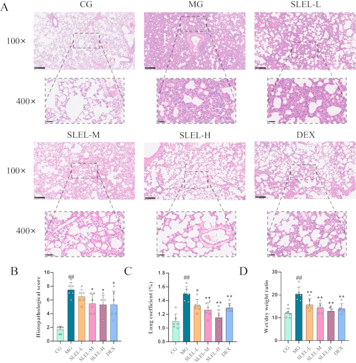
Figure 1: SLEL attenuates PQ-induced histopathological lung injury in rats. (A) Representative hematoxylin and eosin (H&E)-stained lung sections from the control group (CG), model group (MG), low-dose SLEL (SLEL-L), medium-dose SLEL (SLEL-M), high-dose SLEL (SLEL-H), and dexamethasone (DEX) groups. Representative images were captured at 100× (upper panels) and 400× (lower panels) magnification. Scale bars: 250 µm (upper panels) and 50 µm (lower panels). (B) Histopathological injury score. (C) Lung coefficient. (D) Lung wet-to-dry (W/D) weight ratio. Data are presented as the mean ± SD (n = 6). ##p < 0.01 versus the CG; *p < 0.05 and **p < 0.01 versus the MG. Please click here to view a larger version of this figure.
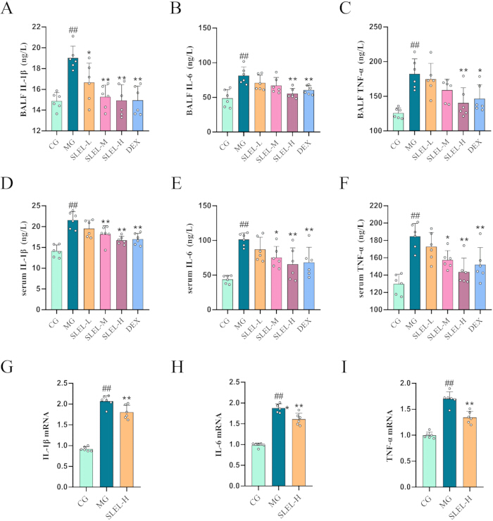
Figure 2: SLEL suppresses inflammatory cytokine production in PQ-induced acute lung injury. (A–C) Levels of IL-1β, IL-6, and TNF-α in bronchoalveolar lavage fluid (BALF) determined by ELISA. (D–F) Levels of IL-1β, IL-6, and TNF-α in serum determined by ELISA. (G–I) Relative mRNA expression levels of IL-1β, IL-6, and TNF-α in lung tissues determined by quantitative real-time PCR (RT-qPCR). Data are presented as the mean ± SD (ELISA, n = 6; RT-qPCR, n = 6). ##p < 0.01 versus the CG; *p < 0.05 and **p < 0.01 versus the MG. Please click here to view a larger version of this figure.
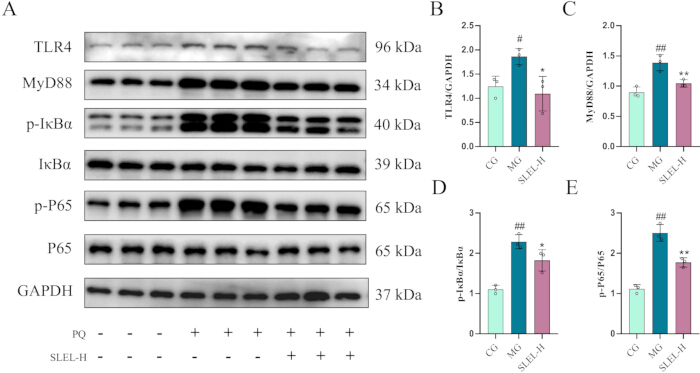
Figure 3: SLEL inhibits activation of the TLR4/NF-κB signaling pathway in lung tissues. (A) Representative Western blot images showing the protein expression of TLR4, MyD88, p-IκBα, IκBα, p-P65, P65, and GAPDH. (B–E) Quantitative analysis of TLR4/GAPDH, MyD88/GAPDH, p-IκBα/IκBα, and p-P65/P65, respectively. Data are presented as the mean ± SD (n = 3). #p < 0.05 and ##p < 0.01 versus the CG; *p < 0.05 and **p < 0.01 versus the MG. Please click here to view a larger version of this figure.
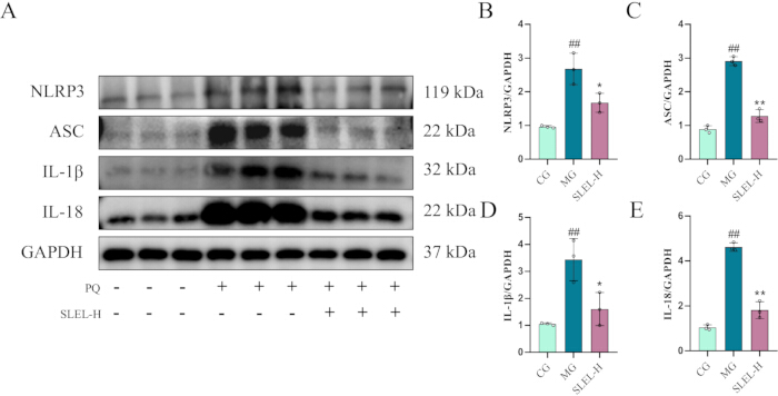
Figure 4: SLEL suppresses NLRP3 inflammasome activation in lung tissues. (A) Representative Western blot images showing the protein expression of NLRP3, ASC, IL-1β, IL-18, and GAPDH. (B–E) Quantitative analysis of NLRP3/GAPDH, ASC/GAPDH, IL-1β/GAPDH, and IL-18/GAPDH, respectively. Data are presented as the mean ± SD (n = 3). ##p < 0.01 versus the CG; *p < 0.05 and **p < 0.01 versus the MG. Please click here to view a larger version of this figure.
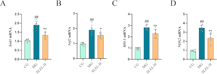
Figure 5: SLEL regulates oxidative stress-related gene expression in lung tissues. (A–D) Relative mRNA expression levels of Sod1, Nrf2, HO-1, and NOX2, respectively, determined by quantitative real-time PCR (RT-qPCR). Data are presented as the mean ± SD (n = 6). ##p < 0.01 versus the CG; *p < 0.05 and **p < 0.01 versus the MG. Please click here to view a larger version of this figure.
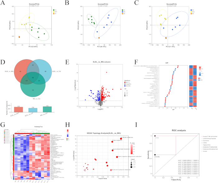
Figure 6: SLEL attenuates PQ-induced metabolic alterations in lung tissues. (A) Principal component analysis (PCA) score plot comparing the CG and MG. (B) PCA score plot comparing the MG and SLEL-H groups. (C) PCA score plot comparing the CG and SLEL-H groups. (D) Venn diagram showing the overlap and the numbers of differential metabolites identified in the three pairwise comparisons. (E) Volcano plot of differential metabolites in the SLEL-H versus MG comparison. (F) Variable importance in projection (VIP) analysis and corresponding expression heatmap of representative differential metabolites. (G) Hierarchical clustering heatmap of differential metabolites. (H) KEGG pathway topology analysis of differential metabolites identified in the SLEL-H versus MG comparison. (I) Receiver operating characteristic (ROC) curve analysis of representative nucleotide metabolism-related metabolites. Data are presented as the mean ± SD (n = 6). Please click here to view a larger version of this figure.
Supplementary Figure 1: UPLC-Q/TOF-MSn profile and chemical structures of the main compounds identified in SLEL. Total ion chromatograms of SLEL acquired in positive and negative ion modes are shown together with the chemical structures of compounds A–R, corresponding to the compounds listed in Supplementary Table 1. Please click here to download this file.
Supplementary Table 1: The main compounds identified in SLEL by UPLC-Q/TOF-MSn. The table lists the retention time, compound name, molecular formula, calculated molecular mass, selected ions, and mass error for each compound identified in SLEL. Please click here to download this file.
Supplementary Table 2: Primer sequences used for RT-qPCR analysis. The table lists the forward and reverse primer sequences used for RT-qPCR analysis of the target genes and the reference gene. Please click here to download this file.
المناقشة
In the present study, a paraquat (PQ)-induced rat model of acute lung injury (ALI) was used to systematically evaluate the protective effects and potential mechanisms of sesquiterpene lactones from Eupatorium lindleyanum DC. (SLEL). SLEL ameliorated histopathological lung injury and pulmonary edema, reduced inflammatory cytokine production, modulated the TLR4/NF-κB signaling pathway and NLRP3 inflammasome, and partially corrected PQ-induced metabolic disturbances. Oxidative stress is a central mechanism of PQ toxicity, in which continuous generation of reactive oxygen species (ROS) through redox cycling promotes inflammatory responses and subsequent lung injury15. In the present study, NOX2, Nrf2, HO-1, and Sod1 were significantly upregulated in the model group, consistent with increased oxidative stress accompanied by a compensatory antioxidant response. The Nrf2/HO-1 pathway is an important antioxidant defense mechanism that promotes cellular responses against excessive ROS12,16. Accordingly, NOX2, Nrf2/HO-1, and Sod1 were examined to reflect ROS generation, antioxidant signaling, and antioxidant defense, respectively. Their reduced expression following SLEL treatment suggests that attenuation of PQ-induced oxidative stress may reduce the requirement for compensatory antioxidant responses.
Inflammation is another major pathological process in PQ-induced lung injury17,18. TLR4/NF-κB signaling plays an important role in inflammatory cytokine production and amplification of inflammatory responses19. PQ activated the TLR4/MyD88/NF-κB pathway, whereas SLEL suppressed pathway activation and reduced IL-1β, IL-6, and TNF-α expression. The NLRP3 inflammasome provides an important link between oxidative stress and inflammation, and its activation is closely associated with NF-κB signaling20. Consistently, SLEL reduced the expression of NLRP3, ASC, IL-1β, and IL-18. These findings suggest that suppression of the TLR4/NF-κB–NLRP3 inflammatory axis may contribute to the protective effects of SLEL against PQ-induced ALI. Compared with LPS-induced ALI, in which inflammatory activation through TLR4 signaling represents a dominant initiating event, PQ-induced ALI is characterized by redox cycling-mediated ROS accumulation and subsequent inflammatory amplification. Therefore, the present study extends previous observations regarding the anti-inflammatory activity of SLEL and further highlights its potential protective effects against chemically induced oxidative lung injury.
Untargeted metabolomics provided additional evidence that the effects of SLEL extend to metabolic regulation. PQ exposure induced marked metabolic alterations in lung tissues, particularly in nucleotide metabolism, whereas SLEL partially reversed these changes. Nucleotide metabolism is closely associated with cellular energy homeostasis, nucleic acid synthesis, inflammatory responses, oxidative stress, and tissue repair21. Beyond serving as substrates for nucleic acid synthesis, nucleotide metabolites, particularly purine metabolites, may participate in inflammatory regulation by affecting ATP availability, ROS generation, and inflammasome activation. Among the differential metabolites, inosine 5′-monophosphate (IMP) showed strong discriminatory performance in the present dataset, suggesting that it may represent a candidate metabolic indicator associated with the response to SLEL treatment. However, because the current analysis was based on untargeted metabolomics, the biological significance of these metabolic alterations requires further validation using targeted quantitative metabolomics approaches. In this study, metabolomic profiling was performed only in the CG, MG, and SLEL-H groups because the high-dose SLEL group exhibited the most pronounced protective effects in the initial efficacy evaluation. Although this strategy facilitated the identification of major metabolic alterations associated with SLEL treatment, the absence of metabolic profiling across different doses limited the assessment of dose-dependent metabolic responses. Taken together, the molecular and metabolomic findings suggest that the protective effects of SLEL involve coordinated regulation of inflammatory, oxidative stress, and metabolic responses rather than a single pathway.
Several methodological considerations are critical for the reproducibility of this protocol. The interval between PQ exposure and SLEL administration should be strictly controlled because variation in treatment timing may influence the severity of lung injury and the observed treatment response. BALF collection should also be standardized to minimize differences in sample recovery and subsequent cytokine measurements, while lung tissues designated for histopathological, molecular, and metabolomic analyses should be collected and processed consistently. In particular, metabolomic samples require rapid and uniform collection, storage, and processing to minimize technical variation. If substantial inter-animal variability occurs, PQ administration, treatment timing, BALF recovery, and sample handling should be examined before downstream analyses. An advantage of this protocol is the integration of histopathological, inflammatory, molecular, and untargeted metabolomic analyses, allowing the effects of SLEL to be evaluated at multiple levels; however, the use of multiple sample types and analytical procedures also increases experimental complexity and requires strict standardization.
Several limitations should be considered when interpreting these findings. First, although changes in Nrf2 and HO-1 expression suggested the involvement of antioxidant responses, further functional validation of Nrf2 activation is warranted. Future studies should confirm Nrf2 activation through approaches such as nuclear/cytoplasmic fractionation followed by Western blot analysis or immunofluorescence staining to evaluate Nrf2 nuclear translocation. Second, Sod1 was selected as a representative antioxidant enzyme in the present study; however, other important antioxidant systems, particularly mitochondrial Sod2 and catalase, were not evaluated. Therefore, the current findings may not fully represent the complexity of antioxidant responses following PQ exposure. Third, although untargeted metabolomics revealed nucleotide metabolism-related alterations, the specific mechanisms linking purine metabolism to inflammatory activation and oxidative stress require further investigation. In addition, the discriminatory performance of IMP should be validated in larger independent datasets before it can be considered a robust biomarker. Fourth, molecular and metabolomic analyses were performed only in the CG, MG, and SLEL-H groups because the high-dose SLEL group showed the strongest protective effects in the initial efficacy evaluation. Although this design enabled the identification of major molecular changes associated with SLEL treatment, the lack of mechanistic analyses across different doses limited the evaluation of dose-dependent responses. Fifth, histopathological evaluation in the present study was based on a composite semi-quantitative scoring system derived from previously reported criteria. Although this approach provides an integrated assessment of overall lung injury severity, future studies incorporating parameter-specific histopathological scoring may provide more detailed information regarding individual pathological alterations. Finally, because this study was conducted exclusively in an animal model, further pharmacokinetic studies, human-equivalent dose evaluation, safety assessment, and validation of candidate metabolic biomarkers in plasma samples from patients with PQ poisoning are required before clinical translation. Overall, the present protocol demonstrates that SLEL attenuates PQ-induced ALI and provides an integrated approach for investigating its associated inflammatory, oxidative stress, and metabolic alterations.
الإفصاحات
The authors declare that they have no conflicts of interest.
Author Contributions:
Li LingHu contributed to the original draft preparation, software, data curation, and methodology. Chen Luo, Keyun Zhou, and Cheng Cheng contributed to the investigation. Yan Yang and Zhaoguo Zhou contributed to data curation. Lian Xia contributed to formal analysis. Chuanxin Liu contributed to methodology. Daike Zou contributed to the original draft preparation. Yi Hou contributed to review, editing, and investigation. Xianqin Luo contributed to review and editing, project administration, investigation, funding acquisition, data curation, and conceptualization. All authors have read and agreed to the published version of the manuscript
شكر وتقدير
This research was funded by Natural Science Foundation Project of Chongqing Municipality (No. cstc2021jcyj-msxmX0365), Science and Technology Research Program of Chongqing Municipal Education Commission (No. KJZD-K202215101, No. KJZD-M202515104), Joint Research Fund for Technological Innovation by Chongqing Bishan District Bureau of Science and Technology and Chongqing University of Chinese Medicine (No. BSZYYLH010).
المواد
| الاسم | الشركة | رقم فهرسي | التعليقات |
|---|---|---|---|
| anti ASC | ImmunoWay | YM8466 | |
| anti GAPDH | ImmunoWay | YN5585 | |
| anti IL-18 | Abmart | TD6252 | |
| anti IL-1β | Abmart | PC6319 | |
| anti IκBα | Cell Signaling Technology | 4812 | |
| anti MyD88 | Abmart | TB2598 | |
| anti NLRP3 | Abmart | TB4673 | |
| anti p-IκBα | Cell Signaling Technology | 2859 | |
| anti p-P65 | Abmart | TP70621 | |
| anti P65 | Abmart | TA5006 | |
| anti TLR4 | Abmart | T61519 | |
| BCA | Beyotime Biotechnology Co., Ltd | P10010 | |
| GraphPad Prism 8.0.1 | San Diego | ||
| IL-1β kit | Xiamen Huijia Biotechnology Co., Ltd | EHJ-20537r | |
| IL-6 kit | Xiamen Huijia Biotechnology Co., Ltd | EHJ-20746r | |
| Micropipette | Eppendorf | ||
| Microplate reader | Gene Co., Ltd | Odyssey Fc | |
| PVDF | Millipore | IPVH00010 | |
| RIPA buffer | Beyotime Biotechnology Co., Ltd | P0013B | |
| Sprague–Dawley (SD) rats | SPF Biotechnology Co., Ltd. | ||
| TNF-α kit | Xiamen Huijia Biotechnology Co., Ltd | EHJ-20039r | |
| TRIzol | Thermo Fisher Scientific | 15596018CN | |
| Ultra-low temperature freezer | SANYO |
المراجع
- Zyoud SH. Investigating global trends in paraquat intoxication research from 1962 to 2015 using bibliometric analysis. Am J Ind Med. 2018;61(6):462-470.
- Zhu Y, et al. Squalene epoxidase promotes paraquat-induced pulmonary toxicity through endoplasmic reticulum-mediated ferroptosis. J Adv Res. 2026;81:673-687.
- Liu S, et al. Genkwanin alleviates paraquat-induced acute lung injury by activating SIRT1-mediated Nrf2/HO-1 signaling pathway. Naunyn Schmiedebergs Arch Pharmacol. 2026;399(3):4215-4229.
- Li Y, et al. Lipoxin A4 protects against paraquat-induced acute lung injury by inhibiting the TLR4/MyD88-mediated activation of the NF-κB and PI3K/AKT pathways. Int J Mol Med. 2021;47(5):86.
- Yuan D, et al. Metformin regulates alveolar macrophage polarization to protect against acute lung injury in rats caused by paraquat poisoning. Front Pharmacol. 2022;13:811372.
- Wang X, et al. Traditional applications, phytochemistry, and pharmacological activities of Eupatorium lindleyanum DC.: A comprehensive review. Front Pharmacol. 2020;11:577124.
- Li R, Yang H, Xue Y, Luo X. Yemazhui (Herba Eupatorii Lindleyani) ameliorates lipopolysaccharide-induced acute lung injury via modulation of the toll-like receptor 4/nuclear factor kappa-B/nod-like receptor family pyrin domain-containing 3 protein signaling pathway and intestinal flora in rats. J Tradit Chin Med. 2024;44(2):303-314.
- Luo C, et al. Eupatorium lindleyanum DC. suppresses cytokine storm by inhibiting NF-κB and PI3K-Akt signaling in sepsis-associated and virus-related acute lung injury. Curr Issues Mol Biol. 2026;48(3):333.
- Luo C, et al. Antiinflammatory effects of sesquiterpene lactones from Eupatorium lindleyanum DC. in rodent models. J Vis Exp. 2026;(229):e69846.
- Luo C, et al. Multi-omics and experimental insights into the protective effects of sesquiterpenoid lactones from Eupatorium lindleyanum DC. in acute lung injury: Regulation of PI3K-Akt and MAPK-NF-κB pathways. Pharmaceuticals (Basel). 2025;18(10):1523.
- Luo C, et al. Study on the protective effect of sesquiterpene lactones of Eupatorium lindleyanum DC. on lipopolysaccharide-induced acute lung injury in rats. J Chongqing Med Univ. 2025;50(7):889-897.
- Abbasifard A, et al. Antioxidative effects of crocin-loaded Noisome in paraquat-induced oxidative stress in lung rat. Cell Biochem Biophys. 2025;83(4):4597-4604.
- Li N, et al. Anthrahydroquinone-2,6-disulfonate attenuates PQ-induced acute lung injury through decreasing pulmonary microvascular permeability via inhibition of the PI3K/AKT/eNOS pathway. Int J Mol Med. 2024;54(1):63.
- Chimenti L, et al. Nebulized heparin attenuates pulmonary coagulopathy and inflammation through alveolar macrophages in a rat model of acute lung injury. Thromb Haemost. 2017;117(11):2125-2134.
- Huang J, Ning N, Zhang W. Effects of paraquat on IL-6 and TNF-α in macrophages. Exp Ther Med. 2019;17(3):1783-1789.
- Xia X, et al. Protective effect of wild polysaccharides extracted from Ulva prolifera on oxidative stress damage in valproic acid-induced neuronal cells. Front Pharmacol. 2026;17:1805470.
- Pu Y, Han S, Chen J, Liu Z. MANF inhibits NLRP3 inflammasome activation by competitively binding to DDX3X in paraquat-stimulated alveolar macrophages. Ecotoxicol Environ Saf. 2024;287:117331.
- Zhang R, et al. Ginkgolide C alleviates acute lung injury caused by paraquat poisoning via regulating the Nrf2 and NF-κB signaling pathways. Oxid Med Cell Longev. 2022;2022:7832983.
- Li S, et al. Released palmitic acid-mediated TLR4/NF-κB activation enhances the virulence of Bordetella pertussis MT28 lineage. Nat Commun. 2026;17(1):5532.
- Yang XR, et al. Role of oxidative stress in sepsis: Mechanisms, pathways, and therapeutic strategies. J Pharm Anal. 2026;16(4):101452.
- Dong W, et al. Luteolin ameliorates Escherichia coli-induced intestinal injury by modulating gut microbiota, metabolites and the TLR4/MyD88/NF-kB signaling pathway. Poult Sci. 2026;105(6):106729.