本研究介绍了一种通过直接培养胶原蛋白、平滑肌细胞和内皮细胞来快速制备活体血管组织的技术。此外,还描述了一种用于工程化血管组织力学表征的新方案。
本文内容
摘要
摘要
合成材料作为血管替代物植入时,已知会引发炎症、狭窄和感染等临床并发症。胶原蛋白因其固有的生物相容性(即抗原性低,炎症反应和细胞毒性反应弱),已被广泛应用于多种生物医学领域,被认为是合成材料的有效替代物。然而,胶原凝胶的力学性能有限,且操作性较差,限制了其在血管组织工程中作为支架材料的应用。因此,本研究的出发点首先是将细胞化的胶原凝胶构建成管状结构,其次增强由平滑肌细胞驱动的胶原基质重组,以获得足够坚韧、便于操作的组织。
本文所述策略基于采用模具技术,在三维圆柱几何结构中直接组装胶原蛋白与平滑肌细胞(构建物)。该过程需要一个成熟期,在此期间,构建物在生物反应器中静态培养(不施加外部动态力学约束)1至2周。“静态生物反应器”为构建物提供受监控和控制的无菌环境(pH、温度、气体交换、营养供给和废物清除)。在培养期间,通过厚度测量评估细胞驱动的胶原基质重塑,并通过测定葡萄糖消耗率和乳酸生成率来监测细胞的代谢活性。最终,对所得管状构建物的力学与黏弹性特性进行评估。为此,开发了特定的实验方案和专门技术(操作、夹持、在水合环境中工作等),以表征工程化组织。
引言
血管组织工程设想了多种旨在构建工程化血管的策略,包括基于合成支架的移植物、基于细胞片的组织工程血管(TEBV)以及基于细胞外基质(ECM)成分的TEBV。在这些方法中,合成聚合物具有良好的机械性能,但存在一个共同的缺点,即缺乏生物活性1。基于细胞片的方法能够制备出具有高机械性能的工程化血管替代物,但制备此类移植物所需时间约为28周2。细胞外基质中的天然生物聚合物,如胶原蛋白、弹性蛋白、纤维蛋白3或其组合,仍是组织工程支架材料的金标准。这主要是因为这些材料通常具有良好的生物相容性,同时能够诱导功能性细胞反应4-5。在这些生物聚合物中,I型胶原是皮肤、血管和肌腱等多种组织中含量最丰富且起主要承重作用的蛋白质。已有大量研究聚焦于胶原蛋白的机械性能6–8,但关于细胞在静态成熟过程中对胶原凝胶进行重塑的研究仍较少。细胞重塑是指细胞诱导的胶原基质结构改变,可能影响胶原原纤维网络的稳定性9。作为一种天然支架,可从大鼠尾腱等不同来源分离、灭菌并储存相对大量的I型胶原10。理解细胞与胶原的相互作用及其相关的细胞化胶原支架(构建物)整体力学行为,是组织构建的关键步骤。基于胶原的TEBV可在凝胶制备过程中直接将细胞与胶原混合,并进一步塑造成特定形状,如管状或平面状11。凝胶内的血管细胞可增殖并对I型胶原进行重塑12。因此,该方法避免了组织工程应用中支架开发所面临的重要问题之一——对特定大孔隙率的需求。然而,胶原凝胶的主要缺点是其机械性能较合成材料低13。
本研究通过一步法将细胞直接与胶原蛋白混合,构建出具有细胞均匀分布的可存活组织。“静态生物反应器”用于细胞化胶原凝胶的静态成熟培养,持续1至2周(未施加外部动态力学约束)。在培养过程中发生了胶原基质的重塑,从而为构建物提供了结构强化。此外,这些构建物可进一步转移至旋转壁式生物反应器,并实现了均匀的内皮层。同时,本研究还提出了一种特定的力学测试方案,为表征管状软组织力学性能提供了适当且新颖的方法。
总之,本研究提出了一种方法用于 体外 快速制备并成熟化具有足够强度的血管组织,不仅可进行生物学和力学表征,还可用于动态生物反应器中的进一步力学训练,后者被认为是组织再生过程中的关键步骤。
方案
1. 静态生物反应器的制备与组装
- 储液池的制备
- 将50 ml离心管准备为生物反应器的培养基储液容器。
- 在储液罐底部和顶部上方20 mm处分别钻两个直径为5 mm的孔,制成两个接口。然后将鲁尔接头插入长度为5 mm的硅胶管中。将带硅胶管的鲁尔接头压入孔内,并使用医用级硅胶胶水密封所有连接处。
- 插入一个0.22 µm 滤膜插入储液罐上端口(图1A).
- 将鲁尔隔垫插入储液罐的下端口(图1A).
- 芯轴-帽组件装配
- 在储液管通气盖的中心钻一个直径为4.5 mm的孔,注意不要损坏覆盖通气孔的滤膜。
- 准备一个搅拌子(直径 = 4.5 mm,长度 = 100 mm)作为构建物的芯轴。
- 准备两个硅胶锥形塞(长度 = 10 mm,中间孔径 = 4.5 mm)。
- 组装芯轴和端盖(芯轴-端盖组件),具体步骤如下所述 图1B.
- 将芯轴压入孔中。将两个堵头套入芯轴,使帽件位于两者之间。调整芯轴的位置,使其有效长度为78 mm。
- 在将瓶盖与硅胶锥形塞连接之前,先在待接触的表面涂抹底漆,然后涂上医用级硅胶胶水。清除瓶盖上多余的胶水。
- 在室温下干燥1至3天。
- 纱布夹的制备
- 准备3根硅胶管(管1:内径 = 6.4 mm,长度 = 5 mm;管2:直径 = 6.4 mm,长度 = 10 mm;管3:直径 = 3.1 mm,长度 = 12 mm)。
- 按照所述方法组装纱布钳 图1C.
- 沿纵向将1号管剪开,并将其展开覆盖在2号管上,用硅胶粘合固定。
- 将无菌手术纱布剪成5 cm × 7 cm的片状,然后沿纱布的长边将纱布紧密地卷在3号管上。将1号管与2号管的复合结构插入纱布卷中。
- 将硅胶粘合剂涂抹于纱布、1号管-2号管复合体以及3号管的连接处以粘合固定。将纱布裁剪至8 mm长度。
- 组装与灭菌
- 组装芯轴-帽组件和纱布夹持器,具体步骤如所述 图2.
- 用医用级润滑脂涂抹芯轴(图 2A)。将纱布夹置于芯轴上(图2B)将夹具间距固定为35 mm。
- 使用台式锯切掉一个10 ml注射器的底部,制备一个管状模具(最终长度 = 8 mm)(图2B).
- 将模具插入带有纱布夹具的芯轴-帽组件(外壳-模具组合体)上,使模具与硅胶塞卡扣配合图2C).
- 对储液池以及外壳-模具复合体进行高压灭菌。
注意:插入模具时,请务必紧紧握住硅胶塞,以防其脱落。
- 组装芯轴-帽组件和纱布夹持器,具体步骤如所述 图2.
2. 平滑肌细胞胶原凝胶构建物的制备及静态成熟
- 构建工程化载体
- 在175 cm²培养瓶中扩增猪主动脉平滑肌细胞(pSMCs)2 装有20 ml完全培养基的培养瓶,培养基成分为添加了10%(v/v)猪血清(PS)、10%(v/v)胎牛血清(FBS)和1%(v/v)青霉素-链霉素(pen-strep)的杜尔贝科改良伊格尔培养基(Dulbecco’s Modified Eagle Medium)。
- 在 ≈90% 汇合度时,弃去 pSMCs 培养瓶中的培养基,加入 5 ml 胰蛋白酶溶液(1x 磷酸盐缓冲液,PBS),37 ℃ 孵育 10 分钟以解离 pSMCs(第 2–4 代) °C,5% CO2,100% 湿度)。
- 将pSMCs重悬至浓度为4 x 10⁶ 细胞/毫升,置于完全培养基中。
- 按照先前所述方法制备胶原蛋白溶液10.
- 从大鼠尾腱中提取并收集胶原纤维束至 PBS 溶液中。
- 将胶原纤维依次转移至丙酮(5 分钟)、70%(v/v)异丙醇(5 分钟)和乙酸(0.02 N,48 小时,4 ℃) °C)溶液。
- 将黏稠溶液混匀并在 -20 ℃ 冻存 °37°C 培养 3 天。
- 将冷冻的溶液进行冻干,以获得胶原海绵。
- 将胶原海绵溶于乙酸溶液(0.02 N)中,浓度为 4 g/L,于 29,581 g 离心力下离心 45 分钟。
- 通过透析法对胶原蛋白溶液进行灭菌,依次使用以下溶液:乙酸(0.02 N,1 小时)、氯仿 1%(v/v,1 小时)和乙酸(0.02 N,无菌溶液每 2 天更换一次,持续 1 周)。
- 在无菌细胞培养罩中收集无菌的胶原蛋白溶液(4 g/L)。
- 如图所示制备含细胞的胶原凝胶 图3.
- 将35 ml DMEM(5×)、4 ml HEPES(1 N)、3 ml NaOH(1 N)与8 ml无菌去离子水混合,配制50 ml无菌缓冲液。
- 将无菌胶原蛋白溶液(4 g/L 乙酸,0.02 N)与缓冲液及完全培养基中的pSMCs悬浮液按体积比50% : 25% : 25%混合,置于冰上的容器中制备细胞与胶原凝胶混合物。
- 测量混合物的pH值,并确保其在7.0至7.4之间。
- 将9 ml细胞与胶原混合物轻柔倒入上述外壳/模具复合体中(步骤1.4.3), 图3A-B).
- 在细胞培养罩内室温静置凝胶1小时(图3B).
- 静态生物反应器中的成熟培养
- 移除模具(图3C)并小心地将构建物转移至含有35 ml培养基的储液槽中(图3D).
- 在 37 ℃ 下孵育该构建体(T = 37) °C,5% CO2,100% 湿度)中垂直静置成熟 1 或 2 周。
- 在培养箱内,将网络摄像头(密封以确保绝缘)安装于构建物前方。
- 每2天更换一次培养基,通过鲁尔隔膜端口吸出旧培养基,并向储液器中补充等量的新鲜培养基。
- 静态培养过程中基于平滑肌细胞-胶原凝胶构建物的厚度与代谢活性的测量
- 将扫描激光干涉仪放入细胞培养罩内,并使用水平仪将其从垂直位置翻转至水平位置。
- 将生物反应器转移至细胞培养操作台,从储液槽中取出构建物。
- 将构建物(仍固定在心轴上)转移至激光束的光路中,并严格使其与光束轴垂直放置(如图所示) 图4).
- 读取扫描激光干涉仪屏幕上显示的数值,该数值对应于构建物的外径。
- 根据构建物的外径和内径计算其壁厚(即 芯轴直径)
注意:在最初的12小时内,每小时重复步骤2.3.1至2.3.5,之后每24小时重复一次。 - 使用1 ml旧培养基(在更换培养基时取样,步骤2.2.4)通过血气分析仪测定乳酸和葡萄糖浓度。
- 使用1 ml新鲜培养基作为葡萄糖和乳酸浓度测定的基线水平14.
注意:每次更换培养基后,每隔2天重复步骤2.3.6和2.3.7。
- 用于进一步力学与生物学表征的构建体收获
- 静置成熟期1至2周后,将静态生物反应器转移至细胞培养操作台。
- 将成熟的构建物轻柔地从其芯轴上转移下来(补充视频 1) 加入含有 40 ml 新鲜培养基的 100 mm 直径培养皿中(图5 和 图7A).
3. 纵向与周向构建物的力学表征
- 安装实验装置,包括配备5 N或10 N载荷传感器的微力学测试仪,以及盛有37 °C PBS的浴槽,以使样品保持在拟生理条件下(图6)。
- 校准载荷传感器和引伸计。
注:校准是微力学测试仪内置的一项功能,指在未安装样品时将显示的伸长值和载荷值归零。该功能用于设定两项测量的参考基准。 - 将管状结构安装至力学装置:轴向方向。
注:直接对完整的管状结构进行轴向疲劳测试。使用自制夹具将结构的纱布夹持端连接至载荷传感器和PBS浴槽底座。- 按照收获步骤(第2.4节),将管状结构安装至夹具上(图7B)。
- 用聚四氟乙烯胶带将夹具与纱布夹持端一起包裹,以防止测试过程中纱布夹持端滑脱。将样品安装至微力学测试仪上(图7C)。
- 将环状结构安装至力学装置:周向方向。
注:对从管状结构切下的环状试样进行周向疲劳测试。使用两根不锈钢棒作为夹具固定试样。- 按照收获步骤(第2.4节),将管状结构安装至带有5 mm间距标记的塑料管作为支撑(图7B)。
- 从管状结构上切割出10 mm宽的环状试样。
- 使用游标卡尺测量试样长度,用于后续分析。
- 将环状试样安装至微力学测试仪的不锈钢棒上(图7C),确保试样位于棒的中心位置。
注:步骤3.4.1中的塑料管以及如图7B所示的切割系统,用于避免切割过程中对结构造成损伤。
- 对结构进行轴向或周向疲劳测试。
- 将结构拉伸至其初始标距长度。
- 在拟生理环境中保持该位置10分钟。
- 以5%/秒的应变速率,对结构施加相当于初始标距长度10%的循环应变(30个循环)。
- 以10%循环应变的增量逐步重复步骤3.5.3,直至样品失效。
注:使用拟生理环境时,需考虑夹持系统所受的浮力和惯性对施加载荷测量的影响。 - 按以下步骤记录背景信号:
- 将载荷框架移动至初始标距长度。
- 在未安装样品的情况下重复步骤3.5.3和3.5.4,同时保持夹具连接至载荷传感器(仅需1个循环)。
4. 构建物的管腔内皮化
注意:在按照收获方案(第2.4节)操作后,这些构建物可耐受操作处理,可用于安装到旋转壁式生物反应器中以进行进一步的内皮化。
- 旋转壁式生物反应器设计
- 在储液管通气盖的中心钻一个直径为4.5 mm的孔,注意不要损坏覆盖通气孔的滤膜。
- 将一根芯轴(直径 = 4.5 mm,长度 = 40 mm)压配合插入孔中,并按照步骤 1.1.2 所述固定芯轴。
- 准备两个C形硅胶支撑架(构建体外径 = 14 mm;内径 = 8 mm)。
- 将一个旋转电机置于旋转壁生物反应器的一端,另一端安装轴承(图8B).
- 管腔内皮化
- 在25 cm²培养瓶中扩增人脐静脉内皮细胞(HUVECs)2 在培养皿中的培养瓶内加入5 ml M199培养基,其中添加10%(v/v)PS、10%(v/v)FBS和1%(v/v)青霉素-链霉素,置于37℃培养箱中培养 °C,5% CO2,100% 湿度)培养至 90% 汇合度。
- 每种构建体准备1.5 ml蛋白包被溶液,将浓缩蛋白混合物用无血清内皮细胞培养基稀释至10.5 ng/ml,以实现最佳细胞黏附。
- 使用游标卡尺测量构件的长度。
- 计算构建物的管腔体积 V 和管腔面积 A 作为: V = D在2 L/4 和 A = D在 L 分别(其中 D在 对应于芯轴直径的内径,以及 L 是构建体的长度。
- 在收获操作后(第2.4节),将构建物置于储液池中央。使用C形硅胶支撑物将构建物两端固定于储液池中图8A).
- 向储液槽中加入 35 ml 培养基。
- 填充构建物计算管腔体积的75%V)加入在步骤 4.2.2 中配制的蛋白包被溶液。封闭构建物的两端以避免蛋白包被溶液泄漏(图8A).
- 在细胞培养罩内组装旋转壁式生物反应器系统。
- 将生物反应器置于37 °C 培养箱中,并以 4.02 × 10 启动生物反应器的旋转-5 离心力下作用1小时,以实现如图所示的管腔涂层 图8B.
- 打开构建物的上端,从管腔中吸出蛋白包被溶液。
- 用移液管吸弃 HUVECs 培养瓶中的培养基,加入 3 ml 胰蛋白酶溶液(1× PBS 配制),消化 5 分钟(T = 37 °C,5% CO2,100% 湿度)。
- 以 4 x 10⁶ 个细胞/mL 的浓度重悬 HUVECs6 细胞/ml,置于添加补充物的M199培养基中。
- 在细胞培养超净台内,将人脐静脉内皮细胞(HUVECs)以每平方厘米1,000个细胞的密度接种至构建物的管腔内。2 15关闭构建物的上端,以防止HUVECs溶液泄漏。
- 孵育构建体(T = 37 °C,5% CO2,100% 湿度)置于旋转壁式生物反应器中(图8B)并在4.02 × 10⁻¹ g恒定旋转条件下培养2天-5 g 力
- 在无菌条件下培养2天后收获构建物,并按照2.4节所述方法进行进一步的生物学表征。
结果
本研究描述了含有血管细胞的工程化管状胶原构建物的制备方法。在胶原凝胶初步凝固仅1小时后,细胞与胶原的混合物即被直接组装成三维管状结构,其外径等于相应模具的直径(约14 mm)。在整个静态成熟过程中,测量结果显示,细胞化管状结构的外径迅速减小,如表1所示。细胞化胶原凝胶在静态培养1天后,其直径收缩至初始值的约60%,7天内收缩近85%(补充视频2)。构建物中的平滑肌细胞(SMCs)是导致上述收缩现象及相关机械强化的原因,因为该现象在无细胞的胶原支架中并不发生。需要注意的是,实验过程中未施加任何类型的梯度(如热力、生化、机械或其他梯度)。由细胞驱动的致密化最终形成一种胶原密度更高、可操作并能承受机械刺激的材料(补充视频3和4)。
为了将细胞驱动的重塑过程与整体的力学和黏弹性特性相关联,对构建物进行了疲劳测试(补充视频 5 和 6)。这些测试包括在不同的恒定应变(分别为初始标距长度的 10%、20% 和 30%)下对构建物进行 30 次循环,并记录构建物在机械刺激下随时间变化的应力响应。其中一个构建物的代表性结果如 图 9 所示。当施加相同的应变范围(30% 应变)时,构建物在纵向方向所能承受的应力(75 kPa)高于环向方向(16 kPa)。同时,在每一次循环中,达到目标最大应变时的应力峰值随时间逐渐降低。这种行为是这些基于胶原的构建物所表现出的高度黏弹性特性的典型特征。
在静态成熟期间评估了细胞化构建物的生物学活性。因此,通过测定静态培养期间的葡萄糖消耗量和乳酸生成量来评价平滑肌细胞(SMCs)的代谢活性。每2天采集一次培养基,使用血气分析仪测定葡萄糖和乳酸浓度。葡萄糖消耗和乳酸生成持续增加,结合构建物显著收缩的现象,表明平滑肌细胞在整个静态培养过程中保持活跃状态。图10).
由于细胞驱动的重塑作用,机械稳定性增强,使得能够对构建物进行操作并进一步实施内皮化。对内皮化构建物进行Masson三色染色显示,内皮层高度均匀。平滑肌细胞(SMCs)呈现纺锤形形态,并在管壁中均匀分布,而人脐静脉内皮细胞(HUVECs)则在管腔侧充分铺展。图11).

图1:静态生物反应器的组成部件。 静态生物反应器由一个经过改造的50 ml离心管(A)和一个带有芯轴的盖子(B)构成。该离心管用作培养基储液槽,配备有一个用于安装0.22 µm滤膜的接口,以实现气体交换,以及一个隔膜,用于培养基的取样和更换。通气盖中的芯轴可用于制备管状结构。纱布夹(C)经过专门设计和加工,用于在芯轴上支撑构建物的凝胶化过程。此外,这些夹具可在静态成熟后便于操作构建物,并将其固定到力学测试装置上。芯轴的外径为4.7 mm。
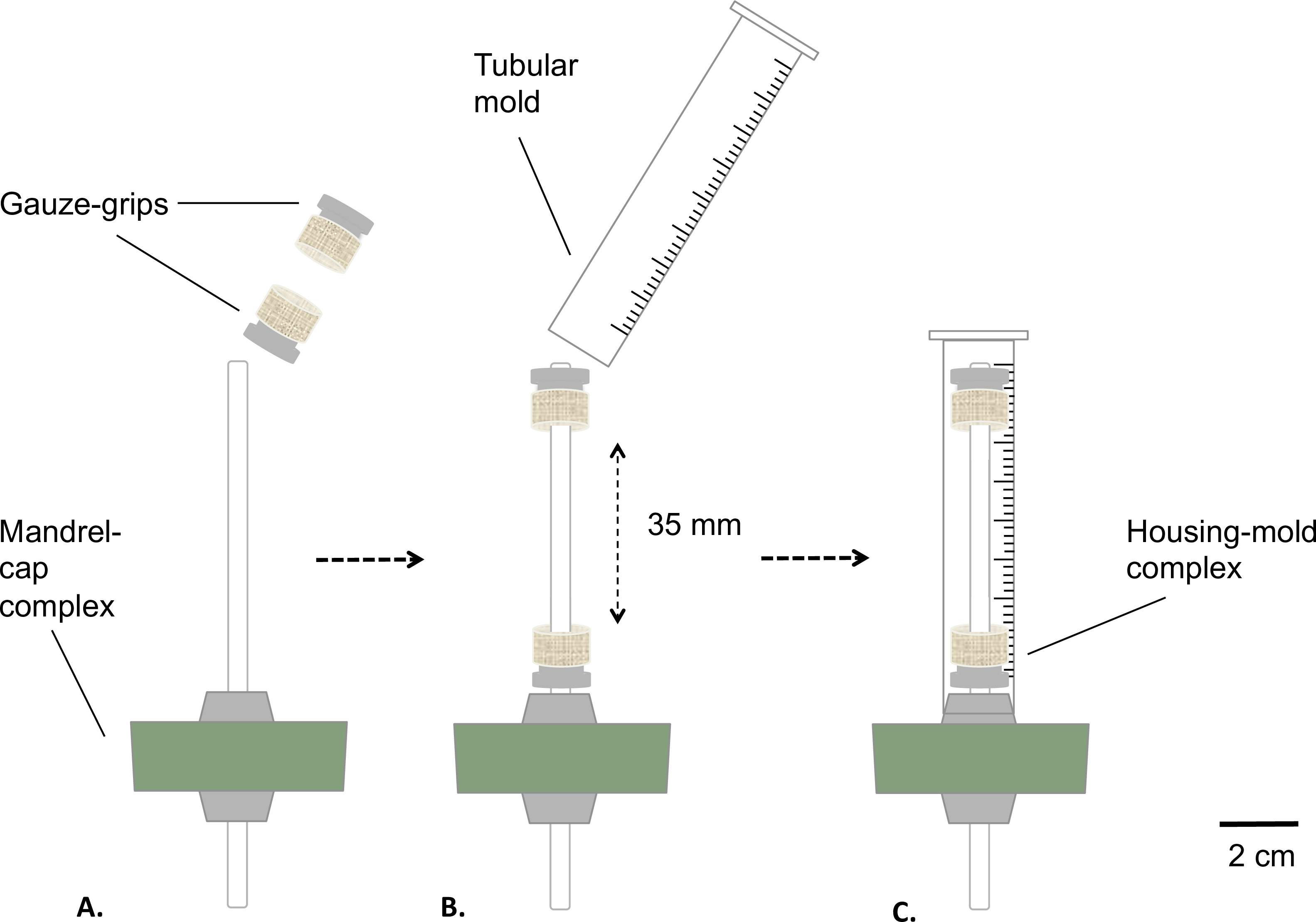
图 2:静态生物反应器的组装。 生物反应器在灭菌前的组装步骤。将纱布夹具以固定间距安装在芯轴上(A);插入模具(B),并将其牢固固定在硅胶塞上(C)。芯轴的外径为 4.7 mm。

图3:在无菌条件下构建组织工程结构。 将细胞与胶原混合物倒入外壳-模具复合体中(A),在室温下静置凝胶化1小时(B)。随后移除模具(C),组装静态生物反应器(D),并将其转移至储液装置中,在培养箱内进行静态成熟培养(T = 37 °C,5% CO2,100%湿度)。芯轴的外径为4.7 mm。
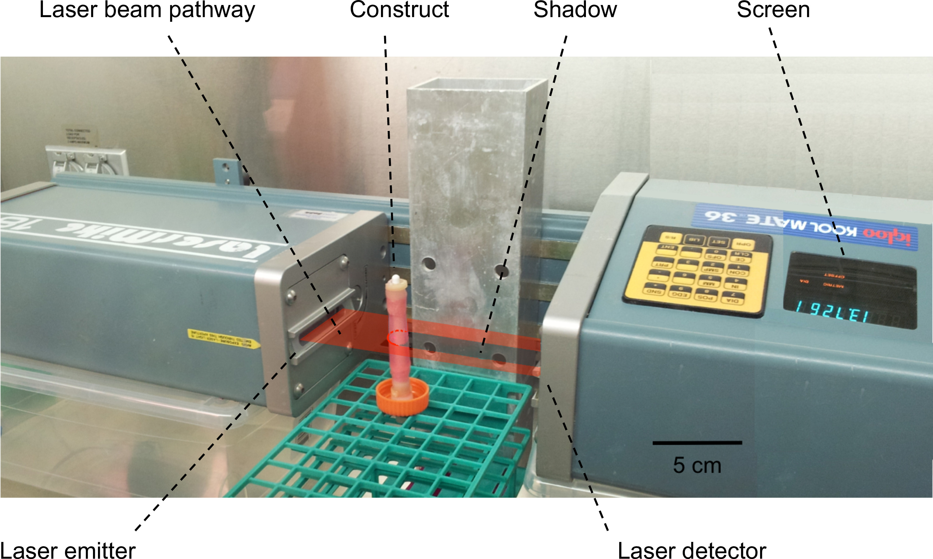
图4: 构建物厚度/外径的测量。 采用激光扫描干涉仪对构建物的外径进行测量。将构建物置于激光束路径中,形成阴影。阴影的宽度对应于构建物的外径,该宽度随后被测量并在屏幕上显示。
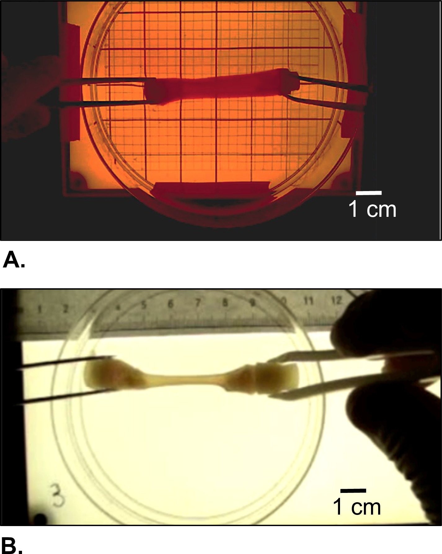
图 5: 收获的构建物的形态学外观。(A)凝胶化后立即观察,以及(B)静态成熟培养 2 周期间由细胞驱动重塑后的形态。
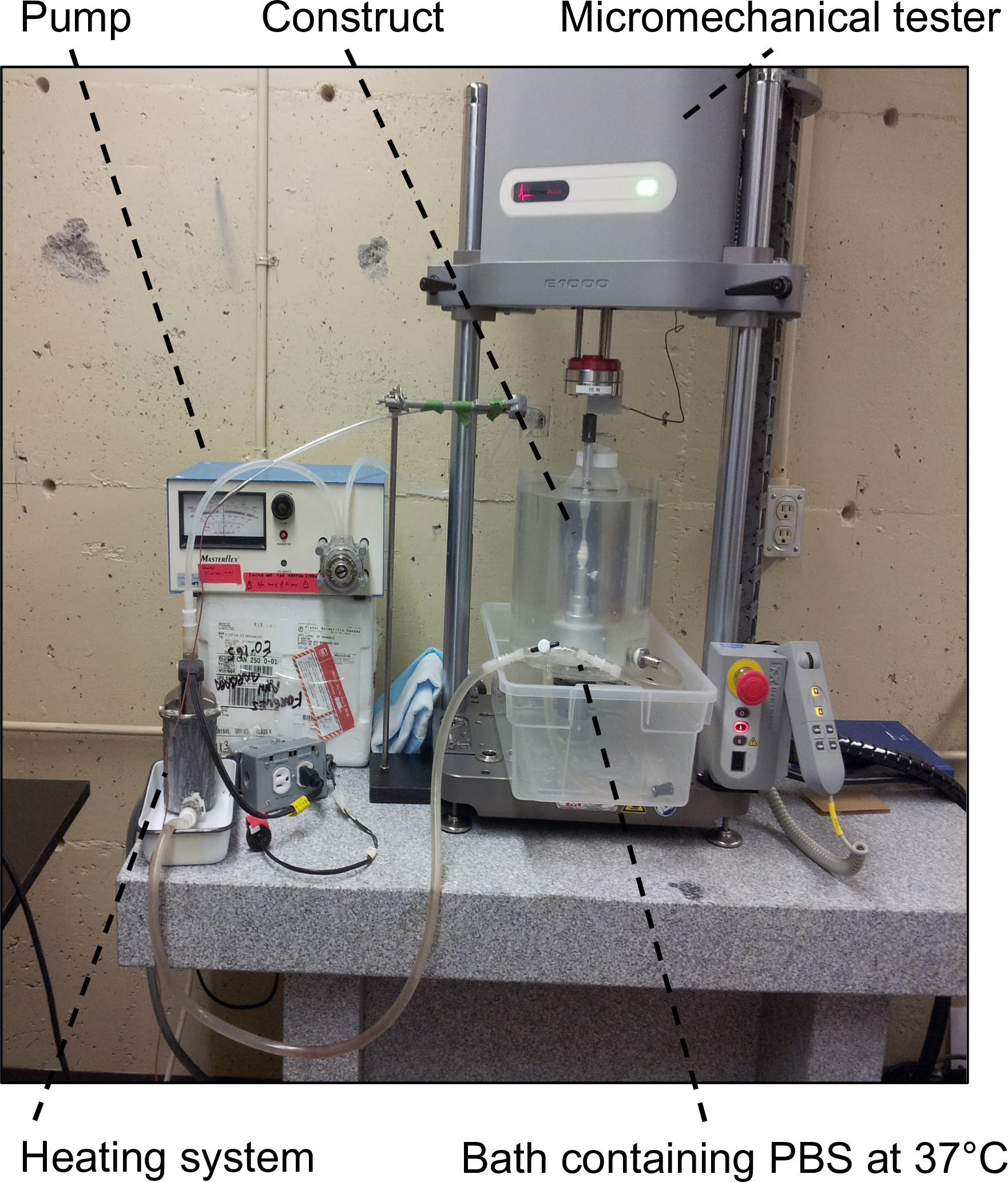
图6: 用于力学表征的实验装置。 该装置包括配备5 N或10 N载荷传感器的微机械测试仪,以及盛有37 °C PBS的浴槽,用于使样品保持在准生理条件下。

图7:用于力学表征的样品制备。 用于在纵向和周向进行疲劳测试的样品采集(A)和制备(B)(C)。芯轴的外径为4.7 mm。
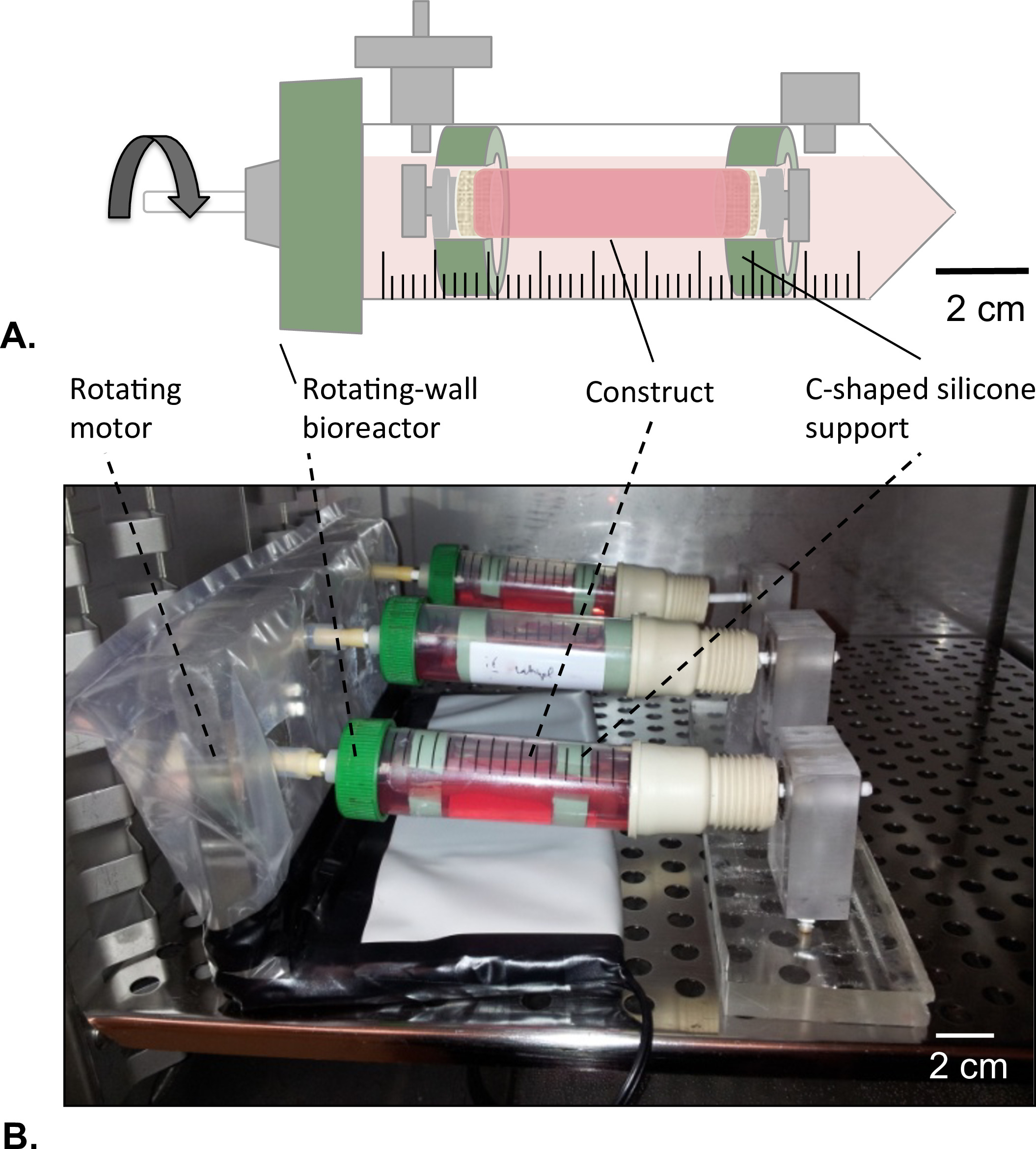
图8:旋转壁式生物反应器。(A)在C形硅胶支撑结构的帮助下,将管状结构组装于储液腔中央。结构两端均封闭,以防止人脐静脉内皮细胞(HUVECs)溶液泄漏。(B)将结构置于培养箱中(温度 = 37 °C,5% CO2,100% 湿度)进行旋转培养,离心力为 4.02 × 10⁻⁵ g,持续2天。

图9: 力学性能表征。 细胞驱动重塑后,沿纵向(A)和周向(B)方向对构建物进行疲劳测试的结果。请点击此处查看该图的放大版本。
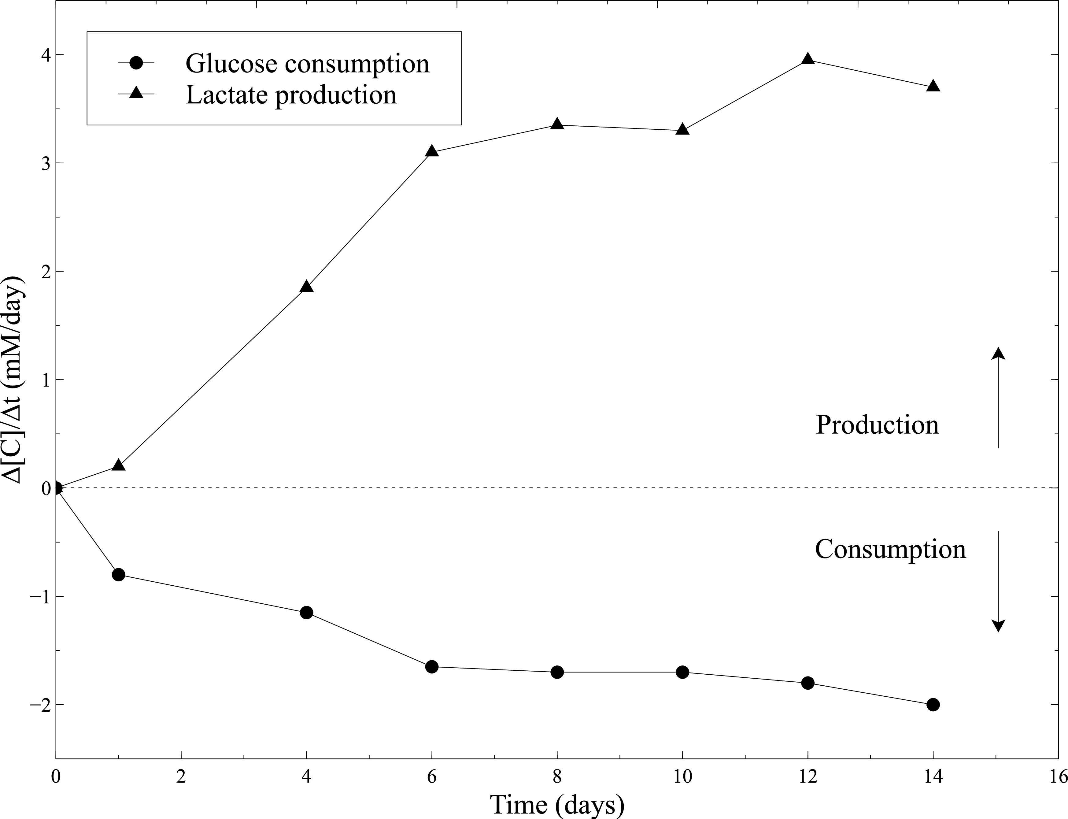
图10:胶原凝胶中平滑肌细胞的代谢活性。使用血气分析仪每2天测定一次葡萄糖消耗速率和乳酸生成速率,在更换培养基后进行。新鲜培养基用作葡萄糖和乳酸浓度测定的基线水平。

图11:管腔内皮化。 管状结构的径向横截面组织学图像。管状结构在静态培养1周(A)和2周(B)后的Masson三色染色结果。管状结构的H&E染色图像(C)。
| 时间 | 厚度 (mm) | 压实率 (%) |
| 0 小时 | 4.83 ± 0.02 | 0 ± 0 |
| 2 小时 | 4.26 ± 0.02 | 12 ± 0 |
| 4 小时 | 4.21 ± 0.03 | 13 ± 1 |
| 6 小时 | 4.06 ± 0.10 | 16 ± 2 |
| 12 小时 | 3.16 ± 0.07 | 35 ± 1 |
| 1 天 | 2.08 ± 0.11 | 57 ± 2 |
| 1 周 | 0.68 ± 0.07 | 86 ± 1 |
| 2 周 | 0.36 ± 0.00 | 93 ± 0 |
表1:静态成熟过程中构建物直径的快速致密化。 管状构建物的壁厚及其在静态培养时间下的致密化速率。通过激光扫描干涉仪(Series 183B, LaserMike 136)测量管状构建物的外径以评估致密化程度。培养24小时后,构建物收缩至其原始模制尺寸的57% ± 2%。数据以均值 ± 标准差表示(n = 3)。此类显著变化的唯一原因是存活且具有活性的平滑肌细胞的存在与活动。
补充视频 1:非重塑管状胶原凝胶的收获。 请点击此处观看该视频。
补充视频 2: 细胞驱动的管状胶原凝胶收缩。请点击此处查看该视频。
补充视频 3: 未重塑管状胶原凝胶的操作。请点击此处查看该视频。
补充视频 4: 细胞重塑的管状胶原凝胶的操作。请点击此处查看该视频。
补充视频 5: 对细胞重塑的管状胶原凝胶进行纵向疲劳测试(30%)。 请点击此处查看该视频。
补充视频 6: 对细胞重塑的管状胶原凝胶进行周向疲劳测试(30% 应变)。请点击此处查看该视频。
讨论
在血管组织工程领域,研究人员已投入巨大努力以重建负责血管机械稳定性的中膜层16。自Weinberg和Bell的开创性工作以来17,胶原蛋白因其良好的生物相容性、无免疫原性以及易获取性,被广泛用作血管组织工程的支架材料。然而,胶原蛋白的使用对研究人员而言仍是一项重大挑战,因为该材料本身缺乏足够的机械刚度,难以操作。在支架制备过程中的任何操作均可能造成支架损伤,从而影响其后续应用。
本研究中描述的技术具有以下优势:i) 可将含细胞的胶原凝胶构建成管状结构;ii) 可在短暂的静态成熟期(1 或 2 周)后获得机械强度足够高、可进行操作的生物组织;iii) 可在两个方向上评估此类管状生物组织的力学和黏弹性特性。凝胶中的细胞在胶原基质重塑过程中发挥关键作用。在成熟期间,收缩型平滑肌细胞(SMCs)促使凝胶发生收缩,从而形成机械稳定性更高的组织构建物,可分别在纵向和环向方向进行力学评估。随后,在构建物腔面接种的人脐静脉内皮细胞(HUVECs)形成了均匀且具有活性的内皮层,从而证明该胶原凝胶适用于血管组织工程应用。
本研究中介绍的生物反应器 是专门为静态成熟过程中细胞 生长提供最佳环境而设计的。 此外,用于表征构建物力学和黏弹性特性的装置在设计时旨在尽量减少操作此类脆弱材料时可能造成的任何损伤。因此,该静态生物反应器的盖子上配备了一个0.22 µm滤膜(步骤1.1.2,图 1A 和 B),可在储液槽内培养基与培养箱之间实现气体交换,同时维持无菌培养环境。底部的鲁尔隔膜用作静态培养期间取样和更换培养基的通道。在构建物制备和表征过程中需注意一些关键步骤。所有可能影响系统无菌性的操作(在步骤2.1.1及后续步骤中进行)均须在无菌生物安全柜中完成。细胞与胶原凝胶混合物的制备需在冰上操作,以延缓凝胶化过程(步骤2.1.4至2.1.7)。在步骤2.1.7中,凝胶化前混合物中截留的气泡可能成为应力集中区域,从而影响构建物的稳定性。因此,需轻微振荡装置或在无菌条件下使用医用真空泵抽气3分钟,以去除这些气泡。最后,夹具经过专门设计,可在凝胶化过程中保持芯轴在管状模具中的中心位置,并便于在收获时(取出芯轴,第2.4节)、内皮化处理以及将构建物安装到力学测试系统(纵向测试)时进行精细操作。
本方案提出了一种基于平滑肌细胞(SMCs)天然内在收缩潜能的、易于操作的胶原凝胶结构增强新方法。目前常用的胶原基质增强技术涉及使用物理和化学交联剂,这些试剂可能对细胞-基质相互作用产生不利影响18–20。本文所介绍的制备技术可引导细胞驱动的基质重塑过程,从而获得具有目标力学性能的组织工程化构建物,且无需任何物理或化学处理。
对水合胶原凝胶的力学和黏弹性性质进行表征是一项重大挑战。从这一角度出发,本方案描述了一种原创的、简单且高效的方法,用于评估管状软组织的力学性能。该表征不仅可在周向进行,也可在轴向直接对完整的管状结构实施。在力学表征过程中,温度、水环境、pH值和离子强度等环境因素已知会显著影响生物组织的力学行为21。因此,本研究提出了一种原创的装置与方案,可在高度可重复的拟生理环境(37 °C、pH 7.4 的生理盐水溶液)中对生物组织进行力学表征。据我们所知,此类表征此前尚未见报道。
总之,本研究提出的技术展示了细胞与胶原蛋白直接混合在血管组织工程应用中的巨大潜力。该方法结合力学表征与内皮化过程,构成了高度多效性的实验方案。因此,在保持基本原理不变的前提下,通过对接口设置和操作流程进行轻微调整,即可满足构建血管类组织的主要需求,例如快速且简便的制备流程(包括内皮化),以及可适用于多种长度和直径的软组织。此外,还可针对不同类型的黏附细胞、细胞外基质(ECM)蛋白及模具几何形状,探索其在多种特定应用中的潜力,如肌腱工程、皮肤移植物、心肌补片、神经组织工程等。尽管目前构建物的力学性能已令人鼓舞,但仍低于天然组织的水平。在此背景下,我们坚信,非常短暂的静态成熟期是迈向生物反应器中动态刺激的关键步骤,从而有助于提高构建物的结构完整性和力学稳定性。然而,本文所述的静态生物反应器能够快速制备适用于力学和组织学分析的、含细胞的胶原基组织工程构建物,因而是一种有用且前景广阔的工具,可用于深入理解细胞与细胞外基质在生长和重塑过程中的相互作用,甚至可作为治疗手段和药物递送系统的模型。
披露
未从可能存有利益冲突的组织或机构获得资助。
致谢
本研究由加拿大自然科学与工程研究理事会、加拿大健康研究院以及魁北克大学医院研究中心(CHU de Québec)资助。
材料
| 姓名 | 公司 | 目录编号 | 评论 |
|---|---|---|---|
| CellTreat 50 ml 生物反应管 | CELLTREAT Scientific Products | 229-475 | 离心管 |
| 带锁环的公鲁尔接头 x 1/8" 软管接头。聚丙烯材质。25个/包 | Cole Parmer | RK-45503-04 | 用于"气体交换口"的鲁尔接头 |
| 母鲁尔接头 x 1/8" 软管接头转换器。聚丙烯材质。25个/包 | Cole Parmer | RK-45500-04 | 用于"培养基取样口"的鲁尔接头 |
| Masterflex 铂金硫化硅胶管 L/S 17,25 英尺 | Cole Parmer | RK-96410-17 | 纱布夹持器用管1和管2 |
| Masterflex 铂金硫化硅胶管 L/S 16,25 英尺 | Cole Parmer | RK-96410-16 | 纱布夹持器用管3 |
| Silastic 医用粘合型硅胶,A型 | Dow Corning | - | 用于静态生物反应器制备的硅胶粘合剂 |
| Polyvent 4 容器通气滤器 | Whatman | 6713-0425 | 用于"气体交换口"的滤器 |
| 搅拌棒。聚丙烯材质。8’’ | Scienceware | 377660008 | 芯轴 |
| 硅橡胶塞 00 号,12个装 | VWR | 59590-084 | 用于将芯轴插入离心管通气盖的塞子 |
| Krytox PFPE/PTFE 润滑脂 | Dupont | GPL 202 | 用于涂覆芯轴的医用级润滑脂 |
| 胰蛋白酶-EDTA(0.5%) | Gibco | 15400-054 | 细胞培养 |
| Xiameter RTV-4130-J 基料与固化剂 | Dow Corning | - | 用于内皮化过程的C型硅胶支撑结构 |
| Dulbecco’s 改良 Eagle 培养基(DMEM),高糖型,含丙酮酸钠 | Gibco (Life Technology) | 11995-065 | 细胞培养 |
| 纯丙酮(99%) | Laboratoire Mat Inc. | AP0102 | 用于胶原蛋白提取的化学试剂 |
| 异丙醇(HPLC级,99.9%) | Fisher Scientific | AC610080040 | 用于胶原蛋白提取的化学试剂 |
| 0.02 N 乙酸(冰乙酸,HPLC级,99%) | Fisher Scientific | FL070494 | 用于胶原蛋白提取的化学试剂 |
| 氯仿溶液(99%) | Laboratoire Mat Inc. | CR 0179 | 用于胶原蛋白提取的化学试剂 |
| Hepes | Sigma-Aldrich | 163716 | 用于构建物制备的化学试剂 |
| NaOH | Laboratoire Mat Inc. | SR-0169 | 用于构建物制备的化学试剂 |
| LaserMike 136 | LaserMike | Series 183B | 扫描激光干涉仪 |
| ElectroPulse MicroTester | Instron Corporation | - | 微力学测试仪 |
| HyClone M199/EBSS 培养基,500 ml | GE Healthcare Life Sciences | SH30253.01 | 细胞培养基组分 |
| 胎牛血清 HI - 500 ml | Gibco | SH 30396.03 | 细胞培养基组分 |
| 猪血清(PS) | Sigma-Aldrich | P9783 | 细胞培养基组分 |
| 青霉素-链霉素 | Gibco | 15140-122 | 细胞培养基组分 |
| 磷酸盐缓冲液(PBS) | Fisher Scientific | BP661-50 | 生理盐水溶液 |
| 组织培养瓶 T17CN,带通气盖,红色 | Sarstedt Inc. | 83.1812.002 | 细胞培养 |
| ColorpHast pH 指示试纸(pH = 6.5-10.0) | EMD | 9583 | pH 测量 |
| Matrigel 基底膜基质(生长因子减少型),5 ml 瓶装 | BD Biosciences - Discovery Labware | 356230 | 用于内皮化过程的浓缩蛋白混合物 |
| LifeCam VX-3000 | Microsoft | - | 厚度测量 |
| 生化分析仪 DxC600 | Beckman Coulter Unicell Synchron | - | 葡萄糖和乳酸浓度测定 |
| 胶原纤维 | 大鼠尾部 | - | 胶原蛋白在实验室中提取 |
| 猪平滑肌细胞(pSMCs) | 猪主动脉 | - | pSMCs 在实验室中分离获得 |
| 人脐静脉内皮细胞(HUVECs) | 人脐静脉 | - | HUVECs 在实验室中分离获得 |
参考文献
- Kim, B. S., Nikolovski, J., Bonadio, J., Smiley, E., Mooney, D. J. Engineered smooth muscle tissues: regulating cell phenotype with the scaffold. Experimental cell research. 251, 318-328 (1999).
- Heureux, N., McAllister, T. N., de la Fuente, H. L. Tissue-engineered blood vessel for adult arterial revascularization. The New England journal of medicine. 357 (14), 1451-1453 (2007).
- Syedain, Z. H., Meier, L. A., Bjork, J. W., Lee, A., Tranquillo, R. T. Implantable arterial grafts from human fibroblasts and fibrin using a multi-graft pulsed flow-stretch bioreactor with noninvasive strength monitoring. Biomaterials. 32 (3), 714-722 (2011).
- Lee, C., Singla, A., Lee, Y. Biomedical applications of collagen. International journal of pharmaceutics. 221 (1-2), 1-22 (2001).
- Couet, F., Rajan, N., Mantovani, D. Macromolecular biomaterials for scaffold-based vascular tissue engineering. Macromolecular bioscience. 7 (5), 701-718 (2007).
- Christiansen, D. L., Huang, E. K., Silver, F. H. Assembly of type I collagen: fusion of fibril subunits and the influence of fibril diameter on mechanical properties. Matrix Biology. 19 (5), 409-420 (2000).
- Eppell, S. J., Smith, B. N., Kahn, H., Ballarini, R. Nano measurements with micro-devices: mechanical properties of hydrated collagen fibrils. Journal of the Royal Society, Interface / the Royal Society. 3 (6), 117-121 (2006).
- Lai, V. K., Lake, S. P., Frey, C. R., Tranquillo, R. T., Barocas, V. H. Mechanical behavior of collagen-fibrin co-gels reflects transition from series to parallel interactions with increasing collagen content. Journal of biomechanical engineering. 134 (1), 011004(2012).
- Seliktar, D., Nerem, R. M., Galis, Z. S. The Role of Matrix Metalloproteinase-2 in the Remodeling of Cell-Seeded Vascular Constructs Subjected to Cyclic Strain. Annals of Biomedical Engineering. 29 (11), 923-934 (2001).
- Rajan, N., Habermehl, J., Coté, M. -F., Doillon, C. J., Mantovani, D. Preparation of ready-to-use, storable and reconstituted type I collagen from rat tail tendon for tissue engineering applications. Nature protocols. 1 (6), 2753-2758 (2006).
- Kumar, V. A., Caves, J. M., et al. Collagen-Based Substrates with Tunable Strength for Soft Tissue Engineering. Biomaterials science. 1 (11), 1193-1202 (2013).
- Li, S., Van Den Diepstraten, C., D’Souza, S. J., Chan, B. M. C., Pickering, J. G. Vascular smooth muscle cells orchestrate the assembly of type I collagen via alpha2beta1 integrin, RhoA, and fibronectin polymerization. The American journal of pathology. 163 (3), 1045-1056 (2003).
- Badylak, S. F., Freytes, D. O., Gilbert, T. W. Extracellular matrix as a biological scaffold material: Structure and function. Acta biomaterialia. 5 (1), 1-13 (2009).
- Engbers-Buijtenhuijs, P., Buttafoco, L., et al. Biological characterisation of vascular grafts cultured in a bioreactor. Biomaterials. 27 (11), 2390-2397 (2006).
- Cheung, A. L. Isolation and culture of human umbilical vein endothelial cells (HUVEC). Current protocols in microbiology. 4, 4B(2007).
- Seifu, D. G., Purnama, A., Mequanint, K., Mantovani, D. Small-diameter vascular tissue engineering. Nature reviews. Cardiology. , (2013).
- Weinberg, C., Bell, E. A blood vessel model constructed from collagen and cultured vascular cells. Science. 231 (4736), 397-400 (1986).
- Meghezi, S., Chevallier, P., Mantovani, D. Why Mechanical Properties of Collagen Scaffolds Should Be Tested in a Pseudo-Physiological Environment. Advanced Materials Research. 409, 158-163 (2011).
- Tirella, A., Liberto, T., Ahluwalia, A. Riboflavin and collagen: New crosslinking methods to tailor the stiffness of hydrogels. Materials Letters. 74, 58-61 (2012).
- Madhavan, K., Belchenko, D., Tan, W. Roles of genipin crosslinking and biomolecule conditioning in collagen-based biopolymer: Potential for vascular media regeneration. Journal of biomedical materials research. Part A. , 16-26 (2011).
- Meghezi, S., Couet, F., Chevallier, P., Mantovani, D. Effects of a pseudophysiological environment on the elastic and viscoelastic properties of collagen gels. International journal of biomaterials. 2012, 319290(2012).
重印与许可
