本文介绍了一种用于模拟生物物理微环境的实验方案,其中由晚期糖基化终末产物(AGEs)诱导的基底膜(BM)交联及其刚度增加具有病理学意义。
本文内容
摘要
摘要
在此,我们描述了一种可用于研究与年龄相关病理及代谢性疾病中基底膜(BM)增厚和硬化相关的生物物理微环境的实验方案。例如 癌症、糖尿病、微血管疾病、视网膜病变、肾病和神经病变)。该模型的核心原理是通过乙醛酸(GLA)处理重建的基底膜(rBM)基质,诱导非酶促交联,经由美拉德反应促进晚期糖基化终末产物(AGE)的生成。文中概述了可用于验证GLA处理后rBM中AGE生成、非酶促交联及基质刚度增加的若干实验室技术,包括制备经磷酸盐缓冲液(PBS)处理的天然rBM和经GLA处理的刚性rBM,并用于测定:通过光度分析和免疫荧光显微镜检测其AGE含量;通过十二烷基硫酸钠-聚丙烯酰胺凝胶电泳(SDS-PAGE)及共聚焦显微镜检测其非酶促交联程度;以及通过流变仪测定其刚度的增加。本文所述方法可使rBM的刚度(弹性模量,E)最高增加至原来的3.2倍,与健康人与前列腺疾病患者组织中的测量值一致。为模拟与前列腺衰老及疾病相关的生物物理微环境,将三种前列腺细胞接种于天然rBM和刚性rBM上:RWPE-1(来源于正常前列腺组织的前列腺上皮细胞,PECs);BPH-1(来源于良性前列腺增生(BPH)组织的PECs);以及PC3(源自前列腺癌骨转移瘤的转移性细胞)。可检测多种参数,包括RWPE-1和BPH-1在天然与刚性rBM上形成的三维腺泡的大小、形态及侵袭特性,以及PC3细胞在相同条件下形成的三维球状体的平均细胞长度、迁移速度和细胞运动的持续性。此外,还可评估细胞信号通路及蛋白质的亚细胞定位。
引言
基底膜(BM)是一种特化的细胞外基质(ECM)结构,通过将上皮细胞层与间质分隔开来,维持稳定的组织边界1。基底膜中相邻的IV型胶原三螺旋之间通过共价交联,形成不规则的超螺旋网络结构,从而稳定其侧向结合2。这些IV型胶原网络作为支架,介导基底膜与层粘连蛋白及其他基底膜组分的相互作用1。基底膜的结构排列为其提供了腺上皮正常发育所必需的机械强度和刚性3。
在衰老和疾病过程中,基底膜(BM)会逐渐增厚并变硬3,4。例如,在正常人群中,眼部基底膜的弹性模量(E)在50至80岁之间增加约3倍,而在糖尿病等代谢性疾病中,这种硬化现象会进一步加剧5。基底膜结构和生物力学特性的改变导致其硬度增加,其机制在于基底膜的细胞外基质(ECM)成分——IV型胶原和层粘连蛋白——在暴露于晚期糖基化终末产物(AGEs)后发生非酶促交联。
本文所述方法的目的是建立一个模型,用于研究由于晚期糖基化终末产物(AGE)暴露导致的基底膜(BM)硬度增加,如何促进前列腺上皮细胞(PEC)和前列腺肿瘤细胞(PTC)在向转移性前列腺癌(PCa)转变过程中的侵袭性。为此,本研究对先前用于在重建的重组基底膜(rBM)凝胶中生成乳腺上皮细胞(MEC)三维腺泡结构的方法6进行了改进,增加了一个额外步骤,即使用乙二醛(GLA)对rBM凝胶进行预处理。文中描述了多种评估GLA诱导rBM凝胶交联和硬化的方法,包括光度分析、十二烷基硫酸钠-聚丙烯酰胺凝胶电泳(SDS-PAGE)、共聚焦显微镜观察以及流变学分析。在预硬化rBM上培养的前列腺细胞类型包括:RWPE-1,来源于正常前列腺组织的PEC7;BPH-1,来源于良性前列腺增生(BPH)组织的PEC8;以及PC3,来源于一名前列腺癌(PCa)患者脊椎骨转移灶的转移性PTC9。
除了推动前列腺腺体病理学的研究外,通过GLA处理使rBM凝胶硬化的方法还可被调整用于研究基底膜(BM)硬度在其他年龄相关性疾病和代谢紊乱中的作用。例如,通过引入适当的细胞类型,该模型可直接应用于研究乳腺、结肠、卵巢和胰腺等器官中基底膜硬度如何诱导转移性癌症的发生。此外,该方案还可被改造用于研究硬化的基底膜如何促进糖尿病相关微血管病变、视网膜病变、肾病和神经病变等疾病进展中的生物力学机制。
方案
1. GLA处理诱导骨髓硬度(非酶促交联)
- 将一管冷冻的基底膜(BM)基质(10 ml)置于4 °C下孵育(在冷室或冰箱中冰上放置),直至管内内容物完全融化(8–16 小时)。
注意:若无冷室/冰箱,需用冰完全覆盖瓶体,以防止BM储存液重新凝固。 - 为便于后续实验并避免反复冻融,从每支新开启的10 ml BM基质中分装成25份、每份0.4 ml的等分试样。将分装后的管子储存于-80 °C,直至制造商标明的过期日期。使用时,在冰上4 °C条件下解冻2小时。
- 准备一个平整的冰面,将8孔腔载玻片置于冰上,以在包被过程中维持4 °C的温度。将一管BM基质在4 °C下解冻。
注:一管0.4 ml的BM基质足以包被整张1×8孔腔载玻片。操作过程中应始终将管子置于冰上,防止BM基质凝固。 - 用剪刀剪去200 µl移液枪头的尖端。将钝化后的200 µl枪头冷却至4 °C,并安装至200 µl移液器上。吸取40 µl冷的BM基质溶液,转移至预冷的8孔腔载玻片的一个孔中。
注:40 µl BM溶液足以覆盖0.8 cm2的表面积。包被过程中应始终保持枪头处于低温状态,以防BM溶液凝固。避免在BM基质溶液中引入气泡,并确保孔内表面均匀包被,边缘无可见弯月面形成。 - 根据所需孔数和腔室数量,重复步骤1.4。
- 包被完成后,将8孔腔载玻片置于37 °C孵育30分钟,以促进BM的聚合。关闭培养箱门时应格外小心,避免扰动液态rBM。孵育时间不得超过30分钟,以防rBM凝胶脱水。
注:所得凝胶为天然重组基底膜(rBM)。该37 °C孵育步骤无需5 % CO2环境,但为操作方便,可在设定为37 °C、5 % CO2(并保持湿度)的组织培养箱中进行。 - 用0.2 M磷酸盐缓冲液(pH 7.8)稀释乙醇醛(GLA)至50 mM。使用50 ml注射器将溶液通过0.22微米注射器滤器过滤,以实现灭菌。
- 为进行终体积250 µl、含50 mM GLA的交联反应,向125 µl的100 mM GLA(2倍储存液)和100 µl的0.2 M磷酸盐缓冲液(pH 7.8)中加入25 µl的0.5 M氰基硼氢化钠或2.5 M氨基胍。使用50 ml注射器将各储存液通过0.22微米注射器滤器过滤灭菌。
注意:操作氰基硼氢化钠时,应在通风橱内穿戴实验服、手套、面罩和呼吸器。
- 为进行终体积250 µl、含50 mM GLA的交联反应,向125 µl的100 mM GLA(2倍储存液)和100 µl的0.2 M磷酸盐缓冲液(pH 7.8)中加入25 µl的0.5 M氰基硼氢化钠或2.5 M氨基胍。使用50 ml注射器将各储存液通过0.22微米注射器滤器过滤灭菌。
- 加入250 µl GLA溶液,完全覆盖已聚合的rBM凝胶,在37 °C孵育6小时以形成半刚性rBM凝胶,或孵育14小时以形成刚性rBM凝胶。
注:所加GLA溶液体积必须足以覆盖聚合后的rBM凝胶,并应相应调整。若使用不同GLA孵育时间,应对rBM凝胶进行分析,以确定其刚性增加的倍数(见步骤2.4)。- 设置阴性对照:将天然rBM凝胶在250 µl无菌磷酸盐缓冲液(PBS)中于37 °C孵育14小时。
- 设置两个额外对照,分别通过添加50 mM氰基硼氢化钠或250 mM氨基胍,抑制交联反应中席夫碱或阿马多里加合物重排的形成。
- 用PBS稀释甘氨酸乙酯(GEE)至1 M。使用50 ml注射器将溶液通过0.22微米注射器滤器过滤灭菌。
- 在指定孵育时间后,小心移除交联rBM凝胶中的GLA溶液、对照rBM凝胶中含抑制剂的GLA溶液以及对照天然rBM凝胶中的PBS。向所有rBM凝胶中加入250 µl GEE溶液,并在37 °C孵育1小时。
注:此步骤用于终止交联反应。 - 用500 µl PBS对所有rBM凝胶清洗10次,以彻底去除GLA和GEE残留。随后在37 °C下用400 µl PBS孵育rBM凝胶过夜,防止其脱水。
- 对rBM凝胶进行AGE积累、非酶促交联及粘弹性特性的分析(步骤2.1–2.4)。若需进行粘弹性分析,应将rBM凝胶制备于克隆环中(步骤2.4)。
- 用于细胞培养时,在接种细胞前用500 µl培养基轻柔冲洗rBM凝胶2次(步骤5和6)。清洗过程中应避免枪头接触凝胶表面。
2. GLA 处理后 rBM 中非酶促交联及硬度的定量分析
- 光度分析
- 采用光度分析法测定经GLA处理及对照组rBM凝胶中的AGE累积量,以确定美拉德反应的程度。
- 完成步骤1.11后,从8孔板中的rBM凝胶中移除PBS,并加入250 µl冰冻的双蒸水。在4 °C下孵育16–24小时,确保基质完全液化。
注意:该溶液中的rBM肽含有具有自荧光特性的AGEs。 - 将液化的BM溶液转移至1.5 ml离心管中,使用分光光度计(激发波长 = 370 nm;发射波长 = 440 nm)测定溶液的荧光发射强度。
- 完成步骤1.11后,从8孔板中的rBM凝胶中移除PBS,并加入250 µl冰冻的双蒸水。在4 °C下孵育16–24小时,确保基质完全液化。
- 采用光度分析法测定经GLA处理及对照组rBM凝胶中的AGE累积量,以确定美拉德反应的程度。
- 溴化氰肽的SDS-PAGE分析
- 将经GLA处理及对照组的rBM凝胶在聚丙烯酰胺凝胶上进行分离,以确认GLA是否诱导了交联及大纤维的形成。
- 将步骤2.1.1.2中收集的液化BM溶液在室温下以10,000 × g离心5分钟。
- 制备含有2 g/ml溴化氰并用乙腈稀释的储备溶液。
警告:操作溴化氰时必须在通风橱内进行,并穿戴实验服、手套、面罩和呼吸器。 - 移除上清液,将BM凝胶沉淀重悬于500 µl含20 mg/ml溴化氰和70% v/v甲酸的溶液中,在室温下孵育过夜。
- 使用1 ml一次性注射器将重悬的BM凝胶沉淀转移至分子量截留为3.5 kDa的透析盒中。
- 将透析盒浸入装有500 ml双蒸水和磁力搅拌子的500 ml玻璃烧杯中,置于磁力搅拌器上,在4 °C(冷藏室中)透析过夜(16小时),以彻底去除残留的溴化氰和甲酸。
- 使用1 ml一次性注射器将透析后的BM溶液从透析盒转移至1.5 ml离心管中。
- 取每种BM样品25 µl,在12% v/v聚丙烯酰胺凝胶上进行分析10,11。SDS-PAGE完成后,对聚丙烯酰胺凝胶进行银染12,以观察溴化氰切割的基质肽的电泳图谱13。
- 将经GLA处理及对照组的rBM凝胶在聚丙烯酰胺凝胶上进行分离,以确认GLA是否诱导了交联及大纤维的形成。
- 免疫荧光显微镜分析
- 对经GLA处理及对照组的rBM凝胶使用抗AGE/戊糖苷素、抗IV型胶原和抗层粘连蛋白抗体进行免疫荧光染色,随后进行共聚焦显微镜观察,以可视化交联rBM凝胶中累积的AGEs以及IV型胶原/层粘连蛋白纤维的结构重排13。
注意:在孵育和洗涤过程中,应使用足量的液体完全覆盖整个rBM凝胶,但避免移液枪头接触rBM表面。有关通过免疫荧光分析三维腺泡培养物的详细信息,请参见参考文献6;有关三维腺泡的共聚焦显微镜分析,请参见参考文献14。- 在8孔板中,用300 µl PBS+(含0.1 mM CaCl2和0.5 mM MgCl2的PBS)在室温下洗涤经GLA处理及对照组的rBM凝胶,每次5分钟,共洗两次。
- 移除PBS+,加入300 µl用PBS+稀释的4% w/v多聚甲醛(PFA),完全覆盖每个rBM凝胶。在室温下孵育30分钟,以固定rBM组分。
- 移除4% w/v PFA溶液,加入300 µl含75 mM NH4Cl和0.5 mM MgCl2的溶液,在室温下孵育5分钟(重复5次),以淬灭固定反应。
- 通过在无菌水中配制以下溶液制备免疫荧光缓冲液(IF缓冲液):130 mM NaCl、7 mM Na2HPO4、3.5 mM NaH2PO4、7.7 mM NaN3、0.1% w/v牛血清白蛋白、0.5% v/v聚乙二醇叔-辛基苯基醚和0.05% v/v聚乙二醇单月桂酸酯。
- 通过在IF缓冲液中添加20% v/v山羊血清制备IF封闭缓冲液。
- 移除淬灭液,向rBM凝胶中加入300 µl IF封闭缓冲液,防止非特异性反应。在摇床上室温孵育2小时。
- 移除IF封闭缓冲液,加入300 µl用IF封闭缓冲液稀释的一抗(1:500小鼠抗戊糖苷素单克隆抗体;1:250兔抗IV型胶原多克隆抗体;1:250兔抗层粘连蛋白A/C多克隆抗体),在4 °C下孵育16小时。
注意:在4 °C下孵育时间超过20小时可能导致rBM液化。 - 移除一抗,在摇床上用300 µl IF缓冲液于室温下洗涤3次,每次10分钟。
- 移除IF缓冲液,加入300 µl用IF封闭缓冲液按1:500稀释的荧光标记二抗(山羊抗兔或抗小鼠IgG [H+L])。在摇床上室温孵育2小时。
- 移除二抗,在室温下用300 µl IF缓冲液孵育10分钟。移除IF缓冲液后,在室温下用300 µl PBS+洗涤3次,每次10分钟。
- 按照上述步骤(2.3.1.2和2.3.1.3)再次进行固定和淬灭。
- 将染色后的rBM凝胶封片于封片剂中,利用落射荧光或共聚焦显微镜分析主要组分致密束的形成。
注意:有关通过免疫荧光分析三维腺泡培养物的详细信息,请参见参考文献6;有关三维腺泡的落射荧光和共聚焦显微镜分析,请参见参考文献14。
- 对经GLA处理及对照组的rBM凝胶使用抗AGE/戊糖苷素、抗IV型胶原和抗层粘连蛋白抗体进行免疫荧光染色,随后进行共聚焦显微镜观察,以可视化交联rBM凝胶中累积的AGEs以及IV型胶原/层粘连蛋白纤维的结构重排13。
- 流变学分析
- 对经GLA处理及对照组的rBM凝胶进行流变学分析,以测量其粘弹性(刚度)。
- 在直径为8 mm的圆形模具中制备厚度为1 mm的rBM凝胶。具体操作为:将一个克隆环(直径8 mm)置于24孔培养板的孔中,并加入按步骤1.3–1.6所述方法制备的BM基质溶液。
注意:为准确重现实验所用的rBM凝胶,用于流变学分析的rBM凝胶应与8孔板中制备的凝胶具有相同的表面积和厚度。图3中分析的rBM凝胶厚度为1 mm,直径为8 mm。 - 按照前述步骤(1.8至1.11),用PBS、GLA处理6小时和GLA处理14小时分别处理克隆环中的rBM凝胶。
- 使用配备8 mm平行板锯齿几何结构的流变仪,在1–3%应变范围内、固定频率1 Hz、温度21 °C条件下,测定直径为8 mm的rBM凝胶的弹性模量(E)。有关ECM凝胶流变学分析的更多细节,请参见参考文献13,15,16。
注意:E值通过剪切储能模量(G')并利用公式E = 2 × G' × (1 + v)计算得出,其中v为泊松比,取值为0.5,详见参考文献13,15,16。
- 在直径为8 mm的圆形模具中制备厚度为1 mm的rBM凝胶。具体操作为:将一个克隆环(直径8 mm)置于24孔培养板的孔中,并加入按步骤1.3–1.6所述方法制备的BM基质溶液。
- 对经GLA处理及对照组的rBM凝胶进行流变学分析,以测量其粘弹性(刚度)。
3. 正常前列腺上皮细胞系 RWPE-1 的培养与处理
- 在无血清角质形成细胞培养基(KSFM)中培养RWPE-1细胞,培养基中添加5 ng/ml表皮生长因子(EGF)、50 µg/ml牛垂体提取物(BPE)以及50 U/ml青霉素和50 µg/ml链霉素(完全KSFM)。
注意:为避免诱导上皮-间质转化(EMT)样转变,请勿将RWPE-1细胞暴露于血清中。从4 °C储存条件取出后,让完全KSFM在室温下平衡30分钟,切勿在37 °C水浴中加热,以免使EGF和BPE失活。 - 吸除汇合度达到100%的10 cm2培养皿中的完全KSFM,用5 ml预热的PBS冲洗一次,然后加入5 ml 0.05 %(v/v)胰蛋白酶溶液,确保完全覆盖所有细胞。
- 将细胞置于设定为标准条件(37 °C、5% CO2,并保持湿度)的组织培养箱中孵育5至10分钟。5分钟后检查胰蛋白酶消化程度,并轻轻敲击培养皿以促进细胞脱落。
注意:RWPE-1细胞对长时间胰蛋白酶消化耐受性差,因此建议每次操作不超过两个培养皿。同时必须将所有细胞从培养皿上完全解离,以避免克隆选择。
- 将细胞置于设定为标准条件(37 °C、5% CO2,并保持湿度)的组织培养箱中孵育5至10分钟。5分钟后检查胰蛋白酶消化程度,并轻轻敲击培养皿以促进细胞脱落。
- 当所有RWPE-1细胞均已解离后,加入5 ml含2 %(v/v)胎牛血清(FCS)的预热PBS以中和胰蛋白酶活性。轻柔吹打混匀,使细胞团块充分分散,随后将细胞悬液转移至离心管中。
- 在25 °C下以125–150 × g离心5分钟,弃去上清液,将细胞沉淀重悬于5 ml完全KSFM中,直至获得单细胞悬液。
- 取1 ml重悬的细胞转移至新管中,加入9 ml完全KSFM,按1:5传代比例扩增培养,用于后续实验。其余细胞使用血细胞计数板计数,用于腺泡形成实验(见第5.1节)。
注意:RWPE-1细胞传代次数不应超过10代,因长期培养后其无法形成具有正确结构的腺泡。 - 每48小时更换一次培养基,以确保EGF和BPE保持活性。
注意:对于任何持续时间超过48小时的处理,均应包含此培养基更换步骤。
4. BPH 细胞系 BPH-1 的培养与处理
- 在补充有5% v/v FCS、50 U/ml青霉素和50 µg/ml链霉素的RPMI 1640培养基中培养BPH-1细胞。使用前将培养基、PBS和0.25% w/v胰蛋白酶-0.53 M EDTA溶液预热至37 °C。
注意:细胞也可在含2.5% v/v FCS的培养基中培养8。 - 从融合生长的10 cm2 BPH-1细胞培养皿中吸除培养基,并用3 ml PBS洗涤细胞2次,以彻底去除可能抑制胰蛋白酶反应的含血清培养基残留。
- 吸除PBS,加入3 ml胰蛋白酶-EDTA溶液覆盖细胞表面。将培养皿置于37 °C、5% CO2(加湿)的培养箱中孵育5分钟。当细胞变圆但仍附着于培养皿时,移除胰蛋白酶-EDTA溶液,并用5 ml PBS洗涤细胞。
- 去除PBS后,加入5 ml培养基,轻轻吹打数次以获得单细胞悬液。将细胞转移至15 ml离心管中。
- 取2 ml细胞悬液加入含8 ml完全培养基的新离心管中,按1:5传代稀释比例将BPH-1细胞接种至10 cm2培养皿中,用于后续实验。其余细胞使用血细胞计数板计数,用于腺泡构建(见第5.2节)。
注意:应记录细胞传代次数,因为传代次数较高的BPH-1细胞无法形成结构正常的腺泡。建议传代次数不超过10代。 - 每72小时更换一次培养基。
5. 在天然和硬化重组基底膜上前列腺腺泡的3D培养
- 若使用 RWPE-1 细胞形成腺泡,将步骤 3.5 中制备的 5,000 个细胞重悬于 300 µl 完全 KSFM 培养基中,该培养基需添加 2% v/v 的 BM 溶液。
- 如果使用 BPH-1 细胞形成腺泡,将第 4.5 步制备的 2,500 个细胞用 300 µl 添加了 2% v/v BM 溶液的 RPMI 1640 培养基进行稀释。
注意:BPH-1 细胞比 RWPE-1 细胞大,因此在培养 6 天后为获得相似的腺泡分布,需使用较少数量的 BPH-1 细胞。 - 将细胞轻柔地接种到天然及AGE硬化处理的rBM上,随后小心地将培养物置于设定为37 °C、5% CO₂的培养箱中2 (需加湿)以确保孔内腺泡均匀分布,且每个细胞分裂形成一个腺泡。
- 每2天用含有2% v/v BM溶液的新鲜培养基替换原培养基,以确保细胞获得正常腺泡发育所需的生长因子 稳态.
- 使用明场显微镜监测培养物生长过程中腺泡形态的变化13.
注意:培养3天后,单个细胞将形成细胞簇 >3个细胞,一周后可观察到直径约为50 µm的前列腺腺泡。 - 按照2.3中所述方案,使用针对细胞-基质黏附、细胞-细胞黏附、顶-基底极性和侵袭性标志物的特异性抗体进行免疫荧光实验13.
- 使用含4',6-二脒基-2-苯基吲哚(DAPI)的封片剂,或在步骤2.3.12之后增加一步:用DAPI孵育5分钟以染色细胞核,随后用PBS+洗涤2次,每次5分钟。
6. 在天然和硬化重组基底膜上进行前列腺肿瘤细胞聚集体的3D培养
- 在含有10% v/v FCS、50 U/ml 青霉素和50 µg/ml 链霉素的RPMI 1640培养基中培养PC3细胞。使用前将培养基、PBS和0.25% w/v胰蛋白酶-0.53 M EDTA溶液预热至37°C。
- 从融合的10 cm2培养皿中吸除PC3细胞的培养基,用3 ml PBS洗涤细胞2次,以去除可能抑制胰蛋白酶反应的FCS残留。
- 吸除PBS,加入3 ml胰蛋白酶-EDTA溶液覆盖细胞,孵育1分钟。
- 当细胞变圆但仍贴附于培养皿时,小心吸除胰蛋白酶-EDTA溶液,并用3 ml PBS洗涤,彻底去除胰蛋白酶残留。
- 去除PBS后,加入5 ml培养基,轻柔吹打数次以获得单细胞悬液,将细胞转移至15 ml离心管中。
- 取1 ml步骤5所得PC3细胞悬液至新的离心管中,加入9 ml培养基稀释(1:10稀释),接种于10 cm2培养皿中以备后续实验使用。用血细胞计数板对剩余细胞进行计数。
- 将第6.6步制备的2,500个PC3细胞加入300 µl RPMI 1640培养基中,该培养基中添加2% v/v BM溶液,以利于培养过程中形成梯度凝胶。
- 轻柔地将细胞接种至天然及AGE硬化rBM上,并小心将培养板放入设定为37°C、5% CO2(带加湿)的培养箱中,以确保细胞球在孔中均匀分布。
- 每72小时更换一次培养基。
- 为研究刚性(富含AGE)rBM对前列腺肿瘤细胞迁移的影响,采用明场视频延时显微镜结合温控/CO2控制及加湿培养室对PC3细胞进行成像17。
注意:PC3细胞在天然rBM上以条索状生长,不形成具有腔体的腺泡结构;但若在天然rBM上培养超过72小时,则会形成三维细胞球。 - 数据采集后,手动追踪PC3细胞,并计算其迁移速度、细胞形态(拉长比)及迁移持续性17-19。
注意:迁移持续性 = D/T,其中D为细胞轨迹起点到终点的直线距离,T为细胞轨迹的总长度。
结果
在刚性重组基底膜上培养的三维前列腺腺泡
培养6天后,来源于正常前列腺组织(RWPE-1)(图1A)和良性前列腺增生组织(BPH-1)(图1B)的前列腺上皮细胞(PECs)在天然(PBS处理)重组基底膜(rBM)上形成腺泡结构,表现为排列均一的上皮细胞球状体。这些腺泡还具有高度有序的前列腺上皮细胞特征,表现出明显的顶-基底极性以及可见的管腔结构13,20。
由正常前列腺组织(RWPE-1)来源的前列腺上皮细胞(PECs)在硬化(AGE富集)的重组基底膜(rBM)(经GLA处理)上形成的腺泡结构(图1A)以及由良性前列腺增生(BPH)组织(BPH-1)来源的PECs形成的腺泡结构(图1B)均表现出结构紊乱(形态由球形转变为多边形,细胞从腺泡向外突出/迁移进入AGE富集的rBM)(图1A)。这些腺泡还表现为PECs高度无序,失去顶-基底极性,腔隙空间极小或缺失13。
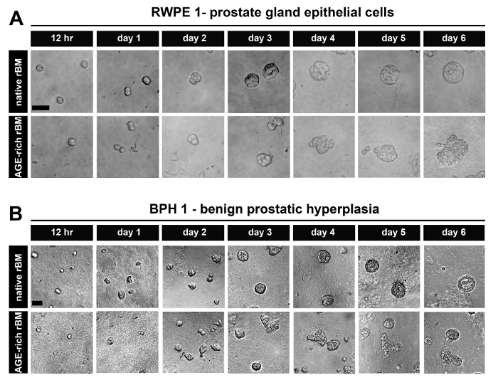
图1:在天然和硬化重组基底膜(rBM)上以三维腺泡结构生长的前列腺上皮细胞。(A)RWPE-1细胞在经PBS处理(天然)或50 mM乙醛酸处理14小时(AGE富集;硬化)的rBM凝胶上培养12小时至6天的明场图像;比例尺 = 50 µm。(B)BPH-1细胞,培养条件同A图;比例尺 = 50 µm;数据代表3次独立实验的结果。请点击此处查看该图的放大版本。
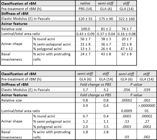
表 1:在天然、中等硬度及高硬度重建基底膜(rBM)上培养的前列腺上皮 RWPE-1 腺泡的特征。 RWPE-1 腺泡在经 PBS 预处理 14 小时(天然)、乙醛酸(GLA)预处理 6 小时(中等硬度)或 GLA 预处理 14 小时(高硬度)的 rBM 上培养。对于腺泡形态,圆形、半多边形和多边形腺泡的百分比(%)± 标准差(SD)来自 5 次独立实验(每种条件下定量 50 个腺泡)。相对腺泡大小基于 3 次独立实验计算(以天然 rBM 为 100%)。对于侵袭性,具有一种或多种突出细胞的腺泡百分比(%)± SD 来自 3 次独立实验。倍数变化通过将中等硬度或高硬度条件下的平均值除以天然条件下的对应值计算得出。P 值采用 Student t 检验计算(α = 0.05)。
AGE 依赖性的骨髓基质刚度增加促进 PC3 前列腺肿瘤细胞迁移
在天然rBM上生长的PC3细胞通过保持持续的细胞间接触进行迁移,而在AGE富集(较硬)rBM上生长的PC3细胞则彼此独立移动(图2A)。培养72小时后,PC3细胞在天然(PBS处理)rBM上形成集落(球状体),而在较硬(AGE富集)rBM上的PC3细胞不形成球状体,并独立迁移(图2B)。在较硬(AGE富集)rBM上的PC3细胞比在天然rBM上生长的PC3细胞更细长(图2C)。在较硬rBM上的PC3细胞比在天然rBM上生长的PC3细胞迁移速度更快(图2D)。在较硬rBM上的PC3细胞表现出比在天然rBM上生长的PC3细胞更低的迁移持续性(图2E)。
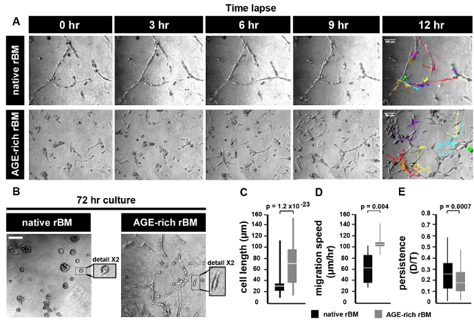
图2:前列腺肿瘤细胞在天然及硬化型重建基底膜(rBM)上的迁移 (A) PBS处理(天然)或50 mM乙醛酸处理14小时(AGE富集、刚性增强)的rBM凝胶上培养的PC3细胞明场图像。使用明场显微镜(10倍物镜)以每小时1帧的采集速率连续成像12小时,并通过细胞追踪生成运动轨迹。图中显示的时间点分别为0、3、6、9和12小时。单个细胞的运动轨迹显示为12小时时间点的结果。比例尺 = 100 µm(B) PC3细胞在天然或硬化的rBM上培养72小时,并按照图示方法进行成像A)。比例尺 = 100 µm. 细节显示所选区域的2倍放大图像。 (C) 平均值 ± 标准差 细胞长度(µm);天然rBM与硬质rBM之间差异显著(p = 1.2 × 10⁻²³). (D) 平均值 ± 标准差 速度(µm/hr)根据细胞轨迹计算得出;天然rBM与僵硬rBM之间差异显著(p = 0.004)。(E) 细胞运动持久性的均值 ± 标准差(D/T 比值,其中 D = 细胞轨迹起点到终点的距离,T = 细胞轨迹总长度);天然 rBM 与僵硬 rBM 之间的差异具有显著性(p = 0.0007)。图 C-E >分析了10个细胞,数据代表3次独立实验的结果。 请点击此处以查看此图的放大版本。
讨论
一种从乳腺上皮细胞在纯重组基底膜凝胶中生成三维腺泡的实验方案6 在之前的一项研究中,通过向rBM基质中添加4 mg/ml I型胶原蛋白对其进行了改良。胶原蛋白的加入使rBM凝胶的弹性模量从175 ± 37 帕斯卡升高至1589 ± 380 帕斯卡。这种刚度的9.1倍增加调节了乳腺上皮细胞(MECs)的生长、存活、迁移和分化21. 通过添加 D-(-)-核糖处理步骤,促进添加至 rBM 凝胶中的 I 型胶原发生非酶促交联,从而再次修改了该方案。结果发现,基质刚度增加了 15 倍,这种刚度的增加与乳腺上皮细胞的癌基因转化协同作用,促进了其侵袭性行为22在rBM凝胶中添加I型胶原的实验方法促进了乳腺上皮细胞与胶原纤维的直接相互作用,这种相互作用仅在人体组织中基底膜(BM)介导的间质与上皮间物理屏障发生蛋白水解降解后才会出现。通过在经GLA预处理的纯rBM凝胶中由PECs生成三维腺泡结构,本方案为研究基底膜刚度如何影响相关生物学过程提供了途径 本身 可触发其侵袭性行为(图3)。本方案所诱导的基底膜(BM)硬度水平具有生理相关性。分别在50 mM GLA中孵育6小时和14小时后,纯重组基底膜(rBM)凝胶的弹性模量增至175 ± 90 和 322 ± 160,而经PBS处理的rBM凝胶为122 ± 55 帕斯卡(Pascals)表1)。这种rBM硬度1.7至3.2倍的增加,重现了恶性前列腺组织相对于正常前列腺或良性前列腺增生(BPH)组织中观察到的2.5至3.4倍的硬度增加。23-26如近期一篇出版物所述13 由AGE积累和rBM刚度增加在PEC腺泡中诱导的形态学变化可被量化,表现为从圆形向多边形形状发生具有统计学意义的转变、管腔/腺泡总面积减小,以及细胞突出并从腺泡迁移 acina 到富含AGE的rBM中(图3)免疫印迹也可用于评估 EMT 的标志物(例如 E-钙黏蛋白缺失13)以及收缩行为(例如 磷酸化肌球蛋白轻链-2,pMLC213) 在正常与较硬的重组基底膜(rBM)中培养的PECs内的表达差异图3)。可进一步采用免疫荧光染色结合共聚焦显微镜进行评估,以观察基底膜(BM)例如 层粘连蛋白、IV型胶原和AGE积聚13),细胞顶-基底极性(例如 EEA1:早期核内体抗原1;以及GM130:130 kDa顺式高尔基体标志物的顶端定位13)以及黏附分子的细胞分布模式(例如 细胞间连接处的 E-cadherin 定位13) (图3).
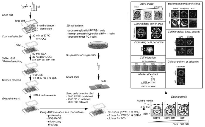
图 3:本文介绍的各种实验方案概览。 该示意图展示了如何使用乙醛酸(美拉德反应)制备并硬化重构成的基底膜(rBM),如何将细胞接种到硬化后的 rBM 上,如何分析硬化 rBM(美拉德反应程度),以及可用于分析 AGE 富集 rBM 所诱导的细胞和分子变化的实验方法。AGE,晚期糖基化终末产物;BM,基底膜;DAPI,4',6-二脒基-2-苯基吲哚;EEA1,早期内体抗原1;GAPDH,甘油醛-3-磷酸脱氢酶;GLA,乙醛酸;GEE,甘氨酸乙酯;GM130,130 kDa 顺式高尔基体标志物;p-MLC2 (Thr18/Ser19),在苏氨酸18和丝氨酸19位点磷酸化的肌球蛋白轻链2;rBM,重构成的基底膜;SDS-PAGE,十二烷基硫酸钠聚丙烯酰胺凝胶电泳。RWPE1 腺泡比例尺 = 10 µm;PC3 肿瘤细胞球体比例尺 = 100 µm。本图经参考文献13修改。 请点击此处查看此图的放大版本。
如果选择D-(-)-核糖作为rBM的交联剂,则可能需要进行故障排除。在方案开发过程中发现,按照先前报道的rBM/胶原凝胶方法22,使用1 M D-(-)-核糖处理72小时会导致rBM凝胶脱水和收缩。评估较低浓度的D-(-)-核糖以及更短的处理时间可能有助于克服这一局限性。
在今后应用该方案时,可能会遇到需要更高rBM硬度的情况,这可能成为一个潜在的局限性。若通过延长孵育时间或提高GLA浓度来增加rBM凝胶的硬度,则有必要评估这些处理条件是否会影响细胞存活与增殖,正如先前研究所描述的那样13。还应注意,血清孵育RWPE-1细胞会诱导表型发生类似上皮-间质转化(EMT)的变化,因此应避免将细胞暴露于血清或含血清的材料中。例如,在进行短干扰RNA(siRNA)寡核苷酸转染实验时,应使用在KSFM培养基中生长的RWPE-1细胞对实验流程进行优化,而不应将细胞更换至低血清转染培养基。这一局限性可能会影响在此模型中使用瞬时siRNA方法所达到的基因沉默效率。对于某些蛋白靶点,建议采用可诱导的shRNA载体以实现可调控的基因沉默,从而获得预期的蛋白水平下调。此外,未来模型还可引入由基质细胞或肿瘤细胞相关赖氨酰氧化酶(LOX)介导的酶促交联策略17。
本方案将有助于未来研究AGE依赖性基底膜硬度在前列腺上皮细胞(RWPE-1、BPH-1)中触发的促侵袭机制,以及在侵袭性前列腺癌细胞(PC3)中评估抗转移治疗靶点。鉴于良性前列腺增生(BPH)被认为是一种代谢性疾病27该方案还为深入理解代谢紊乱与前列腺癌风险增加之间的关联奠定了基础。鉴于AGEs暴露引起的基质硬度增加可能触发其他类型癌症的侵袭性,利用该方案建立包含其他器官正常上皮细胞和肿瘤细胞的类似模型将具有重要意义。例如 乳腺、结肠、卵巢、胰腺
方案中的关键步骤及其对应时间在图4中进行了总结。在初始步骤中,必须将rBM储备液置于4 °C条件下解冻,以防止其发生聚合。移液枪头在使用前必须预冷至4 °C,方可插入rBM储备液中。下一步中,还必须确保腔室载玻片在用rBM溶液包被前已平衡至4 °C。一旦rBM溶液温度升至4 °C以上,便会不可逆地发生聚合形成凝胶。在聚合阶段,必须避免扰动rBM,以确保其形成适合细胞培养和显微分析的均匀表面。使用GLA孵育(无论是否添加美拉德反应抑制剂,如氰基硼氢化钠和氨基胍)的时间长短,将决定rBM凝胶的硬度。若需半硬条件,建议采用6小时孵育;若需硬条件,则建议采用14小时孵育(表1)。若需要不同硬度水平,也可使用其他孵育时间或GLA浓度;在此情况下,必须将rBM凝胶的流变学分析作为关键步骤纳入实验流程。在通过GEE孵育淬灭美拉德反应并随后用PBS洗涤后,rBM凝胶可立即使用,或在4 °C下保存最多48小时后再用于细胞培养。细胞培养建立后,应每两天更换一次培养基(包括任何处理因素)。建议根据所研究的参数,将三维细胞培养维持3至12天。对于三维PEC腺泡结构,建议在培养6天后进行分析;而对于三维PTC类球体,建议在培养3天后首先进行分析。
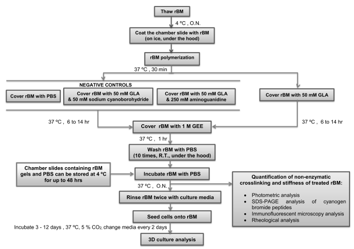
图 4:本实验方案的简要概述,标注了关键步骤和时间点。 该流程图展示了如何利用乙醇醛(美拉德反应)制备并硬化重组基底膜(rBM),并标明了关键步骤和操作时间。图中还标示了实验流程可暂停以及 rBM 凝胶可储存的时间节点。rBM,重组基底膜;GLA,乙醇醛;GEE,甘氨酸乙酯;O.N.,过夜;PBS,磷酸盐缓冲液;R.T.,室温。 请点击此处查看该图的放大版本。
披露
作者无任何利益冲突需要披露。
致谢
我们感谢范德比尔特大学医学中心的 Simon Hayward 提供 BPH-1 细胞;感谢哥本哈根大学生物技术研究与创新中心的 Thomas Cox 和 Janine Erler 在流变学测量方面的协助。MR-T 获得世界癌症研究基金会(原国际癌症研究协会,资助号 08-0803,授予 JS)、乌拉圭蒙得维的亚英国大使馆以及国家研究与创新署(资助号 UK_RH_2015_1_2,授予 MR-T)的资助。MC 获得前列腺癌英国基金会的支持(资助号 S14-017,授予 JS 和 GS)。KW 获得中国国家留学基金管理委员会的资助。MAM 获得沙特阿拉伯文化局的资助。
材料
| 姓名 | 公司 | 目录编号 | 评论 |
|---|---|---|---|
| 一 - 单层培养材料 | |||
| BPH-1 | CaP 细胞系数据库 | PCaCL-132 | 联系人:simon.hayward@mcmail.vanderbilt.edu |
| 完全型无血清角质形成细胞培养基 | ThermoFisher Scientific | 17005-075 | 使用前勿在 37 ºC 预热 |
| 胎牛血清 | First Link UK Ltd | 02-00-850 | 以分装形式储存于 -20 ºC |
| PC3 | 美国模式培养物集存库 | CRL-1435 | |
| 青霉素/链霉素 | ThermoFisher Scientific | 15070-063 | |
| 磷酸盐缓冲液(Dulbecco A)片剂 | Oxoid | BR0014G | |
| RPMI 1640 培养基 | Sigma-Aldrich | R5886 | 使用前在 37 ºC 水浴中预热 |
| RWPE-1 | 美国模式培养物集存库 | CRL-11609 | |
| 胰蛋白酶-EDTA 溶液 | Sigma-Aldrich | T4049 | |
| 名称 | 公司 | 目录编号 | 备注 |
| 二 - 三维培养材料 | |||
| 乙腈 | Sigma-Aldrich | 271004 | |
| 氨基胍盐酸盐 | Sigma-Aldrich | 396494 | 对眼睛、呼吸系统和皮肤有刺激性 |
| 8 孔腔室载玻片 | Thermo Scientific Nunc Lab-Tek | TKT-210-816M | |
| 培养基质重建基底膜(rBM)低生长因子提取物 | AMS Biotechnology | 3445-005-01 | 将基底膜(BM)以分装形式储存于 -80 ºC |
| 溴化氰 | Sigma-Aldrich | C91492 | 接触皮肤和吸入有毒 |
| 甲酸 | Sigma-Aldrich | 695076 | |
| 甘氨酸乙酯盐酸盐(GEE) | Sigma-Aldrich | 50060 | 对眼睛有刺激性 |
| 乙醇醛二聚体(GLA) | Sigma-Aldrich | G6805 | |
| 氰基硼氢化钠 | Sigma-Aldrich | 71435 | 高度易燃;接触皮肤和吸入有毒 |
| 0.22 微米注射器滤器 | Appleton Woods | BC680 | |
| 名称 | 公司 | 目录编号 | 备注 |
| 三 - 美拉德反应定量材料 | |||
| 4’,6-二脒基-2-苯基吲哚(DAPI) | ThemoFisher Scientific | D3571 | 避光,以分装形式储存于 -20 ºC |
| 克隆环 | Sigma-Aldrich | C1059 | |
| 山羊抗小鼠 IgG(H+L)Alexa Fluor 488 偶联物 | ThemoFisher Scientific | A-11001 | 避光 |
| 山羊抗兔 IgG(H+L)Alexa Fluor 488 偶联物 | ThemoFisher Scientific | A-11034 | 避光 |
| 山羊血清 | Abcam | ab7481 | 以分装形式储存于 -20 ºC |
| Vectashield 封片剂 | Vector Laboratories | H-1000 | |
| 小鼠抗戊糖苷胺单克隆抗体(克隆 PEN-12 mAb) | TransGenic Inc | KH012 | |
| 多聚甲醛(PFA) | Sigma-Aldrich | F8775 | 以分装形式储存于 -20 ºC |
| 兔抗人 IV 型胶原多克隆抗体 | Acris Antibodies | R1041 | 以分装形式储存于 -20 ºC |
| 兔抗层粘连蛋白 A/C 多克隆抗体 | Santa Cruz Biotechnology Inc | sc-7292 | 以分装形式储存于 -20 ºC |
| 聚乙二醇叔辛基苯基醚(Triton-X100) | Sigma-Aldrich | T9284 | |
| 聚乙二醇山梨醇单月桂酸酯(Tween-20) | Sigma-Aldrich | P1379 | |
| 透析盒 Slide-A-Lyzer | ThemoFisher Scientific | 66333 | |
| 名称 | 公司 | 目录编号 | 备注 |
| 四 - 设备 | |||
| ARG2 控制应变旋转流变仪 | T.A. Instruments | ||
| Axiovert S100(20X 放大倍数)显微镜 | Zeiss | ||
| 用于 Zeiss Axio S100 显微镜的 CO2 控制加湿培养室 | Solent Scientific | ||
| 共聚焦 Axiovert 200M(40X、63X 放大倍数)显微镜 | Zeiss | ||
| 配备数字相机的 Olympus LH50A 显微镜,使用相差 | Olympus | ||
| PHERAstar Plus 酶标仪分光光度计 | BMG Labtech | ||
| 名称 | 公司 | 目录编号 | 备注 |
| 五 - 软件 | |||
| Image J 1.47v | 美国国立卫生研究院 | ||
| MetaXpress | Molecular Devices |
参考文献
- Timpl, R., Brown, J. C. Supramolecular assembly of basement membranes. Bioessays. 18 (2), 123-132 (1996).
- Yurchenco, P. D., Ruben, G. C. Type IV collagen lateral associations in the EHS tumor matrix. Comparison with amniotic and in vitro networks. Am J Pathol. 132 (2), 278-291 (1988).
- Halfter, W., et al. Protein composition and biomechanical properties of in vivo-derived basement membranes. Cell Adh Migr. 7 (1), 64-71 (2013).
- Candiello, J., Cole, G. J., Halfter, W. Age-dependent changes in the structure, composition and biophysical properties of a human basement membrane. Matrix Biol. 29 (5), 402-410 (2010).
- To, M., et al. Diabetes-induced morphological, biomechanical, and compositional changes in ocular basement membranes. Exp Eye Res. 116, 298-307 (2013).
- Debnath, J., Muthuswamy, S. K., Brugge, J. S. Morphogenesis and oncogenesis of MCF-10A mammary epithelial acini grown in three-dimensional basement membrane cultures. Methods. 30 (3), 256-268 (2003).
- Bello, D., Webber, M. M., Kleinman, H. K., Wartinger, D. D., Rhim, J. S. Androgen responsive adult human prostatic epithelial cell lines immortalized by human papillomavirus 18. Carcinogenesis. 18 (6), 1215-1223 (1997).
- Hayward, S. W., et al. Establishment and characterization of an immortalized but non-transformed human prostate epithelial cell line: BPH-1. In Vitro Cell Dev Biol Anim. 31 (1), 14-24 (1995).
- Kaighn, M. E., Narayan, K. S., Ohnuki, Y., Lechner, J. F., Jones, L. W. Establishment and characterization of a human prostatic carcinoma cell line (PC-3). Invest Urol. 17 (1), 16-23 (1979).
- Laemmli, U. K. Cleavage of structural proteins during the assembly of the head of bacteriophage T4. Nature. 227 (5259), 680-685 (1970).
- Towbin, H., Staehelin, T., Gordon, J. Electrophoretic transfer of proteins from polyacrylamide gels to nitrocellulose sheets: procedure and some applications. Proc Natl Acad Sci U S A. 76 (9), 4350-4354 (1979).
- Rabilloud, T. Mechanisms of protein silver staining in polyacrylamide gels: a 10-year synthesis. Electrophoresis. 11 (10), 785-794 (1990).
- Rodriguez-Teja, M., et al. AGE-modified basement membrane cooperates with Endo180 to promote epithelial cell invasiveness and decrease prostate cancer survival. J Pathol. 235 (4), 581-592 (2015).
- Graf, B. W., Boppart, S. A. Imaging and analysis of three-dimensional cell culture models. Methods Mol Biol. 591, 211-227 (2010).
- Yao, N. Y., Larsen, R. J., Weitz, D. A. Probing nonlinear rheology with inertio-elastic oscillations. J Rheology. 52 (4), 1013-1025 (2008).
- Baker, A. M., Bird, D., Lang, G., Cox, T. R., Erler, J. T. Lysyl oxidase enzymatic function increases stiffness to drive colorectal cancer progression through FAK. Oncogene. 32 (14), 1863-1868 (2013).
- Caley, M. P., et al. Tumor-associated Endo180 requires stromal-derived LOX to promote metastatic prostate cancer cell migration on human ECM surfaces. Clin Exp Metastasis. 3 (2), 151-165 (2016).
- Sturge, J., Wienke, D., East, L., Jones, G. E., Isacke, C. M. GPI-anchored uPAR requires Endo180 for rapid directional sensing during chemotaxis. J Cell Biol. 162 (5), 789-794 (2003).
- Sturge, J., Wienke, D., Isacke, C. M. Endosomes generate localized Rho-ROCK-MLC2-based contractile signals via Endo180 to promote adhesion disassembly. J Cell Biol. 175 (2), 337-347 (2006).
- Rodriguez-Teja, M., et al. Survival Outcome and EMT Suppression Mediated by a Lectin Domain Interaction of Endo180 and CD147. Mol Cancer Res. 13 (3), 538-547 (2015).
- Paszek, M. J., et al. Tensional homeostasis and the malignant phenotype. Cancer Cell. 8 (3), 241-254 (2005).
- Levental, K. R., et al. Matrix crosslinking forces tumor progression by enhancing integrin signaling. Cell. 139 (5), 891-906 (2009).
- Carson, W. C., et al. Material characterization of ex vivo prostate tissue via spherical indentation in the clinic. Med Eng Phys. 33 (3), 302-309 (2011).
- Hoyt, K., et al. Tissue elasticity properties as biomarkers for prostate cancer. Cancer Biomark. 4 (4-5), 213-225 (2008).
- Krouskop, T. A., Wheeler, T. M., Kallel, F., Garra, B. S., Hall, T. Elastic moduli of breast and prostate tissues under compression. Ultrason Imaging. 20 (4), 260-274 (1998).
- Zhang, M., et al. Quantitative characterization of viscoelastic properties of human prostate correlated with histology. Ultrasound Med Biol. 34 (7), 1033-1042 (2008).
- Corona, G., et al. Benign prostatic hyperplasia: a new metabolic disease of the aging male and its correlation with sexual dysfunctions. Int J Endocrinol. , Article ID:329456 (2014).
重印与许可
