本文介绍了一种制备聚五氟苯基丙烯酸酯(poly(PFPA))接枝二氧化硅微球的方案。该聚(PFPA)功能化表面可固定抗体,并已成功用于通过免疫沉淀实现蛋白质分离。
需要JoVE订阅才能观看此内容。 请登录或开始免费试用
本文内容
勘误通知
Important: There has been an erratum issued for this article. View Erratum Notice
摘要
摘要
我们展示了一种简便的方法,用于制备聚(五氟苯基丙烯酸酯)(poly(PFPA))接枝的二氧化硅微球,以实现抗体固定化并进一步应用于免疫沉淀(IP)。poly(PFPA)接枝表面通过一个简单的两步法构建。第一步,将3-氨基丙基三甲氧基硅烷(APTMS)作为连接分子沉积在二氧化硅表面。第二步,通过可逆加成-断裂链转移(RAFT)聚合合成的poly(PFPA)均聚物,通过聚合物上的五氟苯基(PFP)单元与APTMS上的氨基之间的交换反应,接枝到连接分子上。APTMS和poly(PFPA)在二氧化硅颗粒表面的沉积通过X射线光电子能谱(XPS)进行确认,并通过动态光散射(DLS)测量的颗粒尺寸变化进行监测。为了提高微球表面的亲水性,还进行了部分poly(PFPA)与氨基功能化的聚乙二醇(amino-PEG)的取代反应。随后,将PEG取代的poly(PFPA)接枝二氧化硅微球用于抗体的固定化,以进行IP应用。作为示例,采用针对RNA激活蛋白激酶(PKR)的抗体,并通过Western blotting测定IP效率。分析结果表明,固定化抗体的微球确实可用于富集PKR,同时非特异性蛋白相互作用极低。
引言
近年来,反应性聚合物刷引起了广泛关注。它们可用于将功能分子固定在有机或无机材料上,从而制备出具有检测和分离等应用的活化表面1,2,3,4,5。在已报道的反应性聚合物中,含有五氟苯基酯单元的聚合物因其对胺类具有高反应活性且耐水解而尤为有用6。其中一种聚合物是聚(PFPA),它可在聚合后通过含有伯胺或仲胺的分子进行简便的功能化修饰7,8,9,10。例如,聚(PFPA)刷可与氨基螺吡喃反应,制备出具有光响应性的表面7。
聚(PFPA)的制备及其应用已在多项先前发表的研究中有所描述6,7,8,9,10,11,12,13,14,15,16,17特别是,Theato 及其同事报道了通过两种方法合成聚(PFPA)刷状聚合物 "嫁接到" 和 "嫁接自" 方法7,8,10,11,12. 在 "嫁接到" 方法,合成了聚(甲基倍半硅氧烷)-聚(五氟苯基丙烯酸酯)(poly(MSSQ-PFPA))杂化聚合物8,10,11,12聚(MSSQ)组分能够与多种不同的有机和无机表面形成强粘附,从而使得聚(PFPA)组分可在涂覆材料表面形成刷状层。在 "嫁接自" 方法,采用表面引发的可逆加成-断裂链转移(SI-RAFT)聚合法制备聚(PFPA)刷状聚合物7在此情况下,首先通过硅-硅烷反应将表面固定化链转移剂(SI-CTA)共价连接至基底表面。随后,固定化的SI-CTA参与PFPA单体的SI-RAFT聚合反应,生成高度密集且通过稳定共价键与基底相连的聚(PFPA)刷状聚合物。
通过利用SI-RAFT聚合方法合成的聚(PFPA)刷,我们近期展示了将抗体固定在聚(PFPA)接枝的二氧化硅微粒上,并将其应用于蛋白质纯化18。研究发现,采用聚(PFPA)刷进行抗体固定可解决当前免疫沉淀(IP)蛋白质分离中存在的若干问题。传统的IP依赖于Protein A/G作为抗体固定的连接分子19,20,21。由于Protein A/G能够使抗体以特定方向结合,因而可实现较高的目标抗原回收效率。然而,Protein A/G的使用存在非特异性蛋白相互作用以及在蛋白回收过程中抗体丢失的问题,这两者均会导致较高的背景噪声。为克服这些缺点,已有研究探索将抗体直接交联至固相载体的方法22,23,24。但由于交联抗体的方向随机,此类技术的效率通常较低。对于聚(PFPA)接枝的基底,抗体的固定通过PFP单元与抗体上的氨基官能团之间的交换反应实现,具有永久性。尽管抗体的取向仍为随机,但该体系具有大量可反应的PFP位点,且其数量可通过聚合度进行调控。此外,我们还证明,通过部分PFP单元被氨基-PEG取代,可调节表面亲水性,从而进一步提高系统的蛋白回收效率18。总体而言,聚(PFPA)接枝的二氧化硅微粒被证实是一种高效且背景信号更洁净的常规IP技术的有效替代方案。
本文报道了一种用于抗体固定化及免疫沉淀(IP)应用的聚(PFPA)接枝表面的替代制备方法。如图1所示,该方法为简单的两步过程:首先将APTMS连接分子沉积到二氧化硅表面,随后通过聚合物上的PFP单元与APTMS上的氨基之间的反应,将聚(PFPA)聚合物共价连接至连接分子上。该制备方法可实现聚(PFPA)与基底表面的永久交联,同时避免了SI-CTA合成及聚(PFPA)刷状聚合物的SI-RAFT聚合过程中相关的诸多复杂问题。此外,仍可对部分PFP单元进行氨基-PEG取代,从而精确调控聚合物刷表面的性质。本文展示了采用该方法制备的聚(PFPA)接枝二氧化硅微球可用于抗体的固定化,并通过IP实现蛋白质富集。文中详细记录了微球制备、抗体固定化及IP检测的完整流程,为希望寻求传统Protein A/G依赖性IP方法替代方案的读者提供参考。
访问受限。请登录或开始试用以查看此内容。
方案
1. 聚(PFPA)均聚物的制备
- 偶氮二异丁腈(AIBN)的重结晶
- 在250 mL烧杯中加入5 g 2,2’-偶氮双(2-甲基丙腈)(AIBN)和25 mL甲醇。将烧杯浸入60 °C油浴中,用磁力搅拌子剧烈搅拌,直至AIBN完全溶解。
- 将温热的溶液通过滤纸(5–8 μm颗粒截留)过滤,并将滤液置于4 °C下静置,使晶体缓慢析出。
- 通过过滤收集重结晶后的AIBN。将收集的产物与25 mL新鲜甲醇混合,重复重结晶过程。
- 将经过两次重结晶的AIBN在室温(RT)下于真空烘箱中干燥过夜。将产物避光保存于< -10 °C条件下。
- 苯甲基二硫代苯甲酸酯的合成25
- 准备一个500 mL三颈圆底烧瓶,配备磁力搅拌子、回流冷凝管、滴液漏斗和橡胶隔膜。通过回流冷凝管将烧瓶连接至氮气管线,用氮气吹扫烧瓶内部空气。将温度计插入隔膜中。通过注射器经同一隔膜加入41 mL(0.041 mol)四氢呋喃(THF)中1 M苯基溴化镁溶液。
- 将苯基溴化镁溶液在油浴中加热至40 °C,然后通过滴液漏斗缓慢加入3.1 g(0.041 mol)二硫化碳,保持溶液温度在40 °C。
- 在15分钟内通过滴液漏斗向所得混合物中加入7.1 g(0.042 mol)溴化苄。将反应温度升至50 °C,并在此温度下继续搅拌45分钟。
- 将反应混合物转移至分液漏斗中,加入15 mL冰水稀释。加入15 mL乙醚萃取产物,并弃去下层水相。再用乙醚重复萃取两次。
- 将合并的有机相用大量水洗涤,随后用饱和食盐水(50% (w/v) NaCl水溶液)洗涤,并用无水硫酸镁干燥。
- 使用旋转蒸发仪在35 °C下减压除去溶剂。
- 采用柱层析法纯化产物,以400 mL硅胶(孔径60 Å,粒径63–200目)为固定相,石油醚为洗脱剂,得到5 g苯甲基二硫代苯甲酸酯(BDB)红色油状物。通过1H NMR(400 MHz,CDCl3)确认产物纯度:δ 8.02–7.99(2H,m),7.55–7.50(1H,m),7.41–7.29(7H,m),4.60(2H,s)。
- 通过RAFT聚合合成聚(PFPA)9,26
- 市售PFPA单体含有少量阻聚剂。在聚合前,将单体通过装有碱性氧化铝的一次性注射器,以去除阻聚剂。
- 向20 mL Schlenk烧瓶中加入0.4 mg(0.0024 mmol)重结晶AIBN、4.3 mg(0.018 mmol)BDB、1012 mg(4.25 mmol)无阻聚剂PFPA以及0.7 mL无水苯甲醚。
- 将烧瓶连接至Schlenk管线,通过至少三次冷冻-抽气-解冻循环对混合物进行脱气。具体操作为:将反应混合物在液氮浴中冷冻,抽真空除去顶空气体,密封烧瓶后移出液氮,使其在室温下解冻。
- 将烧瓶置于70 °C油浴中,在N2气氛下反应4小时。
- 为终止反应,将烧瓶从油浴中取出,使反应体系暴露于空气中。
- 将聚合物在冷甲醇中沉淀,然后将回收的聚合物在40 °C真空烘箱中干燥过夜。
- 使用凝胶渗透色谱法(GPC)测定聚合物分子量。以THF为流动相,温度35 °C,流速1 mL/min,采用单分散聚苯乙烯标准品绘制校准曲线。进行GPC测定时,将聚合物溶于THF(1–2 mg/mL),经0.2 μm一次性聚四氟乙烯(PTFE)滤膜过滤。取100 μL样品注入GPC仪器,利用聚苯乙烯校准曲线将测得的样品保留时间转换为分子量。
2. 聚(PFPA)功能化SiO2微球的制备
- 使用 APTMS 处理 SiO2 微球
- SiO2 颗粒以 5% (w/v) 的水悬浮液形式提供。将 0.8 mL 的 SiO2 悬浮液与 40 mg APTMS 和 8 mL 甲醇加入一个装有搅拌子的 20 mL 闪烁瓶中。
- 在室温下剧烈搅拌,使反应进行 5 小时。
- 将溶液转移至锥形管中。为分离 APTMS 功能化的 SiO2 微球,以 10,000 × g 离心 5 分钟,然后弃去上清液。通过将微球重新分散于 3 mL 新鲜甲醇中进行洗涤。手动振荡混匀,必要时可在水浴中超声数秒以改善分散。以 10,000 × g 离心 5 分钟,弃去上清液,并重复洗涤步骤一次。
- 将经甲醇洗涤的 SiO2 微球与 3 mL 二甲基亚砜(DMSO)混合。手动振荡混合物,必要时可超声数秒,直至微球在 DMSO 中完全分散。以 10,000 × g 离心 5 分钟,弃去上清液。重复该步骤一次,以确保溶剂从甲醇完全置换为 DMSO。
注:最终悬浮液包含分散于 4 mL DMSO 中的 APTMS 功能化 SiO2 微球。 - 为检测粒径分布,进行动态光散射(DLS)分析。取步骤 2.1.4 制备的悬浮液一滴,置于一次性紫外比色皿中。用新鲜 DMSO 稀释样品至比色皿体积的 2/3。将样品插入样品架以开始数据采集。粒径测量参数设置如下:温度:25 °C;平衡时间:120 秒;测量时长:自动。
- 为检测表面组成,进行 X 射线光电子能谱(XPS)分析。将步骤 2.1.4 制备的悬浮液中少量样品在 40 °C 真空烘箱中干燥过夜。取干燥后的样品均匀装填至 0.5 cm × 0.5 cm 的样品台上。将样品载入高真空腔室(10-8 torr)并开始数据采集。针对所用的特定 XPS 仪器,采用单色 Al Kα X 射线源(工作条件为 15 kV 和 6.7 mA)激发光电子,使用混合模式放大,分析器通能设为 50 eV 以获取高分辨率谱图,100 eV 以进行元素全谱扫描。
- 将聚(PFPA)接枝到 APTMS 功能化的 SiO2 微球上
- 在 20 mL 闪烁瓶中,将 20 mg 聚(PFPA) 溶解于 2 mL DMSO 中,制备聚(PFPA) 溶液。
注:本研究中使用相对低分子量的聚(PFPA)(20 kg/mol)。因此,尽管聚合物浓度较高(10 mg/mL),仍未观察到交联现象。若使用更高分子量的聚合物,则可能需要调整聚合物溶液浓度,以避免潜在的交联。 - 将 1 mL 以 DMSO 悬浮的 APTMS 功能化 SiO2 微球(来自步骤 2.1.4)加入聚(PFPA) 溶液中。在室温下剧烈搅拌反应 1 小时。
- 以 10,000 × g 离心 5 分钟,分离聚(PFPA) 接枝的 SiO2 微球,随后弃去上清液。加入 3 mL DMSO 洗涤微球,通过手动振荡或数秒超声混合。以 10,000 × g 离心 5 分钟,弃去上清液。用 DMSO 对聚(PFPA) 接枝的 SiO2 微球重复洗涤两次。
- 再用三蒸水(TDW)洗涤微球两次。此步骤中,将微球与 3 mL 三蒸水混合,通过手动振荡或数秒超声混匀。以 10,000 × g 离心 5 分钟,弃去上清液。
- 为检测粒径分布,按照步骤 2.1.5 所述程序进行 DLS 分析。为检测表面化学组成,按照步骤 2.1.6 所述程序进行 XPS 分析。
- 在 20 mL 闪烁瓶中,将 20 mg 聚(PFPA) 溶解于 2 mL DMSO 中,制备聚(PFPA) 溶液。
3. 制备接枝了PEG取代的聚(PFPA)的SiO2微球
- 配制聚(PFPA)溶液:将20 mg聚(PFPA)溶解于20 mL小瓶中的2 mL DMSO中。
- 配制PEG溶液:将氨基功能化的PEG溶解于1 mL DMSO中。所用PEG的确切用量由所需的PFP取代度决定,计算公式如下:
氨基-PEG用量(g/g-聚(PFPA))= (N_聚(PFPA) × % PEG-取代) × (MW_PEG / MW_聚(PFPA))
其中,N_聚(PFPA) = 聚(PFPA)的聚合度
% PEG-取代 = PEG取代百分比
MW_PEG = 氨基-PEG的分子量
MW_聚(PFPA) = 聚(PFPA)的分子量 - 将PEG溶液转移至聚(PFPA)溶液中,在室温下剧烈搅拌反应1小时。
- 配制APTMS功能化的SiO2微球DMSO悬浮液时,参照步骤2.1中的相同操作。取1 mL该微球悬浮液加入步骤3.3中制备的PEG取代聚(PFPA)溶液中。在室温下剧烈搅拌反应1小时,使聚(PFPA)与APTMS功能化的SiO2微球之间发生接枝反应。
- 在10,000 × g离心5分钟以分离微球,随后弃去上清液。加入3 mL DMSO洗涤微球,通过手动振荡或短时超声处理混匀。再次在10,000 × g离心5分钟,弃去上清液。重复DMSO洗涤两次。
- 再用TDW洗涤微球两次。每次加入3 mL TDW,通过手动振荡或短时超声处理混匀。每次洗涤后在10,000 × g离心5分钟,弃去上清液。
- 将微球置于真空烘箱中,40 °C下干燥过夜。
4. 聚(PFPA)接枝SiO2微球上的抗体固定化
注意:无论聚(PFPA)的PEG取代率如何,均使用相同的步骤。通过将PBS片剂溶解于TDW中来制备磷酸盐缓冲液(PBS)。通过向PBS中加入1/1000体积的Tween-20来制备含0.1%(v/v)Tween-20的磷酸盐缓冲液(PBST)。
- 将5 mg聚(PFPA)接枝的SiO2微球加入1.5 mL微量离心管中。
- 加入800 µL PBS洗涤微球,并通过涡旋充分混匀。在室温下以10,000 × g离心1分钟,弃去上清液,重复洗涤步骤三次。
- 加入350 µL新鲜PBS、50 µL 0.1% (v/v) PBST和6.67 µg抗体。在4 °C下旋转孵育约20小时。
- 洗涤微球以去除未结合的抗体。在4 °C下以400 × g离心1分钟,弃去上清液,小心加入400 µL裂解缓冲液。用移液器轻轻吹打5次,使微球重新悬浮。
注意:用于洗涤微球的裂解缓冲液应与细胞裂解和免疫沉淀(IP)过程中使用的缓冲液相同,但二硫苏糖醇和蛋白酶抑制剂的添加为可选步骤(见步骤5)。 - 重复此洗涤步骤三次。最后一次洗涤后,尽可能彻底去除上清液。
5. 细胞裂解与免疫沉淀
- 裂解缓冲液和洗涤缓冲液的制备
- 配制裂解缓冲液(50 mM Tris-HCl (pH 8.0),100 mM KCl,0.5% (v/v) NP-40,10% (v/v) 甘油,1 mM 二硫苏糖醇 (DTT) 和蛋白酶抑制剂混合物)。
- 配制洗涤缓冲液(50 mM Tris-HCl (pH 8.0),100 mM KCl,0.1% (v/v) NP-40,10% (v/v) 甘油)。
- 将缓冲液储存于 4 °C。
- 细胞的准备
- 在免疫沉淀实验前一至两天接种细胞(HeLa 细胞),并在 37 °C、5% CO2 条件下培养。
- 用细胞刮收集约 1.4 × 10⁷ 个细胞,转移至 15 mL 圆锥管中。在室温下以 380 × g 离心 3 分钟。弃去上清液,用 1 mL 冷 PBS 重悬,并转移至 1.5 mL 微量离心管中。
- 在 4 °C 下以 10,000 × g 离心 30 秒。彻底弃去上清液。去除上清液后,细胞沉淀可在 -80 °C 保存。
- 细胞裂解物的制备
- 用 400 µL 裂解缓冲液重悬细胞沉淀。使用超声波破碎仪对细胞进行超声处理。
- 超声处理后,短暂涡旋混匀,然后在 4 °C 下以 20,000 × g 离心 10 分钟。
- 将上清液转移至新的 1.5 mL 离心管中。
- 免疫沉淀
- 将 300 µL 细胞裂解物转移至预先与抗体孵育的聚(PFPA)接枝 SiO2 微球中。另取 30 µL 细胞裂解物保留于新的微量离心管中作为输入样品。将输入样品置于 4 °C 保存。
注意:细胞裂解物中的总蛋白量应约为 4 mg。 - 将裂解物与微球的混合物在 4 °C 下于旋转仪上孵育 3 小时。
- 在 4 °C 下以 400 × g 离心 1 分钟。弃去上清液,小心加入 400 µL 洗涤缓冲液。通过移液枪轻柔吹打约五次,使微球重新悬浮。
- 重复此洗涤步骤三次。最后一次洗涤后,尽可能彻底去除上清液。
- 配制 2× 十二烷基硫酸钠 (SDS) 上样缓冲液(25% (v/v) 甘油,0.1% (w/v) 溴酚蓝 (BPB),60 mM Tris-HCl (pH 6.8),2% (w/v) SDS,2.75 mM 巯基乙醇)。将 2× SDS 上样缓冲液储存于 -20 °C。向微球及保存的输入样品中各加入 30 µL 2× SDS 上样缓冲液,并在 95 °C 加热 10 分钟。
- 加热后,使用 Western 印迹法27 分析样品,或将样品储存于 -20 °C。
- 将 300 µL 细胞裂解物转移至预先与抗体孵育的聚(PFPA)接枝 SiO2 微球中。另取 30 µL 细胞裂解物保留于新的微量离心管中作为输入样品。将输入样品置于 4 °C 保存。
访问受限。请登录或开始试用以查看此内容。
结果
聚(PFPA)接枝SiO2微球的制备示意图(可选是否进行PEG取代)如图1所示。为监测APTMS和聚(PFPA)的接枝过程,采用动态光散射(DLS)(图2)和X射线光电子能谱(XPS)(图3)对裸SiO2微球、APTMS功能化的SiO2微球以及聚(PFPA)接枝的SiO2微球进行了表征。微球的免疫沉淀效率通过Western blotting测定。图4展示了使用1% PEG取代的聚(PFPA)接枝微球进行免疫沉淀的Western blotting结果,其中微球分别与无抗体、非特异性抗体或抗PKR抗体共同孵育。图5展示了使用0%和1% PEG取代的聚(PFPA)接枝微球进行免疫沉淀的Wester...
访问受限。请登录或开始试用以查看此内容。
讨论
聚(PFPA)接枝SiO2微球的合成过程如图1所示。通过使用APTM作为连接分子,可通过一个简单的两步法在SiO2基底上共价接枝聚(PFPA)刷状聚合物。尽管部分PFP单元在与APTM反应中被消耗,但仍预期有大量PFP单元可保留下来,用于后续与氨基-PEG或抗体的反应。已知PFP基团会形成低表面能,因此聚(PFPA)刷在水中溶胀性较差28。在免疫沉淀(IP)应用中,抗体需固定在聚(PFPA)刷上,该置换反应在水相缓冲溶液中进行,以保持抗体的活性。正如我们先前发表的研究所述,用亲水性分子(如氨基功能化的PEG)部分取代PFP单元,可提高表面亲水性,从而提升抗体固定效率18。在本研究中,还制备了部分PEG取代的聚(PFPA),再通过相同的APTM连接分子将其接枝到SiO2表面。总体而言, 图1所示的方法可用于制备具有不同PEG取代程度的聚(PFPA)接枝...
访问受限。请登录或开始试用以查看此内容。
披露
作者无任何利益冲突需要披露。
致谢
本工作由国防发展局(资助编号:UD170039ID)提供支持。
访问受限。请登录或开始试用以查看此内容。
材料
| 姓名 | 公司 | 目录编号 | 评论 |
|---|---|---|---|
| 2,2-偶氮二异丁腈,99% | Daejung Chemicals | 1102-4405 | |
| 用于高效液相色谱的甲醇,99.9% | Duksan Pure Chemicals | d62 | |
| 苯基溴化镁 THF 溶液,1.0 M | Sigma-Aldrich | 331376 | |
| 无水二硫化碳,≥99% | Sigma-Aldrich | 335266 | |
| 溴化苄,98% | Sigma-Aldrich | B17905 | |
| 石油醚,90% | Samchun Chemicals | P0220 | |
| 乙醚,99% | Daejung Chemicals | 4025-4404 | |
| 无水硫酸镁,粉末, 99% | Daejung Chemicals | 5514-4405 | |
| 五氟苯基丙烯酸酯 | Santa Cruz Biotechnology | sc-264001 | 含抑制剂 |
| 氧化铝,活化,碱性,Brockmann I | Sigma-Aldrich | 199443 | |
| 氯化钠 (NaCl) | Daejung Chemicals | 7548-4400 | |
| 无水苯甲醚,99.7% | Sigma-Aldrich | 296295 | |
| 二氧化硅纳米颗粒 | Microparticles GmbH | SiO2-R-0.7 | 5% w/v 水悬浮液 |
| 3-氨基丙基三甲氧基硅烷,>96.0% | Tokyo Chemical Industry | T1255 | |
| 用于高效液相色谱的二甲基亚砜,≥99.7% | Sigma-Aldrich | 34869 | |
| 氨基封端的聚(乙二醇)甲醚 | Polymer Source | P16082-EGOCH3NH2 | |
| 磷酸盐缓冲液片剂 | Takara | T9181 | |
| 吐温-20 | Calbiochem | 9480 | |
| Tris-HCl (pH 8.0) | Invitrogen | AM9855G | |
| KCl | Invitrogen | AM9640G | |
| NP-40 | VWR | E109-50ML | |
| 甘油 | Invitrogen | 15514-011 | |
| 二硫苏糖醇 | Biosesang | D1037 | |
| 蛋白酶抑制剂 | Merck | 535140-1MLCN | |
| 溴酚蓝 | Sigma-Aldrich | B5525-5G | |
| Tris-HCl (pH 6.8) | Biosolution | BT033 | |
| 十二烷基硫酸钠 | Biosolution | BS003 | |
| 2-巯基乙醇 | Gibco | 21985-023 | |
| PKR 抗体 | Cell Signaling Technology | 12297S | |
| GAPDH 抗体 | Santa Cruz Biotechnology | sc-32233 | |
| 正常兔 IgG | Cell Signaling Technology | 2729S | |
| HeLa | 韩国细胞库 | 10002 | |
| 超声破碎仪 | DAIHAN Scientific | WUC-D10H | |
| 超声波处理器 | BMBio | BR2006A | |
| 离心机 I | Eppendorf | 5424 R | |
| 离心机 II | LABOGENE | 1736R | |
| 旋转混合仪 | FINEPCR | ROTATOR/AG | |
| 真空干燥箱 | DAIHAN Scientific | ThermoStable OV-30 | |
| 凝胶渗透色谱(THF) | Agilent Technologies | 1260 Infinity II | |
| X射线光电子能谱仪 | Thermo VG Scientific | Sigma Probe | |
| 动态光散射仪 | Malvern Instruments | ZEN 3690 |
参考文献
- Johnsson, B., Löfås, S., Lindquist, G. Immobilization of proteins to a carboxymethyldextran-modified gold surface for biospecific interaction analysis in surface plasmon resonance sensors. Analytical Biochemistry. 198 (2), 268-277 (1991).
- Kurzawa, C., Hengstenberg, A., Schuhmann, W. Immobilization method for the preparation of biosensors based on pH shift-induced deposition of biomolecule-containing polymer films. Analytical Chemistry. 74 (2), 355-361 (2002).
- You, C. C., et al. Detection and identification of proteins using nanoparticle-fluorescent polymer 'chemical nose' sensors. Nature Nanotechnology. 2 (5), 318-323 (2007).
- Roberts, M. W., Ongkudon, C. M., Forde, G. M., Danquah, M. K. Versatility of polymethacrylate monoliths for chromatographic purification of biomolecules. Journal of Separation Science. 32 (15-16), 2485-2494 (2009).
- Sandison, M. E., Cumming, S. A., Kolch, W., Pitt, A. R. On-chip immunoprecipitation for protein purification. Lab on a Chip. 10 (20), 2805-2813 (2010).
- Das, A., Theato, P. Activated Ester Containing Polymers: Opportunities and Challenges for the Design of Functional Macromolecules. Chemical Reviews. 116 (3), 1434-1495 (2016).
- Choi, J., et al. Functionalization and patterning of reactive polymer brushes based on surface reversible addition and fragmentation chain transfer polymerization. Journal of Polymer Science Part A: Polymer Chemistry. 50 (19), 4010-4018 (2012).
- Kessler, D., Jochum, F. D., Choi, J., Char, K., Theato, P. Reactive surface coatings based on polysilsesquioxanes: universal method toward light-responsive surfaces. ACS Applied Materials & Interfaces. 3 (2), 124-128 (2011).
- Son, H., et al. Penetration and exchange kinetics of primary alkyl amines applied to reactive poly(pentafluorophenyl acrylate) thin films. Polymer Journal. 48 (4), 487-495 (2016).
- Kessler, D., Roth, P. J., Theato, P. Reactive surface coatings based on polysilsesquioxanes: controlled functionalization for specific protein immobilization. Langmuir. 25 (17), 10068-10076 (2009).
- Kessler, D., Theato, P. Reactive surface coatings based on polysilsesquioxanes: defined adjustment of surface wettability. Langmuir. 25 (24), 14200-14206 (2009).
- Kessler, D., Nilles, K., Theato, P. Modular approach towards multi-functional surfaces with adjustable and dual-responsive wettability using a hybrid polymer toolbox. Journal of Materials Chemistry. 19 (43), 8184-8189 (2009).
- Eberhardt, M., Mruk, R., Zentel, R., Theato, P. Synthesis of pentafluorophenyl(meth)acrylate polymers: new precursor polymers for the synthesis of multifunctional materials. European Polymer Journal. 41 (7), 1569-1575 (2005).
- Jochum, F. D., Forst, F. R., Theato, P. PNIPAM copolymers containing light-responsive chromophores: a method toward molecular logic gates. Macromolecular Rapid Communications. 31 (16), 1456-1461 (2010).
- Schattling, P., Pollmann, I., Theato, P. Synthesis of CO2-responsive polymers by post-polymerization modification. Reactive & Functional Polymers. 75, 16-21 (2014).
- He, L., Szameit, K., Zhao, H., Hahn, U., Theato, P. Postpolymerization modification using less cytotoxic activated ester polymers for the synthesis of biological active polymers. Biomacromolecules. 15 (8), 3197-3205 (2014).
- Arnold, R. M., McNitt, C. D., Popik, V. V., Locklin, J. Direct grafting of poly(pentafluorophenyl acrylate) onto oxides: versatile substrates for reactive microcapillary printing and self-sorting modification. Chemical Communications. 50 (40), 5307-5309 (2014).
- Son, H., Ku, J., Kim, Y., Li, S., Char, K. Amine-Reactive Poly(pentafluorophenyl acrylate) Brush Platforms for Cleaner Protein Purification. Biomacromolecules. 19 (3), 951-961 (2018).
- Cullen, S. E., Schwartz, B. D. An improved method for isolation of H-2 and Ia alloantigens with immunoprecipitation induced by protein A-bearing staphylococci. The Journal of Immunology. 117 (1), 136-142 (1976).
- Sisson, T. H., Castor, C. W. An improved method for immobilizing IgG antibodies on protein A-agarose. Journal of Immunology Methods. 127 (2), 215-220 (1990).
- Peritz, T., et al. Immunoprecipitation of mRNA-protein complexes. Nature Protocols. 1 (2), 577-580 (2006).
- Zhang, Z., Chen, S., Jiang, S. Dual-functional biomimetic materials: nonfouling poly (carboxybetaine) with active functional groups for protein immobilization. Biomacromolecules. 7 (12), 3311-3315 (2006).
- Yao, Y., et al. NHS-ester functionalized poly(PEGMA) brushes on silicon surface for covalent protein immobilization. Colloids and Surfaces B: Biointerfaces. 66 (2), 233-239 (2008).
- Ma, J., et al. Facile fabrication of microsphere-polymer brush hierarchically three-dimensional (3D) substrates for immunoassays. Chemical Communications. 51 (31), 6749-6752 (2015).
- Chong, Y., et al. Thiocarbonylthio compounds [SC (Ph) S− R] in free radical polymerization with reversible addition-fragmentation chain transfer (RAFT Polymerization). Role of the free-radical leaving group (R). Macromolecules. 36 (7), 2256-2272 (2003).
- Jochum, F. D., Theato, P. Temperature- and Light-Responsive Polyacrylamides Prepared by a Double Polymer Analogous Reaction of Activated Ester Polymers. Macromolecules. 42 (16), 5941-5945 (2009).
- JoVE Science Education Database. Basic Methods in Cellular and Molecular Biology. The Western Blot. JoVE. , Cambridge, MA. (2018).
- Chua, G. B. H., Roth, P. J., Duong, H. T. T., Davis, T. P., Lowe, A. B. Synthesis and Thermoresponsive Solution Properties of Poly[oligo(ethylene glycol) (meth)acrylamide]s: Biocompatible PEG Analogues. Macromolecules. 45 (3), 1362-1374 (2012).
访问受限。请登录或开始试用以查看此内容。
重印与许可
勘误
Formal Correction: Erratum: Preparation of Poly(pentafluorophenyl acrylate) Functionalized SiO2 Beads for Protein Purification
Posted by JoVE Editors on 4/30/2019. Citeable Link.
An erratum was issued for: Preparation of Poly(pentafluorophenyl acrylate) Functionalized SiO2 Beads for Protein Purification. Throughout the article, the term "3-aminopropyltriethoxysilane" has been replaced with "3-aminopropyltrimethoxysilane", and "APTES" with "APTMS".
The Keywords were updated from:
Poly(pentafluorophenyl acrylate), 3-aminopropyltriethoxysilane, reactive polymer brush, post-polymerization functionalization, antibody immobilization, immunoprecipitation
to:
Poly(pentafluorophenyl acrylate), 3-aminopropyltrimethoxysilane, reactive polymer brush, post-polymerization functionalization, antibody immobilization, immunoprecipitation
The Abstract was updated from:
We demonstrate a simple method to prepare poly(pentafluorophenyl acrylate) (poly(PFPA)) grafted silica beads for antibody immobilization and subsequent immunoprecipitation (IP) application. The poly(PFPA) grafted surface is prepared via a simple two-step process. In the first step, 3-aminopropyltriethoxysilane (APTES) is deposited as a linker molecule onto the silica surface. In the second step, poly(PFPA) homopolymer, synthesized via the reversible addition and fragmentation chain transfer (RAFT) polymerization, is grafted to the linker molecule through the exchange reaction between the pentafluorophenyl (PFP) units on the polymer and the amine groups on APTES. The deposition of APTES and poly(PFPA) on the silica particles are confirmed by X-ray photoelectron spectroscopy (XPS), as well as monitored by the particle size change measured via dynamic light scattering (DLS). To improve the surface hydrophilicity of the beads, partial substitution of poly(PFPA) with amine-functionalized poly(ethylene glycol) (amino-PEG) is also performed. The PEG-substituted poly(PFPA) grafted silica beads are then immobilized with antibodies for IP application. For demonstration, an antibody against protein kinase RNA-activated (PKR) is employed, and IP efficiency is determined by Western blotting. The analysis results show that the antibody immobilized beads can indeed be used to enrich PKR while non-specific protein interactions are minimal.
to:
We demonstrate a simple method to prepare poly(pentafluorophenyl acrylate) (poly(PFPA)) grafted silica beads for antibody immobilization and subsequent immunoprecipitation (IP) application. The poly(PFPA) grafted surface is prepared via a simple two-step process. In the first step, 3-aminopropyltrimethoxysilane (APTMS) is deposited as a linker molecule onto the silica surface. In the second step, poly(PFPA) homopolymer, synthesized via the reversible addition and fragmentation chain transfer (RAFT) polymerization, is grafted to the linker molecule through the exchange reaction between the pentafluorophenyl (PFP) units on the polymer and the amine groups on APTMS. The deposition of APTMS and poly(PFPA) on the silica particles are confirmed by X-ray photoelectron spectroscopy (XPS), as well as monitored by the particle size change measured via dynamic light scattering (DLS). To improve the surface hydrophilicity of the beads, partial substitution of poly(PFPA) with amine-functionalized poly(ethylene glycol) (amino-PEG) is also performed. The PEG-substituted poly(PFPA) grafted silica beads are then immobilized with antibodies for IP application. For demonstration, an antibody against protein kinase RNA-activated (PKR) is employed, and IP efficiency is determined by Western blotting. The analysis results show that the antibody immobilized beads can indeed be used to enrich PKR while non-specific protein interactions are minimal.
The fourth paragraph of the Introduction was updated from:
In this contribution, we report an alternative method to prepare poly(PFPA) grafted surface for antibody immobilization and IP application. In a simple two-step process, as illustrated in Figure 1, an APTES linker molecule is first deposited onto the silica surface, then the poly(PFPA) polymer is covalently attached to the linker molecule through the reaction between the PFP units on the polymer and the amine functions on APTES. This preparation method allows for the permanent crosslinking of poly(PFPA) to a substrate surface, but avoids the many complications associated with SI-CTA synthesis and SI-RAFT polymerization of poly(PFPA) brushes. Partial substitution of the PFP units with amino-PEG can still be performed, allowing fine-tuning of the polymer brush surface properties. We show the poly(PFPA) grafted silica beads thus prepared can be immobilized with antibodies and used for protein enrichment via IP. The detailed bead preparation procedure, antibody immobilization, and IP testing are documented in this article, for readers interested in seeking an alternative to conventional Protein A/G based IP.
to:
In this contribution, we report an alternative method to prepare poly(PFPA) grafted surface for antibody immobilization and IP application. In a simple two-step process, as illustrated in Figure 1, an APTMS linker molecule is first deposited onto the silica surface, then the poly(PFPA) polymer is covalently attached to the linker molecule through the reaction between the PFP units on the polymer and the amine functions on APTMS. This preparation method allows for the permanent crosslinking of poly(PFPA) to a substrate surface, but avoids the many complications associated with SI-CTA synthesis and SI-RAFT polymerization of poly(PFPA) brushes. Partial substitution of the PFP units with amino-PEG can still be performed, allowing fine-tuning of the polymer brush surface properties. We show the poly(PFPA) grafted silica beads thus prepared can be immobilized with antibodies and used for protein enrichment via IP. The detailed bead preparation procedure, antibody immobilization, and IP testing are documented in this article, for readers interested in seeking an alternative to conventional Protein A/G based IP.
Step 2.1 of the Protocol was updated from:
Treatment of SiO2 beads with APTES
to:
Treatment of SiO2 beads with APTMS
Step 2.1.1 of the Protocol was updated from:
SiO2 particles are available in the form of a 5% (w/v) aqueous suspension. Combine 0.8 mL of SiO2 suspension with 40 mg of APTES and 8 mL of methanol in a 20 mL scintillation vial equipped with a stir bar.
to:
SiO2 particles are available in the form of a 5% (w/v) aqueous suspension. Combine 0.8 mL of SiO2 suspension with 40 mg of APTMS and 8 mL of methanol in a 20 mL scintillation vial equipped with a stir bar.
Step 2.1.3 of the Protocol was updated from:
Transfer the solution to a conical tube. To isolate the APTES functionalized SiO2 beads, centrifuge the solution at 10,000 x g for 5 min, then remove the supernatant. Wash the beads by re-dispersing them in 3 mL of fresh methanol. Shake the tube by hand for mixing, but if necessary, improve the dispersion by sonication in a water bath for a few seconds. Centrifuge the beads at 10,000 x g for 5 min. Remove the supernatant and repeat the wash step one more time.
to:
Transfer the solution to a conical tube. To isolate the APTMS functionalized SiO2 beads, centrifuge the solution at 10,000 x g for 5 min, then remove the supernatant. Wash the beads by re-dispersing them in 3 mL of fresh methanol. Shake the tube by hand for mixing, but if necessary, improve the dispersion by sonication in a water bath for a few seconds. Centrifuge the beads at 10,000 x g for 5 min. Remove the supernatant and repeat the wash step one more time.
Step 2.1.4 of the Protocol was updated from:
Combine the methanol washed SiO2 beads with 3 mL of dimethyl sulfoxide (DMSO). Shake the mixture by hand, or if necessary sonicate for a few seconds, until the beads are fully dispersed in DMSO. Centrifuge the beads at 10,000 x g for 5 min, then remove the supernatant. Repeat the step to ensure complete solvent exchange from methanol to DMSO.NOTE: The final suspension contains the APTES functionalized SiO2 beads dispersed in 4 mL of DMSO.
to:
Combine the methanol washed SiO2 beads with 3 mL of dimethyl sulfoxide (DMSO). Shake the mixture by hand, or if necessary sonicate for a few seconds, until the beads are fully dispersed in DMSO. Centrifuge the beads at 10,000 x g for 5 min, then remove the supernatant. Repeat the step to ensure complete solvent exchange from methanol to DMSO.
NOTE: The final suspension contains the APTMS functionalized SiO2 beads dispersed in 4 mL of DMSO.
Step 2.2 of the Protocol was updated from:
Grafting poly(PFPA) to APTES functionalized SiO2 beads
to:
Grafting poly(PFPA) to APTMS functionalized SiO2 beads
Step 2.2.2 of the Protocol was updated from:
Add 1 mL of APTES functionalized SiO2 beads suspended in DMSO (from Step 2.1.4) to the poly(PFPA) solution. React at RT for 1 h with vigorous stirring.
to:
Add 1 mL of APTMS functionalized SiO2 beads suspended in DMSO (from Step 2.1.4) to the poly(PFPA) solution. React at RT for 1 h with vigorous stirring.
Step 3.4 of the Protocol was updated from:
To prepare APTES functionalized SiO2 beads suspended in DMSO, follow the same steps shown in Step 2.1. Transfer 1 mL of the bead suspension into the PEG-substituted poly(PFPA) solution prepared in Step 3.3. Allow the grafting between poly(PFPA) and APTES functionalized SiO2 beads to proceed at RT for 1 h with vigorous stirring.
to:
To prepare APTMS functionalized SiO2 beads suspended in DMSO, follow the same steps shown in Step 2.1. Transfer 1 mL of the bead suspension into the PEG-substituted poly(PFPA) solution prepared in Step 3.3. Allow the grafting between poly(PFPA) and APTMS functionalized SiO2 beads to proceed at RT for 1 h with vigorous stirring.
The first paragraph of the Representative Results was updated from:
A schematic for the preparation of poly(PFPA) grafted SiO2 beads, with or without PEG substitution is shown in Figure 1. To monitor the APTES and poly(PFPA) grafting process, bare SiO2 beads, APTES functionalized SiO2 beads, and poly(PFPA) grafted SiO2 beads are characterized by both DLS (Figure 2) and XPS (Figure 3). IP efficiencies of the beads are determined by Western blotting. Figure 4 shows the Western blotting results for IP using 1% PEG-substituted poly(PFPA) grafted beads, where the beads are incubated with no antibody, a non-specific antibody, or anti-PKR antibody. Figure 5 shows the Western blotting results for IP using 0% PEG-substituted poly(PFPA) grafted beads and 1% PEG-substituted poly(PFPA) grafted beads, both incubated with anti-PKR antibodies.
to:
A schematic for the preparation of poly(PFPA) grafted SiO2 beads, with or without PEG substitution is shown in Figure 1. To monitor the APTMS and poly(PFPA) grafting process, bare SiO2 beads, APTMS functionalized SiO2 beads, and poly(PFPA) grafted SiO2 beads are characterized by both DLS (Figure 2) and XPS (Figure 3). IP efficiencies of the beads are determined by Western blotting. Figure 4 shows the Western blotting results for IP using 1% PEG-substituted poly(PFPA) grafted beads, where the beads are incubated with no antibody, a non-specific antibody, or anti-PKR antibody. Figure 5 shows the Western blotting results for IP using 0% PEG-substituted poly(PFPA) grafted beads and 1% PEG-substituted poly(PFPA) grafted beads, both incubated with anti-PKR antibodies.
Figure 1 was updated from:
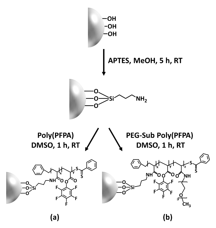
Figure 1: Schematic for the preparation of poly(PFPA) grafted SiO2 beads using APTES as a linker molecule. (a) Poly(PFPA) grafted beads. (b) Partially PEG-substituted poly(PFPA) grafted beads.
to:
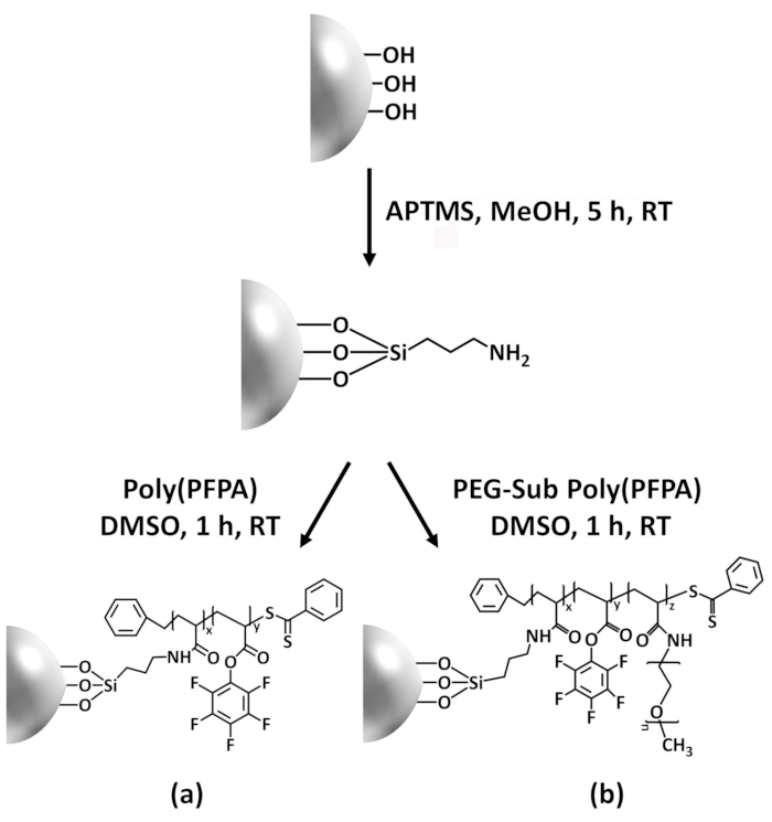
Figure 1: Schematic for the preparation of poly(PFPA) grafted SiO2 beads using APTMS as a linker molecule. (a) Poly(PFPA) grafted beads. (b) Partially PEG-substituted poly(PFPA) grafted beads.
Figure 2 was updated from:
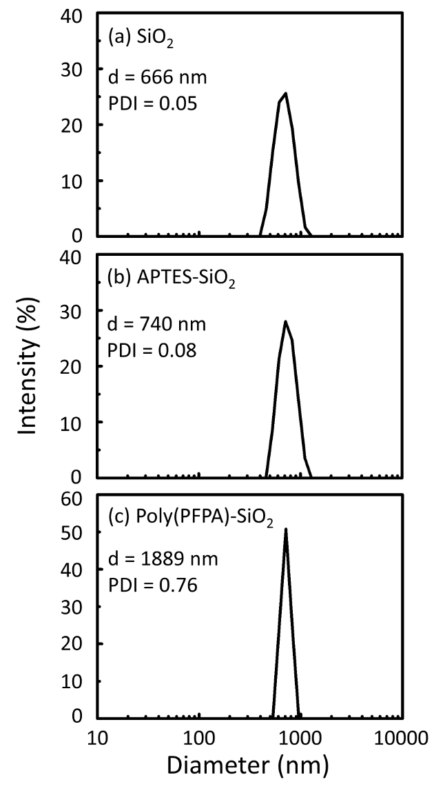
Figure 2: DLS measurements for (a) bare SiO2 beads (SiO2), (b) APTES functionalized SiO2 beads (APTES-SiO2), and (c) poly(PFPA) grafted SiO2 beads (poly(PFPA)-SiO2), dispersed in DMSO. The Z-average diameter (d) and polydispersity index (PDI) of each sample are reported.
to:
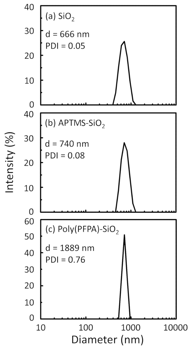
Figure 2: DLS measurements for (a) bare SiO2 beads (SiO2), (b) APTMS functionalized SiO2 beads (APTMS-SiO2), and (c) poly(PFPA) grafted SiO2 beads (poly(PFPA)-SiO2), dispersed in DMSO. The Z-average diameter (d) and polydispersity index (PDI) of each sample are reported.
Figure 3 was updated from:
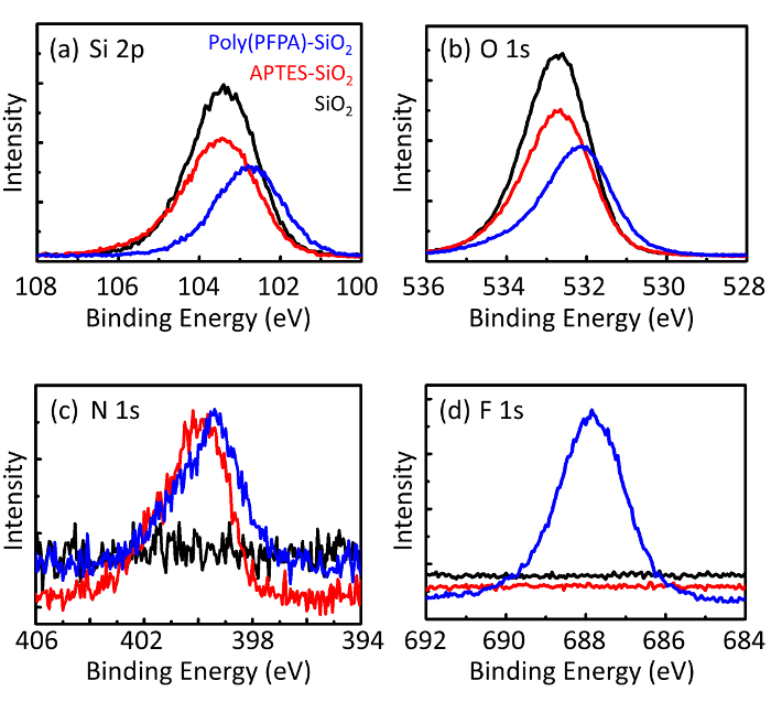
Figure 3: XPS spectra for bare SiO2 beads (SiO2), APTES functionalized SiO2 beads (APTES-SiO2), and poly(PFPA) grafted SiO2 beads (poly(PFPA)-SiO2). The peaks examined correspond to (a) Si 2p, (b) O 1s, (c) N 1s, and (d) F 1s.
to:
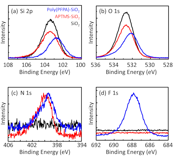
Figure 3: XPS spectra for bare SiO2 beads (SiO2), APTMS functionalized SiO2 beads (APTMS-SiO2), and poly(PFPA) grafted SiO2 beads (poly(PFPA)-SiO2). The peaks examined correspond to (a) Si 2p, (b) O 1s, (c) N 1s, and (d) F 1s.
The first and second paragraphs of the Discussion were updated from:
The synthesis of poly(PFPA) grafted SiO2 beads is illustrated in Figure 1. By employing APTES as a linker molecule, poly(PFPA) brushes covalently grafted to SiO2 substrate can be prepared via a simple two-step process. Although some of the PFP units are sacrificed for the reaction with APTES, a large number of the PFP units are expected to remain available for later reaction with either amino-PEG or antibodies. The PFP groups are known to form low energy surfaces so poly(PFPA) brushes do not solvate well in water28. For IP application, the antibodies need to be immobilized on the poly(PFPA) brushes, and this exchange reaction is done in aqueous buffer solution in order to preserve the activity of the antibodies. As reported in our previous publication, partial substitution of the PFP units with hydrophilic molecules such as amine-functionalized PEG can improve surface hydrophilicity, leading to increased antibody immobilization efficiency18. In this study, partially PEG substituted poly(PFPA) is also prepared, then grafted to the SiO2 surface using the same APTES linker molecule. Overall, the methods illustrated in Figure 1 allow the preparation of poly(PFPA) grafted surfaces with different degrees of PEG substitution. These polymer brushes with tunable surface properties provide an ideal platform for antibody immobilization and subsequent IP application.
The bead preparation process is monitored by both DLS and XPS. The DLS results for various functionalized SiO2 beads in DMSO are summarized in Figure 2. The bare SiO2 beads exhibit hydrodynamic diameter of 666 nm, in agreement with the manufacturer reported bead size (0.676 μm; SD = 0.03 μm). After APTES treatment, the bead diameter increases to 740 nm; and with poly(PFPA) treatment, the bead diameter further increases to 1889 nm. It is important to point out that the polydispersity index (PDI) for the poly(PFPA) grafted beads is rather large (PDI = 0.76), which is indicative of poor quality sample containing large aggregates. Although the DLS curve only shows one nano-sized peak, small amount of aggregates may be present in the suspension. The functionalized SiO2 beads are also examined by XPS to determine surface composition (Figure 3). Following APTES treatment, N 1s peak associated with the amine groups on APTES is detected. And, following poly(PFPA) treatment, F 1s peak associated with the PFP units on the polymer is detected. Together these data show the successful functionalization of the SiO2 surface, first with APTES, then with poly(PFPA).
to:
The synthesis of poly(PFPA) grafted SiO2 beads is illustrated in Figure 1. By employing APTMS as a linker molecule, poly(PFPA) brushes covalently grafted to SiO2 substrate can be prepared via a simple two-step process. Although some of the PFP units are sacrificed for the reaction with APTMS, a large number of the PFP units are expected to remain available for later reaction with either amino-PEG or antibodies. The PFP groups are known to form low energy surfaces so poly(PFPA) brushes do not solvate well in water28. For IP application, the antibodies need to be immobilized on the poly(PFPA) brushes, and this exchange reaction is done in aqueous buffer solution in order to preserve the activity of the antibodies. As reported in our previous publication, partial substitution of the PFP units with hydrophilic molecules such as amine-functionalized PEG can improve surface hydrophilicity, leading to increased antibody immobilization efficiency18. In this study, partially PEG substituted poly(PFPA) is also prepared, then grafted to the SiO2 surface using the same APTMS linker molecule. Overall, the methods illustrated in Figure 1 allow the preparation of poly(PFPA) grafted surfaces with different degrees of PEG substitution. These polymer brushes with tunable surface properties provide an ideal platform for antibody immobilization and subsequent IP application.
The bead preparation process is monitored by both DLS and XPS. The DLS results for various functionalized SiO2 beads in DMSO are summarized in Figure 2. The bare SiO2 beads exhibit hydrodynamic diameter of 666 nm, in agreement with the manufacturer reported bead size (0.676 μm; SD = 0.03 μm). After APTMS treatment, the bead diameter increases to 740 nm; and with poly(PFPA) treatment, the bead diameter further increases to 1889 nm. It is important to point out that the polydispersity index (PDI) for the poly(PFPA) grafted beads is rather large (PDI = 0.76), which is indicative of poor quality sample containing large aggregates. Although the DLS curve only shows one nano-sized peak, small amount of aggregates may be present in the suspension. The functionalized SiO2 beads are also examined by XPS to determine surface composition (Figure 3). Following APTMS treatment, N 1s peak associated with the amine groups on APTMS is detected. And, following poly(PFPA) treatment, F 1s peak associated with the PFP units on the polymer is detected. Together these data show the successful functionalization of the SiO2 surface, first with APTMS, then with poly(PFPA).
