无细胞表达系统是用于重要蛋白质高通量合成与筛选的高效且经济的工具。本文介绍了利用Vibrio natriegens制备无细胞蛋白质表达系统的方法,可用于以质粒DNA、线性DNA和mRNA为模板快速生产蛋白质。
方法文章
利用快速生长的细菌 Vibrio natriegens 进行无细胞蛋白表达
* These authors contributed equally
本文内容
摘要
摘要
海洋细菌Vibrio natriegens因其快速生长的特性,作为新兴的微生物宿主在生物技术领域受到广泛关注。本文描述了一种使用常规实验室设备制备V. natriegens粗提细胞提取物的通用方案。该方案产率高,且特别优化以提高用户的可操作性并降低成本。无细胞蛋白合成(CFPS)可在96孔或384孔板中进行小规模(10 μL)批次反应,3小时内可重复地获得浓度超过> 260 μg/mL的超折叠绿色荧光蛋白(sfGFP)。总体而言,单个操作者可在1−2天内完成粗提细胞提取物的制备及无细胞蛋白合成。该方案可轻松整合到现有的蛋白合成流程中,从而推动生物制造和合成生物学应用的发展。
引言
无细胞蛋白质合成是一种多功能且经济高效的方法,用于表达有价值的蛋白质或肽1,2,3,4。传统上,无细胞蛋白质合成采用Escherichia coli表达系统进行;然而,近年来越来越多的研究开始利用具有新颖特性的非模式生物作为无细胞表达的底盘系统5,6,7,8,9,10,11,12。具有独特代谢特征的生物是替代E. coli无细胞系统的理想候选者。例如,海洋细菌Vibrio natriegens是目前已知生长最快的生物之一,其倍增时间少于10分钟13。这一特性使V. natriegens在科研与生物技术领域作为新兴的微生物宿主受到广泛关注14,15,16,17,18。鉴于V. natriegens的快速生长速率与其高效的蛋白质合成能力和代谢效率密切相关19,20,21,利用其细胞机制进行无细胞合成有望显著拓展快速蛋白质生产与高通量筛选的技术工具箱。
最近已证明一种无细胞的 V. natriegens 表达系统能够利用T7启动子在3小时内产生浓度超过> 260 μg/mL的超折叠绿色荧光蛋白(sfGFP)9。开发该方法的总体目标是为用户提供一种高度易得、成本效益高、可重复且产率高的无细胞蛋白表达系统,该系统可在短时间内使用常规实验室设备制备。本方案采用1 L摇瓶培养、脉冲式超声破碎细胞裂解,以及在96孔或384孔板中进行小规模批次反应,以实现最大程度的并行化和高通量筛选。通过添加3-磷酸甘油酸(3-PGA)作为能量来源,可实现长时间持续的蛋白质表达8,22,23。成功完成本方案后,用户将能够利用V. natriegens粗细胞提取物以无细胞形式表达目标蛋白或一组蛋白。
从甘油保存菌种开始,收集光密度在 600 nm 处(OD600)为 1.0 的 V. natriegens 菌体,制备粗细胞提取物。1 L 培养液可获得约 2−3 mL 提取物,足以进行超过 800 次含 25% 粗细胞提取物的无细胞反应。可使用质粒 DNA、线性 DNA 或 mRNA 模板表达蛋白质;然而,在使用野生型 V. natriegens 无细胞体系时,内源核酸酶对线性 DNA 模板的降解仍是主要缺点9。从 V. natriegens 培养物开始,单个操作者可在 1−2 天内完成可用于下游应用的蛋白质制备。
方案
1. V. natriegens 粗提细胞提取物的制备——细菌培养
- 根据表1配制V. natriegens细菌培养用LB-V2培养基。通过高压灭菌法对培养基进行灭菌处理,待其冷却至室温(RT)。多余的培养基储存于室温下。
注意:操作高压灭菌器时,请穿戴适当的个人防护装备(PPE),并查阅实验室特定的操作说明。 - 使用野生型V. natriegens甘油菌种接种3 mL LB-V2培养基,于30 °C、225 rpm振荡条件下过夜培养。
- 取1 mL过夜培养物,在台式离心机上以10,600 × g离心1分钟进行洗涤。小心吸除上清液,避免扰动形成的菌体沉淀,并将沉淀重悬于1 mL新鲜LB-V2培养基中。
- 在已灭菌的4 L带挡板锥形瓶(带无菌封口)中加入1 L新鲜LB-V2培养基,用1 mL洗涤后的过夜培养物接种(稀释比例为1:1,000),于30 °C、225 rpm振荡条件下培养。
注意:V. natriegens培养体系可按比例放大或缩小,同时保持1:1,000的稀释比例。例如,将250 μL洗涤后的过夜培养物加入装有250 mL新鲜LB-V2培养基的2 L带挡板锥形瓶(带无菌封口)中。 - 使用分光光度计监测培养物在600 nm处的光密度(OD600)。当OD600达到1.0 ± 0.2时,在4 °C条件下以3,500 × g离心20分钟收集菌体,并将菌体沉淀置于冰上。
注意:V. natriegens生长迅速,因此需密切监测培养过程。在1:1,000稀释比例下,培养至OD600 = 1.0大约需要1.5–2小时。 - 吸除上清液后,立即将所得细菌沉淀储存在-80 °C,或直接进入第2节所述的细胞裂解步骤。
注意:此步骤适合暂停操作;但为获得最佳效果,建议尽快处理沉淀,或在1–2天内完成后续操作。
2. V. natriegens 粗提细胞提取物的制备——细胞裂解
- 按如下方法配制 S30 裂解缓冲液 表1 在无菌去离子水(DI 水)中溶解,并用冰醋酸调节 pH 至 7.7。
警告: 调节pH时应使用适当的个人防护装备(PPE)处理冰醋酸。 - 将S30裂解缓冲液冷却至约4 °在开始细胞裂解程序之前,将样品置于冰箱或冰上保存。
注意: 为快速降温,可将裂解缓冲液置于 -20 °C. 不要让缓冲液结冰。 - 将细胞沉淀置于冰上 10−20 分钟,或直至完全解冻。使用 10 mL 冷的 S30 裂解缓冲液重悬来自相同 1 L 培养物的所有沉淀,然后将悬液转移至 50 mL 离心管中。若沉淀未在 −80 °C 冰箱中冷冻保存 °在步骤1.6中,直接进行重悬。
注意: 根据需要增加用于初始重悬沉淀的 S30 裂解缓冲液体积。 - 3,500 x g 离心悬浮液 g 4℃下离心10分钟 °C. 吸出上清液,注意不要扰动沉淀。用10 mL预冷的S30裂解缓冲液将沉淀再洗涤一次。将沉淀置于冰上。
- 在低温室中,加入500 μ将冷的S30裂解缓冲液加入50 mL离心管中的沉淀中。使用宽口移液器吸头重悬沉淀,并小心地将全部沉淀悬液转移至2 mL离心管中。
注意: 若无宽口移液器,可使用剪刀剪去1 mL移液枪头末端以增大孔径,便于沉淀转移。- 尽可能转移大部分沉淀,同时不显著增加体积;但应使用足量液体重悬沉淀,以便进行超声处理,以2 mL离心管中形成均一悬液为准。切勿过度填充2 mL离心管,悬液体积不应超过1.5 mL;若必要,可分装至多个离心管中。
- 将细胞沉淀置于冰上,并在冷室中操作。取一个600 mL烧杯装满冰,将2 mL离心管架放在冰上。.
注意: 将试管架放置在烧杯的一侧有助于观察冰中沉淀重悬的情况,以便监测超声处理的进程(参见 图1). - 在2 mL离心管中短暂涡旋悬浮菌体沉淀以混匀细胞,轻弹管壁去除管盖底部附着的细胞,随后将离心管放入管架中并打开管盖。将超声破碎仪探头插入菌液悬浮液中,使其刚好位于液面下方。
- 按照图示准备超声破碎装置 图2 使用超声波破碎仪和探头 ⅛英寸尖端直径。将以下参数输入超声波破碎仪控制器:20 kHz 频率,50% 振幅,脉冲开启时间:10 s,脉冲关闭时间:60 s。
- 运行脉冲超声处理程序,共三个循环。如果菌体沉淀悬浮液的总体积为 > 500 μL,脉冲超声处理程序运行六次。
注意: 通常,3−6 次脉冲足以裂解 V. natriegens 沉淀;根据超声破碎仪的不同,可能需要额外的脉冲处理。在超声过程中,使用平台上的调节旋钮上下移动探头,以裂解可能沉降到管底的沉淀物。可在脉冲间隔将离心管从支架中取出,短暂涡旋以重新均质化悬浮液。关于超声后细胞的理想均一程度,请参见下文讨论。
注意: 超声波破碎仪工作时,请佩戴适当的听力保护装置。 - 超声处理后,将粗提细胞提取物在16,000 x g 4°C 下 30−45 分钟 °C,或直至裂解物中无任何细胞碎片,如图所示 图3.
- 在低温室中,分装 50 μ将所得上清液转移至新的2 mL离心管中,注意勿扰动沉淀。
注意: 任何意外转移至新2 mL离心管中的碎片都会显著降低提取物高效合成蛋白质的能力。- 可选步骤:留出 10 μ在另一支2 mL离心管中留取细胞裂解后用于蛋白质定量的细胞提取物(见下文步骤2.13)。
- 通过将试管放入带有浸入式绳索的试管架中,如图所示,快速冷冻粗制细胞提取物 图4将离心管浸入装有液氮的杜瓦瓶中,随即转移至-80℃超低温冰箱中冷冻 °直至使用前均置于 C 保存。
警告: 使用液氮时应穿戴适当的个人防护装备,包括实验服、护目镜、面罩、防冻围裙和手套。 - 可选地,使用10 μ将步骤 2.11.1 中保留的 L 样品用 1× 磷酸盐缓冲液(PBS)按 1:100 比例稀释,并采用任何标准的总蛋白定量检测方法(如 Bradford 法、二辛可宁酸法)进行测定 测定(BCA)等
3. 无细胞反应组分的制备
- 通用反应组分
- 使用无菌去离子水在50 mL离心管中分别配制浓度为100 mM和2000 mM的Mg-谷氨酸盐和K-谷氨酸盐工作液。
- 通过向250 mL烧杯中加入100 mL无菌去离子水来配制50% (w/v) 聚乙二醇(PEG)-8000工作液。将一个小磁力搅拌子放入烧杯中。称取50 g PEG-8000并加入烧杯中的100 mL水中。
- 将烧杯置于设定为100 °C的加热磁力搅拌器上,以250 rpm搅拌直至PEG-8000完全溶解。待PEG-8000溶液冷却后再转移至50 mL离心管中。
- 能量溶液主混合液
- 通过将140 g KOH颗粒加入500 mL无菌去离子水中配制5 M KOH溶液。
注意:该溶液应在化学通风橱中配制,操作强碱时需佩戴个人防护装备(PPE)。溶液会放热,因此瓶盖应略微松开以防止压力积聚。使用前需让KOH溶液恢复至室温。 - 通过将20.85 g HEPES加入100 mL瓶中配制1750 mM HEPES-KOH缓冲液。缓慢加入无菌去离子水至体积达40 mL。涡旋振荡使HEPES溶解。使用5 M KOH溶液调节pH至8.0,然后加水定容至50 mL。
注意:调节pH时应佩戴适当的个人防护装备操作KOH。 - 按照表2所示浓度,用无菌去离子水配制其余10倍能量主混合液储备液组分,并将每种储备液置于冰上。在室温下解冻100 mM ATP、GTP、CTP和UTP储备液,随后置于冰上。
注意:如有需要,可使用设定为37 °C和350 rpm的温控混匀仪溶解试剂。切勿过热或将试剂长时间置于温控混匀仪上。 - 在15 mL离心管中,按照表2指定的顺序和体积加入各能量溶液主混合液组分。每加入一种组分后均需涡旋振荡混合。此步骤可制备5 mL的10倍能量溶液主混合液。
- 将10倍能量溶液主混合液分装至2 mL离心管中,每管200 μL。按照步骤2.12的方法对每份分装液进行速冻。立即放入-80 °C冰箱中保存备用。
- 通过将140 g KOH颗粒加入500 mL无菌去离子水中配制5 M KOH溶液。
- 氨基酸主混合液
- 为配制新鲜的4倍氨基酸主混合液,首先在室温下解冻各氨基酸储备液,然后置于冰上。使用涡旋振荡器和/或设定为37 °C和350 rpm的温控混匀仪,确保所有氨基酸储备液完全溶解。
注意:半胱氨酸可能无法完全溶解;可将其以悬浮液形式加入氨基酸主混合液中。切勿过热或将试剂长时间置于温控混匀仪上。 - 在15 mL离心管中,按以下顺序向无菌去离子水中加入适量体积的氨基酸,使每种氨基酸的终浓度为8 mM:ALA、ARG、ASN、ASP、GLN、GLU、GLY、HIS、IIE、LYS、MET、PHE、PRO、SER、THR、VAL、TRP、TYR、LEU和CYS。每加入一种氨基酸后均需涡旋振荡混合主混合液。表2所列体积可配制2.4 mL氨基酸主混合液。
- 将4倍氨基酸主混合液分装至2 mL离心管中,每管200 μL。按照步骤2.12的方法对每份分装液进行速冻。立即放入-80 °C冰箱中保存备用。
- 为配制新鲜的4倍氨基酸主混合液,首先在室温下解冻各氨基酸储备液,然后置于冰上。使用涡旋振荡器和/或设定为37 °C和350 rpm的温控混匀仪,确保所有氨基酸储备液完全溶解。
- 制备可用于反应的质粒DNA模板
注意:本系统的无细胞蛋白表达已使用超折叠绿色荧光蛋白(GFP)表达载体T7-pJL1-sfGFP(材料表)进行了优化。建议使用该质粒作为无细胞反应效率的对照,并使用pJL1骨架进行其他蛋白序列的克隆与表达。也可使用其他质粒DNA模板;但需注意,转录由T7启动子序列和T7 RNA聚合酶的存在所控制。以下描述了一种从转化的E. coli大规模制备任意质粒DNA模板的简单方案。- 按照生产商说明书使用质粒纯化试剂盒(材料表)纯化目标载体。
注意:建议尽可能浓缩质粒DNA模板,以满足无细胞反应严格的体积限制。通常,工作液浓度应达到750−1500 ng/μL。
- 按照生产商说明书使用质粒纯化试剂盒(材料表)纯化目标载体。
- 制备可用于反应的mRNA模板
注意:本节为可选步骤。已使用所列T7 RNA聚合酶(材料表)对质粒T7-pJL1-sfGFP进行体外转录生成mRNA模板,并测试了蛋白表达。- 使用表3所列的体外转录反应组分,从编码目标蛋白的质粒DNA制备mRNA模板。在PCR仪中37 °C孵育反应1小时。
注意:建议并行进行8−10次此类反应,以获得足够量的产物。 - 合并所有转录反应产物。按照生产商说明书使用mRNA纯化与浓缩试剂盒(材料表)进行纯化。用无菌去离子水洗脱mRNA模板。将mRNA模板储存于-80 °C备用。
- 使用表3所列的体外转录反应组分,从编码目标蛋白的质粒DNA制备mRNA模板。在PCR仪中37 °C孵育反应1小时。
4. 使用 V. natriegens 粗提物进行无细胞蛋白表达反应
- 使用质粒或线性DNA模板进行无细胞蛋白表达
- 从-80 °C冰箱中取出10x能量溶液主混合液和4x氨基酸主混合液的分装样品,在室温下解冻后置于冰上。从-20 °C冰箱中取出T7 RNA聚合酶和RNase抑制剂储备液并置于冰上。将DNA模板在室温下解冻后置于冰上。
- 按照表4所示,在冰上的2 mL离心管中依次加入以下组分,配制无细胞反应主混合液:氨基酸主混合液、能量溶液主混合液、Mg-谷氨酸盐、K-谷氨酸盐、DNA模板、PEG-8000、T7 RNA聚合酶和RNase抑制剂。每次加入后轻轻弹击离心管以混匀。
注意:若使用线性模板进行无细胞蛋白表达,与质粒模板相比,需增加5−10倍的模板用量,以获得可观的蛋白产量。 - 从-80 °C冰箱中取出步骤2.12制备的V. natriegens粗细胞裂解液,置于冰上10−20分钟直至完全解冻。根据表4加入相应体积的粗细胞提取物至无细胞反应主混合液中,并通过轻弹或吹打方式温和混匀。
- 使用PCR仪进行终点法无细胞蛋白表达
- 将10 μL无细胞反应主混合液移至96孔或384孔PCR板的孔底。每次向PCR板转移后,均需轻轻弹击主混合液管以充分混匀。
注意:无细胞反应主混合液应始终保持充分混匀,以确保所有样品的反应重复性和蛋白表达水平最大化。
- 将10 μL无细胞反应主混合液移至96孔或384孔PCR板的孔底。每次向PCR板转移后,均需轻轻弹击主混合液管以充分混匀。
- 将PCR板短暂离心(1,000 x g,10秒),使附着在孔壁上的液体沉至孔底。用板密封膜封住各孔以防止蒸发,然后将PCR板放入预设温度为26 °C、加热盖温度为105 °C的PCR仪中。
注意:PCR仪均匀的热分布及加热盖可显著提高蛋白表达产量。 - 将无细胞反应体系孵育至少3小时。孵育结束后,可对表达的蛋白进行纯化、定量,并用于后续实验。
注意:表达的蛋白可通过用户选择的方法直接在无细胞反应体系中定量。例如,荧光蛋白可通过外标曲线法进行定量,或在无细胞反应中使用放射性标记氨基酸时测定放射活性。通常不建议在未经过初步纯化的情况下,直接使用紫外-可见光谱法或总蛋白检测试剂盒测量无细胞反应中的蛋白产量。 - 或者,使用酶标仪监测无细胞蛋白表达动力学
- 将10 μL无细胞反应主混合液移至底部为透明玻璃的黑色384孔检测板的孔底。在添加主混合液过程中,保持检测板置于冰上或在低温环境中操作,以确保获得完整的动力学曲线。用透明板密封膜封住各孔以防止蒸发,然后将检测板放入设定温度为26 °C的酶标仪中。
- 在3−6小时内持续孵育无细胞反应体系,同时监测与目标表达蛋白相对应的适当荧光激发/发射波长。例如,sfGFP的检测波长为Ex/Em = 485 nm/528 nm。
- 使用mRNA模板进行无细胞蛋白表达
- 将步骤3.5.2制备的mRNA模板在室温下解冻后置于冰上。
- 参照前述针对线性或质粒DNA模板的操作,执行步骤4.1.1至步骤4.1.6,并依据表4配制适用于mRNA模板的另一种无细胞反应主混合液。
5. 使用 sfGFP 对 V. natriegens 无细胞反应进行校准
注意:本节为可选内容。由于细胞裂解条件的不同,每次制备的粗提物在无细胞反应中的最佳离子浓度可能略有差异。如果反应产量显著低于预期(浓度 < 1.0 μg/mL),建议使用sfGFP进行下述可选的无细胞反应离子校准实验。
- 根据步骤 3.1.1 中制备的 100 mM Mg-谷氨酸盐和 2,000 mM K-谷氨酸盐储备液,在无菌去离子水中配制 表 5 所示的 Mg2+ 和 K+ 离子校准溶液。将校准溶液置于冰上,待用。
- 按照表 6 中的校准图,用移液器将 1 μL Mg-谷氨酸盐和 2 μL K-谷氨酸盐离子校准溶液加入 384 孔 PCR 板的相应孔中。
- 本步骤的操作顺序如下:先将 Mg-谷氨酸盐离子校准溶液加入各孔底部,再将 K-谷氨酸盐离子校准溶液沿孔壁加入,避免接触孔中已存在的液体。
- 在准备校准无细胞反应主混合液期间,用粘性封膜覆盖板面以防止蒸发。轻轻敲击 384 孔板以混匀 Mg-和 K-谷氨酸盐校准溶液。
注意: 强烈建议使用多通道重复移液器以确保该步骤的可重复性和操作速度。
- 在 2 mL 离心管中,按照 表 5 所示配方,使用 T7-pJL1-sfGFP 质粒 DNA 模板配制校准用无细胞反应主混合液。按以下顺序依次加入各组分:氨基酸主混合液、能量溶液主混合液、T7-pJL1-sfGFP DNA 模板、PEG-8000、T7 RNA 聚合酶和 RNase 抑制剂。每加入一种组分后,轻轻弹击管壁以混匀。
注意: 校准用无细胞主混合液中不得添加 Mg2+ 或 K+。 - 移去板上的封膜。小心地将 7 μL 校准用无细胞反应主混合液沿孔壁加入,避免接触孔中已混合的离子校准溶液。重新用封膜密封各孔,并轻轻敲击 384 孔 PCR 板,使无细胞主混合液与离子校准溶液充分混匀。
注意: 强烈建议使用多通道重复移液器以确保该步骤的可重复性和操作速度。 - 将板短暂离心(1,000 × g,10 秒),使可能附着在孔壁上的液体沉至孔底。将 384 孔板放入设定为 26 °C、热盖温度为 105 °C 的 PCR 仪中。
- 孵育无细胞反应体系 3 小时。孵育结束后,使用多通道移液器将各孔内容物转移至底部为玻璃材质的黑色 384 孔检测板中,用酶标仪在激发/发射波长 485 nm/528 nm 条件下测定 sfGFP 荧光强度,以确定产生蛋白量最高的离子浓度组合。
结果
本方案描述了利用V. natriegens无细胞表达系统进行蛋白质生产的操作流程,单个操作者从培养物接种到获得可用于下游应用的蛋白质,可在1−2天内完成。粗提细胞提取物和主混合物储备液的制备占用了其中大部分时间;然而,一旦制备完成,大多数大量试剂可长期储存(Table 7),并按需使用,从而缩短完成该实验流程所需的时间。
所述表达系统最适合使用质粒DNA模板或通过体外转录反应生成的mRNA。尽管线性DNA也可用作蛋白质生产的模板,但其产生的蛋白量显著较低。例如,在26℃的最佳反应条件下 °C,单个 10 μL 无细胞反应可产生 > 260 μg/mL 使用0.3 pmol质粒DNA模板或相当量的sfGFP在3小时内 > 125 μg/mL 使用14 pmol的mRNA转录本对sfGFP进行图5A,B)。然而,0.3 pmol 的线性 DNA 产生的蛋白质量将显著减少(< 20 μg/mL)。对于每种模板类型,大部分蛋白质将在1−1.5小时内生成;然而,建议反应时间至少持续3小时。通过使用纯化的sfGFP标准品曲线,在激发/发射波长Ex/Em = 485 nm/528 nm下测量荧光,利用线性回归确定无细胞反应中sfGFP的浓度。
蛋白质的产率显著受到钾离子和镁离子(K+ 和 Mg2+)浓度的影响。在优化条件下,发现最佳的 Mg2+ 和 K+ 离子浓度分别为 3.5 mM 和 80 mM(图 6A,B)。偏离最佳离子浓度可能导致无细胞表达系统产生蛋白质的能力下降,从而降低产量。如果反应产率显著低于预期,可能需要进行额外的校准。第 5 节描述了一种可选的离子校准方案,可用于补偿因不同细胞裂解设备和条件导致的粗提细胞提取物的差异性。
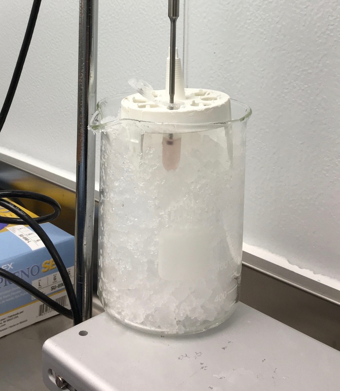
图1:超声平台上的烧杯和试管架的特写图。 请点击此处查看该图的放大版本。
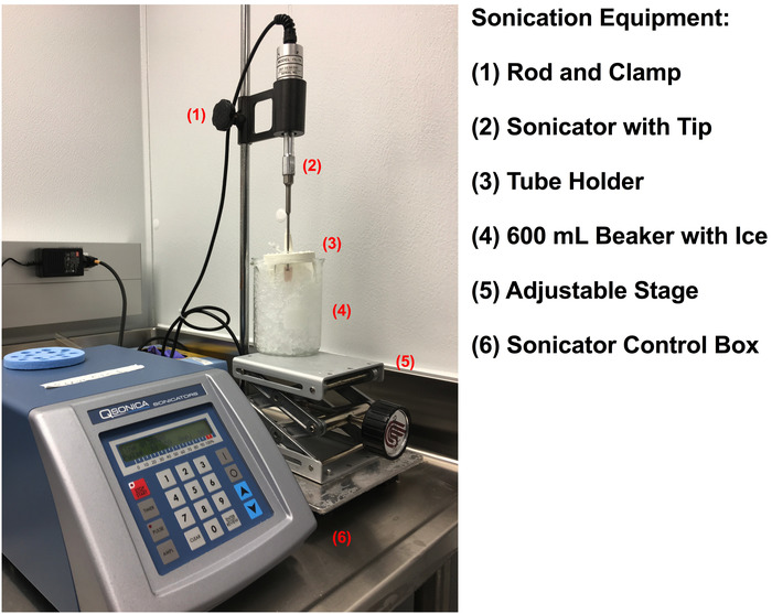
图2:制备粗制细胞提取物的超声破碎设备装置。 请点击此处查看该图的放大版本。
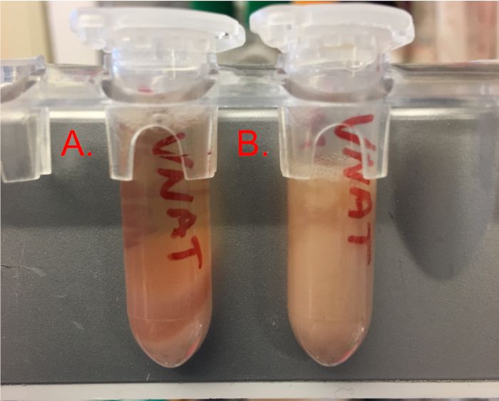
图3:超声处理和离心后沉淀物的视图。 请点击此处查看此图的放大版本。
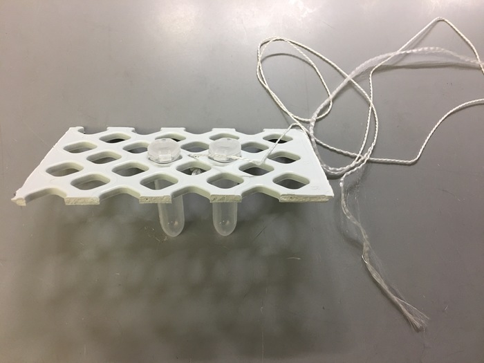
图 4:用于提取物储存的快速冷冻浸渍装置。 请点击此处查看该图的放大版本。
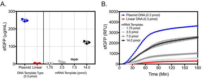
图5:使用不同类型模板的V. natriegens无细胞蛋白质合成的代表性结果。 (A)使用等摩尔浓度的质粒和线性DNA模板(0.3 pmol)以及递增浓度的mRNA模板进行sfGFP生成的终点检测。在26 °C、10 μL反应体积、最佳反应条件下孵育180分钟后,通过在Ex/Em = 485 nm/528 nm处测定纯化sfGFP的标准曲线,确定无细胞体系中sfGFP的浓度。(B)使用等摩尔浓度的质粒和线性DNA模板以及递增浓度的mRNA模板进行sfGFP生成的动力学检测。在总孵育时间180分钟内,每3分钟在Ex/Em = 485 nm/528 nm处测定一次sfGFP,反应条件同上,体积为10 μL。终点和动力学检测均使用除模板外其余组分完整的无细胞反应作为空白对照进行校正。结果显示为平均值及标准偏差(n = 3)。请点击此处查看该图的放大版本。
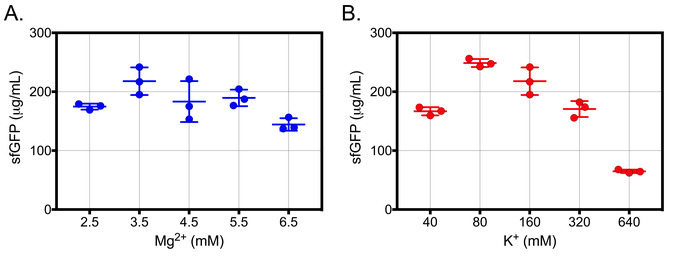
图6:离子浓度校准的代表性结果。 (A) V. natriegens 无细胞反应中添加了浓度递增的 Mg2+ 并在以下条件下孵育: 26 °C 180 分钟 10 μL 反应。K的总浓度+ 为所有 Mg 的 160 mM2+ 浓度。无细胞 sfGFP 浓度通过在激发/发射波长 485 nm/528 nm 处测定的纯化 sfGFP 标准曲线确定。B) V. natriegens 无细胞反应中添加了浓度递增的 K+ 并在以下条件下孵育: 26 °C 180 分钟 10 μL 反应。Mg的总浓度2+ 对所有 K 均为 3.5 mM+ 浓度。两次校准中,均使用不含模板但包含所有其他组分的无细胞反应作为空白对照对样品进行校正。图中显示了均值和标准偏差(n = 3)。该图改编自 Wiegand 等人。9. 经许可转载自 Wiegand, D.J., Lee, H.H., Ostrov, N., Church, G.M. 建立无细胞的 Vibrio natriegens 表达系统。 ACS合成生物学. 7 (10), 2475−2479 (2018)。版权所有 2018 美国化学会。 请点击此处以查看此图的放大版本。
| 表 1 A | LB-V2 细菌培养基的制备 | ||
| 组分 | 用量 (g) | 终浓度 (mM) | 终体积 (L) |
| LB Broth (Miller) | 25 | 1 | |
| NaCl | 11.69 | 200 | |
| MgCl2 | 2.20 | 23.1 | |
| KCl | 0.31 | 4.2 | |
| 表 1 B | S30A 裂解缓冲液的制备 | ||
| 组分 | 用量 (g) | 终浓度 (mM) | 终体积 (L) |
| Tris 溶液 (pH 8.0) - 1 M (mL)* | 25 | 50 | 0.5 |
| Mg-glutamate | 2.72 | 14 | |
| K-glutamate | 6.10 | 60 | |
| 二硫苏糖醇 (DTT) - 1 M (mL)* | 1 | 2 | |
表1:制备1 L LB-V2细菌培养基和0.5 L S30A细胞裂解缓冲液的试剂。请点击此处下载Excel文件。
| 10x 能量溶液母液的配制 | ||||
| 组分 | 储备浓度 (mM) | 终浓度 (mM) | 体积 (µL) | 终体积 (µL) |
| HEPES-KOH pH 8 | 1750 | 500 | 1428.57 | 5000 |
| ATP | 100 | 15 | 750.00 | |
| GTP | 100 | 15 | 750.00 | |
| CTP | 100 | 9 | 450.00 | |
| UTP | 100 | 9 | 450.00 | |
| 来自E. coli MRE 600 的 tRNA (mg/mL)* | 100 | 2 | 100.00 | |
| 辅酶 A 水合物 | 200 | 2.6 | 65.00 | |
| NAD | 200 | 3.3 | 82.50 | |
| cAMP | 650 | 7.5 | 57.69 | |
| 亚叶酸 | 100 | 0.7 | 35.00 | |
| 亚精胺 | 1600 | 10 | 31.25 | |
| 3-PGA | 2000 | 300 | 750.00 | |
| 无菌去离子水 | 49.99 | |||
| 4x 氨基酸母液的配制 | ||||
| 组分 | 储备浓度 (mM) | 终浓度 (mM) | 体积 (µL) | 终体积 (µL) |
| ALA | 168 | 8 | 114.3 | 2400 |
| ARG | 168 | 8 | 114.3 | |
| ASN | 168 | 8 | 114.3 | |
| ASP | 168 | 8 | 114.3 | |
| GLN | 168 | 8 | 114.3 | |
| GLU | 168 | 8 | 114.3 | |
| GLY | 168 | 8 | 114.3 | |
| HIS | 168 | 8 | 114.3 | |
| IIE | 168 | 8 | 114.3 | |
| LYS | 168 | 8 | 114.3 | |
| MET | 168 | 8 | 114.3 | |
| PHE | 168 | 8 | 114.3 | |
| PRO | 168 | 8 | 114.3 | |
| SER | 168 | 8 | 114.3 | |
| THR | 168 | 8 | 114.3 | |
| VAL | 168 | 8 | 114.3 | |
| TRP | 168 | 8 | 114.3 | |
| TYR | 168 | 8 | 114.3 | |
| LEU | 140 | 8 | 137.1 | |
| CYS | 168 | 8 | 114.3 | |
| 无菌去离子水 | 91.4 | |||
表2:配制5 mL 10x能量溶液母液和2.4 mL 4x氨基酸母液所需的试剂。 请点击此处下载Excel文件。
| T7 RNA 聚合酶体外转录反应 | ||||
| 组分 | 储存液浓度 (mM) | 终浓度 (mM) | 单次反应体积 (µL) | 50 次反应总体积 (µL) |
| 10x RNAPol 反应缓冲液 | 1.00 | 50 | ||
| ATP | 100 | 0.5 | 0.10 | 5 |
| GTP | 100 | 0.5 | 0.10 | 5 |
| CTP | 100 | 0.5 | 0.10 | 5 |
| UTP | 100 | 0.5 | 0.10 | 5 |
| DNA 模板 (ng/µL)* | 1000 | 500 | 0.50 | 25 |
| T7 RNA 聚合酶 | 200 | 2.6 | 2.00 | 100 |
| 小鼠来源 RNase 抑制剂 | 200 | 3.3 | 0.50 | 25 |
| 无菌去离子水 | 15.60 | 780 | ||
| 反应体系总体积 (µL): | 20 | |||
表3:体外转录生成mRNA的组分。 请点击此处下载该Excel文件。
| 无细胞反应主混合液 | ||||
| 组分 | 储存浓度 (mM) | 终浓度 (mM) | 单次反应用量 (µL) | 50次反应用量 (µL) |
| 提取物 (%) * | 25 | 2.50 | 125.00 | |
| Mg-谷氨酸盐 | 100 | 3.5 | 0.35 | 17.50 |
| K-谷氨酸盐 | 2000 | 80 | 0.40 | 20.00 |
| 4x 氨基酸主混合液 | 8.0 | 2 | 2.50 | 125.00 |
| 10x 能量溶液主混合液 | 1.00 | 50.00 | ||
| 质粒DNA (ng/µL) * | 1000 | 500 | 0.50 | 25.00 |
| 50% PEG-8000 (%) * | 50 | 2 | 0.40 | 20.00 |
| T7 RNA聚合酶 | 1.00 | 50.00 | ||
| 鼠源RNase抑制剂 | 0.10 | 5.00 | ||
| 无菌去离子水 | 1.25 | 62.50 | ||
| 反应体积 (µL): | 10 | |||
| 以mRNA为模板的无细胞反应主混合液(替代方案) | ||||
| 组分 | 储存浓度 (mM) | 终浓度 (mM) | 单次反应用量 (µL) | 50次反应用量 (µL) |
| 提取物 (%) * | 25 | 2.50 | 125.00 | |
| Mg-谷氨酸盐 | 100 | 3.5 | 0.35 | 17.50 |
| K-谷氨酸盐 | 2000 | 80 | 0.40 | 20.00 |
| 4x 氨基酸主混合液 | 8.0 | 2 | 2.50 | 125.00 |
| 10x 能量溶液主混合液 | 1.00 | 50.00 | ||
| mRNA模板 (ng/µL) * | 2000 | 4000 | 2.00 | 100.00 |
| 50% PEG-8000 (%) * | 50 | 2 | 0.40 | 20.00 |
| 鼠源RNase抑制剂 | 0.10 | 5.00 | ||
| 无菌去离子水 | 0.75 | 37.50 | ||
| 反应体积 (µL): | 10 | |||
表4:用于DNA模板和mRNA模板的优化V. natriegens无细胞反应主混合液的组分。 请点击此处下载Excel格式文件。
| Mg2+ 校准 | 单次反应用量 (µL) | 单次反应用量 (µL) | 100 次反应用量 (µL) | 100 次反应用量 (µL) | |
| 终浓度 (mM) | 储备液 | 100 mM Mg-Glu | diH20 | 100 mM Mg-Glu | 去离子 H2O |
| 2.5 | 0 | 0.00 | 1.00 | 0 | 100 |
| 3.5 | 1 | 0.10 | 0.90 | 10 | 90 |
| 4.5 | 2 | 0.20 | 0.80 | 20 | 80 |
| 5.5 | 3 | 0.30 | 0.70 | 30 | 70 |
| 反应体积 (µL): | 10 | ||||
| K+ 校准 | 单次反应用量 (µL) | 单次反应用量 (µL) | 100 次反应用量 (µL) | 100 次反应用量 (µL) | |
| 终浓度 (mM) | 储备液 | 2000 mM K-Glu | diH20 | 2000 mM K-Glu | 去离子 H2O |
| 40 | 30 | 0.15 | 1.85 | 12 | 148 |
| 80 | 70 | 0.35 | 1.65 | 28 | 132 |
| 160 | 150 | 0.75 | 1.25 | 60 | 100 |
| 320 | 310 | 1.55 | 0.45 | 124 | 36 |
| 反应体积 (µL): | 10 | ||||
| 离子校准无细胞反应主混合液 | |||||
| 组分 | 储备液浓度 (mM) | 终浓度 (mM) | 单次反应用量 (µL) | 50 次反应用量 (µL) | |
| 提取物 (%) * | 25 | 2.50 | 125.00 | ||
| Mg-谷氨酸盐 | 可变 | 可变 | 1.00 | 50.00 | |
| K-谷氨酸盐 | 可变 | 可变 | 2.00 | 100.00 | |
| 4x 氨基酸主混合液 | 8.0 | 2 | 2.50 | 125.00 | |
| 10x 能量溶液主混合液 | 1.00 | 50.00 | |||
| 质粒 DNA (ng/µL) * | 1000 | 250 | 0.25 | 12.50 | |
| 50% PEG-8000 (%) * | 50 | 2 | 0.40 | 20.00 | |
| T7 RNA 聚合酶 | 1.00 | 50.00 | |||
| 小鼠 RNase 抑制剂 | 0.10 | 5.00 | |||
| 无菌去离子水 | 0.00 | 0.00 | |||
| 反应体积 (µL): | 10 | ||||
表5:离子浓度校准母液混合物。 请点击此处以Excel格式下载该文件。
| CF 离子校准图谱 | 40 mM K+ | 80 mM K+ | 160 mM K+ | 320 mM K+ | |||||||||
| 1 | 2 | 3 | 4 | 5 | 6 | 7 | 8 | 9 | 10 | 11 | 12 | ||
| 2.5 mM Mg2+ | A | ||||||||||||
| 3.5 mM Mg2+ | B | ||||||||||||
| 4.5 mM Mg2+ | C | ||||||||||||
| 5.5 mM Mg2+ | D | ||||||||||||
表6:离子校准图。 请点击此处下载Excel格式文件。
| CF 组分的保存条件与保质期 | ||
| 组分 | 保存位置 | 保质期 |
| 无菌 LB-V2 培养基 | 4 °C | 3-6 个月 |
| V. natriegens 粗提细胞提取物 | -80 °C | 1-3 周 |
| 100 mM Mg-Glutamate | 室温 | 6 个月 |
| 2000 mM K-Glutamate | 室温 | 6 个月 |
| 50% PEG-8000 | 室温 | 6 个月 |
| 10x 能量溶液母液 | -80 °C | 3-6 个月 |
| 4x 氨基酸母液 | -80 °C | 3-6 个月 |
| 质粒/线性 DNA 模板 | -20 °C | 6-12 个月 |
| mRNA 模板 | -80 °C | 3-4 周 |
表7:无细胞物质的储存条件与保质期。 请点击此处下载Excel文件。
讨论
本方案已针对野生型 V. natriegens 及含有V2盐的LB培养基(材料表)进行了优化。其他 V. natriegens 菌株也可采用类似方法培养,以制备用于无细胞反应的粗细胞提取物;但使用这些菌株时需要对该方案进行进一步优化。此外,本无细胞蛋白表达系统已采用3-磷酸甘油酸(3-PGA)作为主要的能量再生来源进行优化。也可使用其他能量再生来源;但可能需要对试剂进行优化和校准,才能实现高效的蛋白表达10,11。
特别注意本方案中的几个关键步骤,可确保获得最大提取效率,从而实现高产率的无细胞蛋白质合成。首先,必须使用处于对数生长中期收获的 V. natriegens 细胞培养物制备粗提细胞提取物;当培养物的 OD600 达到 1.0 ± 0.2 时,蛋白质产量最高。尽管在不同光密度下收获的细胞均可用于无细胞蛋白质合成,但我们先前的研究发现,处于对数生长期的细胞所获得的蛋白质产量显著更高9。光密度对粗提细胞提取物性能的影响与在分批培养条件下生长的其他细胞来源的无细胞表达系统中报道的结果一致1。由于 V. natriegens 生长速度较快,必须密切监测培养物的光密度。通常情况下,采用本方案时,V. natriegens 培养物可在 1−1.5 小时内稳定达到 OD600 为 1.0;然而,个体培养条件(如使用带挡板与不带挡板的锥形瓶,这会影响通气效果,或空气浴与水浴培养,这会影响培养温度的升高速率和稳定性)可能会改变其生长时间。此外,通常建议在 1 L 带挡板锥形瓶中培养至少 250 mL 的 V. natriegens,以确保收获时获得足够大的细胞沉淀,便于后续操作和转移。这将显著提高粗提细胞提取物制备的成功率,并增加从单次沉淀中获得的提取物体积。在进行小规模制备时,可相应调整培养条件和试剂用量。对于大规模发酵,则可能需要进一步优化培养条件。最后,为确保高蛋白质产量,细胞沉淀必须在收获后立即处理,或在 -80 °C 下保存 1−2 天内完成处理。
通过脉冲超声处理对细胞沉淀进行充分裂解,对无细胞蛋白表达的成功至关重要,对于初次使用者而言,这通常是本实验方案中最困难的部分。通常情况下,裂解充分的沉淀会产生大量无碎片的澄清液体提取物。提取物应略带黏性,但在分装至储存管并投入液氮速冻前,仍可轻松地用移液器转移。图3展示了裂解良好(图3A)与裂解不充分(图3B)的沉淀在裂解后离心步骤中的对比示意图。细胞裂解完全的一个重要指标是,通过总蛋白测定法(步骤2.13)测得的粗细胞提取物总蛋白浓度> 20 mg/mL。过度超声或粗提取物温度过高会损伤细胞内的分子机器,而此类损伤若不进行无细胞反应则无法察觉。因此,在将大量时间和精力投入下游蛋白表达应用之前,使用对照反应检测提取物的活性极为重要。尽管不同超声设备可能需要进一步优化条件,但本文所述的脉冲超声步骤在我们的实验中具有高度可重复性。
利用PCR扩增、限制性内切酶消化或商业基因合成获得的线性DNA模板,可显著提高无细胞表达系统中蛋白质的高通量和快速生产能力24。尽管已有利用PCR扩增的线性模板进行蛋白质生产的报道,但在等摩尔浓度条件下,其产量比使用质粒DNA模板的反应低约13.5倍9。这主要是由于线性DNA模板不稳定,可能被V. natriegens粗细胞提取物中存在的内源性核酸酶降解所致。虽然此前已使用λ噬菌体蛋白GamS来保护线性DNA模板24,25,但研究发现其与V. natriegens提取物不兼容9。此外,尽管提高线性DNA模板的浓度可能有助于获得更高的蛋白产量,但其在粗细胞提取物中快速降解的问题仍是一个主要障碍。
克服线性DNA模板降解的一种方法可能是在线性DNA体外转录生成的mRNA模板基础上补充无细胞反应体系。另一方面,使用RNase抑制剂可显著防止mRNA转录本的降解,并可在10 μL无细胞反应体系中获得可观的蛋白质产量(图5A,B)。通过TA连接、TOPO克隆、Golden Gate组装或其他重组方法将线性DNA克隆至环状模板中,也可避免模板降解。然而,要利用线性DNA模板实现高效的蛋白质表达,仍需进一步开发抑制核酸酶活性的方法。
迄今为止,已有多种不同的方法被提出用于制备无细胞蛋白表达所需的粗提物9,10,11,26。在开发本实验方案时,我们力求最大限度地提高用户的可及性,降低总体成本,并减少耗时的操作步骤。例如,本方法仅通过简单的两步超声-离心处理即可获得较高的蛋白产量,无需使用细胞匀浆器、长时间透析步骤或跑脱反应。该方法可在短时间内轻松完成,且不需要高水平的实验室操作技能。因此,该方案有助于推动无细胞表达成为转化型学术研究和工业流程设计中的常规技术。
本方案拓展了可用于研究和利用V. natriegens(一种具有独特生物学特性的非模式生物)的工具集。通过采用半连续或全连续的无细胞反应体系,可实现能量再生、氨基酸的持续补充以及代谢废物的清除,从而获得更高的蛋白质产量3,5,27。此外,对野生型V. natriegens进行基因改造,构建DNA酶或RNA酶缺陷型菌株,去除有害且竞争性的代谢通路,并表达额外的tRNA,可显著提升该系统中蛋白质的表达水平28,29。随着对其快速生长机制相关生物学基础的深入解析,V. natriegens无细胞系统的进一步开发有望加速生物制造能力,并实现治疗性多肽、小分子及合成材料的高效表达。
披露
DJW、NO 和 GMC 已就本研究提交了专利申请。
致谢
本工作由美国普通医学科学研究所(1U01GM110714-01)和美国能源部(DE-FG02-02ER63445)资助。作者谨此感谢 Richard Kohman 博士、Jenny Tam 博士和 Edgar Goluch 博士在构建本文实验方案部分提供的有益建议。
材料
| 姓名 | 公司 | 目录编号 | 评论 |
|---|---|---|---|
| 15 mL 离心管 | Corning | 352196 | |
| 2 mL 离心管 | Eppendorf | 22363352 | |
| 384 孔黑色检测板 | Corning | 3544 | |
| 384 孔 PCR 板 | Eppendorf | 951020702 | |
| 50 mL 离心管 | Corning | 352070 | |
| 96 孔 PCR 板 | Eppendorf | 30129300 | |
| 3',5'-环腺苷酸单钠盐一水合物 | Sigma | A6885 | |
| 5'-三磷酸腺苷 - 100 mM | NEB | N0450L | |
| Applied Biosystems Veriti 384 孔热循环仪 | ThermoFisher | 4388444 | |
| 检测板封板膜 | BioRad | MSB1001 | |
| β-烟酰胺腺嘌呤二核苷酸水合物(NAD) | Sigma/Roche | 10127965001 | |
| 辅酶 A 水合物 | Sigma | C4283 | |
| 5'-三磷酸胞苷 - 100 mM | NEB | N0450L | |
| D-(-)-3-磷酸甘油酸二钠盐(3-PGA) | Sigma | P8877 | |
| 杜瓦瓶 - 4 L | ThermoScientific | 10-194-100C | |
| 二硫苏糖醇溶液 - 1 M | Sigma | 42816 | |
| 亚叶酸钙盐水合物 | Sigma | 47612 | |
| 冰乙酸 | Sigma | A6283 | |
| 5'-三磷酸鸟苷 - 100 mM | NEB | N0450L | |
| HEPES | Sigma | H3375 | |
| L-谷氨酸半镁盐四水合物 | Sigma | 49605 | |
| L-谷氨酸钾盐一水合物 | Sigma | G1149 | |
| LB 培养基(Miller) | Sigma | L3522 | |
| 氯化镁(MgCl2) | Sigma | M8266 | |
| 质粒 pJL1-sfGFP | Addgene | 69496 | |
| Plasmid Plus Maxi 试剂盒 | Qiagen | 12963 | |
| 聚乙二醇(PEG)-8000 | Sigma | 89510 | |
| 氯化钾(KCl) | Sigma | P9333 | |
| 氢氧化钾颗粒 | Sigma/Roche | 1050121000 | |
| Q125 超声波破碎仪及带 1/2 英寸探头的 CL-18 探针 | Qsonica | 4422 | |
| RNA 纯化与浓缩试剂盒 | Zymo | R1013 | |
| 鼠源 RNase 抑制剂 | NEB | M0314 | |
| RTS 氨基酸筛选试剂盒 | Biotechrabbit | BR1401801 | |
| 氯化钠(NaCl) | Sigma | S7653 | |
| 亚精胺 | Sigma | S0266 | |
| T7 RNA 聚合酶 | NEB | M0251 | |
| Tris 溶液(pH 8.0)- 1 M | Invitrogen | AM9856 | |
| 来自 E. coli MRE 600 的 tRNA | Sigma/Roche | 10109541001 | |
| 5'-三磷酸尿苷 - 100 mM | NEB | N0450L | |
| Vibrio natriegens (野生型)冻干菌种 | ATCC | 14048 |
参考文献
- Kwon, Y. C., Jewett, M. C. High-throughput preparation methods of crude extract for robust cell-free protein synthesis. Scientific Reports. 5, 8663(2015).
- Schoborg, J. A., et al. A cell-free platform for rapid synthesis and testing of active oligosaccharyltransferases. Biotechnology and Bioengineering. 115 (3), 739-750 (2018).
- Caschera, F., Noireaux, V. Synthesis of 2.3 mg/ml of protein with an all Escherichia coli cell-free transcription-translation system. Biochimie. 99, 162-168 (2014).
- Henrich, E., Hein, C., Dötsch, V., Bernhard, F. Membrane protein production in Escherichia coli cell-free lysates. FEBS Letters. 589 (15), 1713-1722 (2015).
- Li, J., Wang, H., Kwon, Y. C. Establishing a high yielding streptomyces‐based cell‐free protein synthesis system. Biotechnology and Bioengineering. 114 (6), 1343-1353 (2017).
- Moore, S. J., Lai, H. E., Needham, H., Polizzi, K. M., Freemont, P. S. Streptomyces venezuelae TX-TL--a next generation cell-free synthetic biology tool. Biotechnology Journal. 12 (4), 1600678(2017).
- Wang, H., Li, J., Jewett, M. C. Development of a Pseudomonas putida cell-free protein synthesis platform for rapid screening of gene regulatory elements. Synthetic Biology. 3 (1), ysy003(2018).
- Kelwick, R., Webb, A. J., MacDonald, J. T., Freemont, P. S. Development of a Bacillus subtilis cell-free transcription-translation system for prototyping regulatory elements. Metabolic Engineering. 38, 370-381 (2016).
- Wiegand, D. J., Lee, H. H., Ostrov, N., Church, G. M. Establishing a Cell-Free Vibrio natriegens Expression System. ACS Synthetic Biology. 7 (10), 2475-2479 (2018).
- Des Soye, B. J., Davidson, S. R., Weinstock, M. T., Gibson, D. G., Jewett, M. C. Establishing a High-Yielding Cell-Free Protein Synthesis Platform Derived from Vibrio natriegens. ACS Synthetic Biology. 7 (9), 2245-2255 (2018).
- Failmezger, J., Scholz, S., Blombach, B., Siemann-Herzberg, M. Cell-Free Protein Synthesis From Fast-Growing Vibrio natriegens. Frontiers in Microbiology. 9, 1146(2018).
- Moore, S. J., MacDonald, J. T., Wienecke, S. Rapid acquisition and model-based analysis of cell-free transcription-translation reactions from nonmodel bacteria. Proceedings of the National Academy of Sciences of the United States of America. 115 (19), E4340-E4349 (2018).
- Eagon, R. G. Pseudomonas natriegens, a marine bacterium with a generation time of less than 10 minutes. Journal of Bacteriology. 83, 736-737 (1962).
- Weinstock, M. T., Hesek, E. D., Wilson, C. M., Gibson, D. G. Vibrio natriegens as a fast-growing host for molecular biology. Nature Methods. 13 (10), 849-851 (2016).
- Lee, H. H., et al. Vibrio natriegens, a new genomic powerhouse. bioRxiv. , 058487(2016).
- Lee, H. H., Ostrov, N., Gold, M. A., Church, G. M. Recombineering in Vibrio natriegens. bioRxiv. , 130088(2017).
- Schleicher, L., et al. Vibrio natriegens as Host for Expression of Multisubunit Membrane Protein Complexes. Frontiers in Microbiology. 9, 2537(2018).
- Fernández-Llamosas, H., Castro, L., Blázquez, M. L., Díaz, E., Carmona, M. Speeding up bioproduction of selenium nanoparticles by using Vibrio natriegens as microbial factory. Scientific Reports. 7 (1), 16046(2017).
- Aiyar, S. E., Gaal, T., Gourse, R. L. rRNA promoter activity in the fast-growing bacterium Vibrio natriegens. Journal of Bacteriology. 184 (5), 1349-1358 (2002).
- Hoffart, E., et al. High substrate uptake rates empower Vibrio natriegens as production host for industrial biotechnology. Applied and Environmental Microbiology. 83, e01614-e01617 (2017).
- Long, C. P., Gonzalez, J. E., Cipolla, R. M., Antoniewicz, M. R. Metabolism of the fast-growing bacterium Vibrio natriegens elucidated by 13C metabolic flux analysis. Metabolic Engineering. 44, 191-197 (2017).
- Shin, J., Noireaux, V. Efficient cell-free expression with the endogenous E. Coli RNA polymerase and sigma factor 70. Journal of Biological Engineering. 4, 8(2010).
- Sitaraman, K., et al. A novel cell-free protein synthesis system. Journal of Biotechnology. 110 (3), 257-263 (2004).
- Sun, Z. Z., Yeung, E., Hayes, C. A., Noireaux, V., Murray, R. M. Linear DNA for rapid prototyping of synthetic biological circuits in an Escherichia coli based TX-TL cell-free system. ACS Synthetic Biology. 3 (6), 387-397 (2014).
- Seki, E., Matsuda, N., Yokoyama, S., Kigawa, T. Cell-free protein synthesis system from Escherichia coli cells cultured at decreased temperatures improves productivity by decreasing DNA template degradation. Analytical Biochemistry. 377 (2), 156-161 (2008).
- Failmezger, J., Rauter, M., Nitschel, R., Kraml, M., Siemann-Herzberg, M. Cell-free protein synthesis from non-growing, stressed Escherichia coli. Scientific Reports. 7 (1), 16524(2017).
- Shirokov, V. A., Simonenko, P. N., Biryukov, S. V., Spirin, A. S. Continuous-Flow and Continuous-Exchange Cell-Free Translation Systems and Reactors. Cell-Free Translation Systems. , 91-107 (2002).
- Michel-Reydellet, N., Woodrow, K., Swartz, J. Increasing PCR fragment stability and protein yields in a cell-free system with genetically modified Escherichia coli extracts. Journal of Molecular Microbiology and Biotechnology. 9 (1), 26-34 (2005).
- Swartz, J. R. Expanding biological applications using cell-free metabolic engineering: An overview. Metabolic Engineering. 50, 156-172 (2018).
重印与许可
