人血脑屏障选择性地阻止亲水性分子和病原体进入大脑。多种病理状态,包括脑膜炎和术后谵妄,均与血脑屏障通透性增加相关。本文中,我们描述了一种内皮细胞培养模型,用于通过微生物跨膜迁移来检测屏障通透性。
本文内容
摘要
摘要
人血脑屏障(BBB)对生物大分子具有极低的通透性,以保护并调控大脑的代谢活动。血脑屏障主要由嵌在富含胶原蛋白IV和纤连蛋白的基底膜中的内皮细胞构成。多种病理状态源于血脑屏障功能障碍,继而导致微生物穿越屏障,引发脑膜炎等疾病。为了检测多种参数(包括不同药物和麻醉剂)对血脑屏障通透性的影响,我们建立了一种新型的人源细胞培养模型,该模型利用人脑微血管内皮细胞模拟血脑屏障。内皮细胞在涂有胶原蛋白IV和纤连蛋白的滤膜装置上培养至形成融合单层后,可接受不同目标化合物的处理。为验证微生物的穿越能力,可在上室(即内皮细胞的顶面)接种细菌。经过一定孵育时间后,取下室样本接种于琼脂平板,计数所形成的菌落,菌落数量与血脑屏障的通透性呈正相关。在此实验体系中,可分析内源性细胞因子,以阐明参与构成血脑屏障的内皮细胞的基本细胞机制。此外,该平台可用于筛选可能影响内皮细胞通透性的化合物。最后,可研究细菌穿越过程,并将其与脑膜炎等不同病理状态相关联。未来有望拓展该模型,进一步解析细菌穿越血脑屏障的具体通路。本文提供了上述方法的详细实验方案,用于研究血脑屏障的通透性。
引言
人类血脑屏障(BBB)是脑组织的一种独特边界,将大脑与血液分隔开来。它严格调控大分子和亲水性分子的通过,阻断细胞旁路扩散,并维持大脑的稳态。同时,它还能保护大脑免受血浆波动、毒素、微生物的侵害,并作为中枢神经系统(CNS)免疫的一部分引导炎性细胞。自一个世纪前被发现以来1,已有大量研究致力于揭示血脑屏障的结构与功能。脑组织与血液之间在细胞、蛋白质和信号分子层面存在的复杂相互作用,仍需进一步研究和模型支持。
人类血脑屏障(BBB)由三种细胞类型组成:脑微血管内皮细胞(BMECs)、周细胞和星形胶质细胞2,3。BMECs 与其他大多数体内的内皮细胞不同,其具有大量紧密连接和黏附连接4、较低的胞饮活性2,5,以及连续的基底膜6,7,以阻止细胞旁路扩散。小分子脂溶性物质可顺着浓度梯度扩散并通过血脑屏障;而大分子或亲水性分子则只能通过极性表达的选择性转运系统进入或离开大脑8。这种调控机制导致跨内皮细胞电阻(TEER)高达 1,500–2,000 Ω·cm2,且与通透性呈负相关9,10。尽管 BMECs 构成了紧密的屏障,但它们仍可响应局部和外周信号11,12。BMECs 与星形胶质细胞之间存在密切相互作用13;星形胶质细胞的终足在血管周围形成一层结构,并诱导紧密连接的形成13,14。它们通过多种因子参与血脑屏障的成熟过程,其中包括转化生长因子-β(TGF-β)15,16。此外,周细胞在血管生成调控17以及细胞分化过程中防止内皮细胞凋亡方面发挥关键作用18(图 1)。周细胞嵌入基底膜中,为血管壁提供结构稳定性19。

图1:血脑屏障的结构示意图。 人类血脑屏障(BBB)具有独特的结构,由三种不同类型的细胞组成。微血管管腔周围环绕着内皮细胞,这些细胞富含紧密连接,且无窗孔结构。内皮细胞嵌于基底膜中,周细胞也是如此。这些细胞对于血管壁的结构稳定性至关重要,并与星形胶质细胞共同参与血脑屏障的发育过程。星形胶质细胞的终足在血管周围形成一层紧密的包覆结构,有助于紧密连接的形成。血脑屏障的所有组分对于其生理功能的实现均具有重要意义。请点击此处查看此图的放大版本。
多种不同的病理状况与血脑屏障(BBB)的破坏有关(例如,脓毒症性脑病)。这些患者脑脊液中的蛋白质水平升高。20,且受累啮齿类动物的脑实质表现出对标记的胶体铁氧化物和氨基酸的摄取增加21,22这些结果表明,BMEC 中胞饮作用增强的同时,血脑屏障(BBB)的通透性也相应增加。21 和内皮细胞活化23另一种与血脑屏障功能异常相关的病理是脑膜炎,这是一种医学急症,表现为复杂的炎症反应并伴随脑水肿,可能导致神经元细胞死亡。循环细菌的主要入侵部位被认为是微血管。24;然而,血脑屏障可阻止细菌进入。血脑屏障的通透性并不总是与实验性血源性脑膜炎的发生增加相关25 且其机制可能是多因素的。脓毒症与术后谵妄(POD)的并发情况26 以及与术前感染的关联27,28 表明需要一种血脑屏障模型,以实现细菌的直接暴露,从而更深入地理解细菌致病机制。
目前在理解与量化微生物穿越血脑屏障(BBB)方面仍存在诸多空白。因此,我们建立了一种模型,可方便地测试不同因素和条件,并直接关联细菌穿越过程与BBB通透性的变化。以往的研究主要关注细胞旁通透性,包括跨内皮电阻(TEER)测量和示踪剂通量检测。此外,还通过偶联分子或抗体分析大分子转运,由此发展出仅使用内皮细胞或联合星形胶质细胞与周细胞的不同模型。由于难以常规获取人体组织,目前广泛采用基于动物的模型。牛源和猪源脑微血管内皮细胞可形成紧密的单层结构,具有较高的TEER值,并表现出良好的顶端-基底极性,适用于研究小分子穿越BBB的转运过程。然而,这些动物来源的蛋白序列与人类同源蛋白存在差异29,30,使得治疗性抗体的研究变得困难。因此,小鼠或人类细胞培养模型可能更为合适。以小鼠或大鼠作为样本来源的优势在于其物种特征明确,但可获得的细胞数量较少,限制了研究应用。这一问题可通过使用永生化的小鼠脑内皮瘤(END)细胞系bEND.3、bEND.5或cEND来克服31,32,33。
人组织来源的原代培养细胞通常难以获取且常规操作困难。因此,目前研究人血脑屏障(BBB)所用的大多数细胞模型均为永生化的内皮细胞系。其中一种已发表的细胞系为人脑微血管内皮细胞系hCMEC/D3,该细胞系适用于药物摄取研究,且易于操作。这些细胞可形成单层结构,并表达血脑屏障特征性的紧密连接蛋白34,但据报道其claudin-5的表达水平低于完整微血管35,许多特异性转运体在转录水平36和蛋白质组学研究中34均已被检测到。然而,其经内皮电阻(TEER)相对较低,通常在30–50 Ω·cm2范围内,仍是一大挑战37。另一种脑内皮细胞来源为人多能干细胞(hPSCs)38以及来源于人脐血的循环内皮祖细胞和造血谱系干细胞39,40。这两种分化方案均可形成紧密的细胞单层,并获得较高的TEER值(例如,共培养体系中可达1,450 Ω·cm2)38。这些干细胞模型在培养过程中需要极为精细的操作,但为研究调控激素41或遗传背景相关疾病42对血脑屏障发育的影响提供了良好机会。
在本研究中,我们建立了一种永生化转染的人脑微血管内皮细胞系 THBMEC43,用于模拟血脑屏障(BBB)并研究细菌穿越过程。在此细胞培养模型中,将细胞接种于滤膜上,并培养至100%汇合。细菌接种于细胞培养室的上层。在示例研究中,我们使用 Escherichia coli(E. coli),因为 E. coli 引起的脑膜炎发病率较高44。已有研究表明,细胞单层的最低通透性出现在接种后第13至第15天之间45。因此,在此时间点之后对 THBMEC 单层进行处理,并随后将细菌接种于单层顶面的培养基中。经过一定孵育时间后,通过将培养基中的细菌涂布于琼脂平板并计数菌落,来定量能够穿越屏障的细菌。菌落数量增加表明细菌穿越血脑屏障的能力增强。该模型的跨上皮电阻(TEER)约为 70 Ω·cm2 46。然而,在本方法描述中无需测量 TEER。尽管 TEER 是评估血脑屏障通透性的公认指标,但其对细菌穿越血脑屏障的过程似乎无显著影响。未处理的细胞在本模型中作为紧密性的对照。先前的研究表明,这些细胞能够响应促炎性细胞因子,并表达典型的紧密连接蛋白47。这使得该模型可用于化合物筛选,并验证大量转运体底物和受体的功能。
方案
1. 缓冲液与试剂的准备
- 通过加入80 g氯化钠(NaCl)、2 g氯化钾(KCl)、14.4 g二水合磷酸氢二钠(Na₂HPO₄·2H₂O)和2.4 g磷酸二氢钾(KH₂PO₄)至800 mL去离子水中,配制10倍浓度磷酸盐缓冲液(10x PBS)。使用盐酸(HCl)将pH调节至7.4,然后将溶液定容至1 L。使用0.22 μm滤器过滤除菌,于室温保存。2HPO4 · 2H2O) 和 2 g 磷酸二氢钾(KH₂PO₄)2PO4) 置于1 L玻璃烧瓶中,加入1 L重蒸馏H2O. 将10x PBS溶液高压灭菌,取100 mL该溶液加入900 mL双蒸水中,稀释成1x PBS。
- 使用高压灭菌器对溶液进行灭菌。将玻璃烧瓶放入灭菌篮中,盖上盖子,在121°C下灭菌15分钟 121 °C 和 98.9 kPa。
注意:本方案在后续步骤中始终用于溶液的高压灭菌。
- 使用高压灭菌器对溶液进行灭菌。将玻璃烧瓶放入灭菌篮中,盖上盖子,在121°C下灭菌15分钟 121 °C 和 98.9 kPa。
- 制备一种 10 µg/mL IV型胶原蛋白和 10 µg/mL 用1×PBS将0.5 mg/mL的纤连蛋白溶液和0.3 mg/mL的IV型胶原蛋白溶液分别稀释至100 mg/µL 1.5 mL 微量离心管中分装等量样品。随后混匀 100 µL 两份等分试样 1,800 µL 1× PBS 溶液中,置于 2 mL 微量离心管内,于 -20 °C.
- 通过向 DMEM/F-12 培养基中添加 4% 胎牛血清、2 mM L-谷氨酰胺和 100 mg/L 青霉素/链霉素配制培养基,并将其储存在 4 °C.
- 在50 mL离心管中,将5 mL 10x浓缩的胰蛋白酶-EDTA溶液与45 mL 1x PBS混合,配制成1x胰蛋白酶-EDTA溶液,并将其储存于 4 °C.
- 在500 mL玻璃烧瓶中称取10 g LB肉汤基础培养基,加入500 mL灭菌水,混匀后高压灭菌。
- 称取10 g LB肉汤基础培养基和7.5 g琼脂-琼脂,加入500 mL玻璃烧瓶中。加入500 mL灭菌水,灭菌前不要盖紧瓶盖。经高压灭菌后,静置冷却至手感温热。
- 通过向 DMEM/F-12 培养基中添加 4% 胎牛血清和 2 mM L-谷氨酰胺,但不添加青霉素/链霉素,制备无抗生素培养基,并将其储存于 4 °C 如步骤1.3所示。
2. 构建血脑屏障模拟细胞的培养
- 组装12孔板时,将细胞培养小室放入各孔中(图2A)。
- 在生物安全柜中拆开培养板及每个小室,并在此环境中进行后续操作。使用灭菌镊子夹住小室的宽基底部以移动小室。
- 用90 µL的10 µg/mL IV型胶原蛋白和10 µg/mL纤连蛋白混合液包被每个小室的多孔膜。将12孔板置于细胞培养箱中,37 °C孵育24小时(图2B)。
- 通过移液器向每个小室加入1 mL的1×PBS,清洗两次,并使用细胞培养用真空泵抽吸溶液。
- 通过移液器在上室加入0.5 mL预热的DMEM/F-12培养基,在下室加入1.5 mL,以平衡膜。将培养板置于含5% CO2气氛的细胞培养箱中,37 °C孵育30分钟(图2C)。
- 将2 × 10⁵个人微血管内皮细胞接种至每个上室,并将12孔板置于细胞培养箱中,37 °C孵育(图2D)。
- 使用细胞培养用真空泵吸除细胞培养瓶中的培养基,并通过移液器加入10 mL的1×PBS清洗单层细胞,随后用真空泵吸除溶液。
- 通过移液器向细胞培养瓶中加入5 mL的1×胰蛋白酶-EDTA溶液,完全覆盖细胞。将培养瓶置于细胞培养箱中,37 °C孵育3–5分钟。
注意:若细胞未解离,可用力轻敲培养瓶底部以帮助细胞脱落。 - 从细胞悬液中取出5 mL分装至15 mL离心管中,并加入5 mL含FCS的培养基以终止酶反应。
- 将悬液在210 × g条件下离心3分钟,用真空泵吸除上清液,并用移液器将沉淀重悬于5 mL培养基中。
- 进行细胞计数时,取10 µL细胞悬液与10 µL 0.4%台盼蓝染液在1.5 mL微量离心管中混匀。取10 µL混合液加入细胞计数板,插入细胞计数仪并开始计数。
- 调节计数仪使细胞边缘呈深蓝色,中心为白色。随后启动相应的细胞计数程序。
- 为计算每个小室所需的接种体积,将每个小室所需的2 × 10⁵个细胞数除以所测得的细胞悬液浓度。
3. 血脑屏障模型的培养
- 将12孔板在37 °C条件下孵育14天。
- 每2-3天更换上室0.5 mL培养基和下室1.5 mL培养基。加入细胞培养瓶前需预热培养基。使用真空泵吸取废液(图2E)。
注意:操作时应小心,避免接触膜。 - 通过显微镜成像观察细胞状态,并评估汇合度。确保在14天后细胞汇合度达到100%。
4. 细菌的制备
- 测量前一天,取一环菌落 E. coli 将菌株 GM2163 接种于 LB 培养基中。将培养管在 37 °C 下孵育 24 小时。 37 °C 以180 rpm在恒温振荡培养箱中孵育(图2F).
- 培养 E. coli 在LB琼脂平板上培养菌株于 4 °C用无菌接种针挑取一个菌落,并将接种针放入含有3 mL LB培养基的已准备好的培养管中。
- 为每个插入片段准备一个LB琼脂平板,使用温热的LB琼脂溶液,将培养皿填充至其总体积的一半。待其凝固后,置于 4 °C.
5. 细胞处理
- 接种后第14天,根据实验计划用化合物处理细胞或测量跨内皮电阻(TEER)(图2G)。
注意:始终保留部分未处理的细胞作为对照。- 若需用目标化合物处理细胞,将化合物用DMEM/F-12培养基稀释至终浓度。向顶室加入0.5 mL该混合液,向底室加入1.5 mL。将培养板置于细胞培养箱中孵育所需时间。
- 随后,使用真空泵抽吸并用移液器更换为不含抗生素的完全培养基(图2H)。
6. 通透性测定
- 为获得稳定的细菌浓度,使用分光光度计在600 nm波长下测定光密度。将过夜培养的细菌溶液用LB培养基在50 mL离心管中稀释至OD600为0.5 ± 0.05。操作过程中保持冰上操作。
- 取1 mL LB培养基加入比色皿中。启动分光光度计,将比色皿有标记的一侧朝前放入仪器中,按下“空白”按钮以测定空白值。
- 将细菌溶液加入比色皿中,放入仪器后按下“样品”按钮测定其密度。在稀释过程中重复测量,直至达到最终所需浓度。
- 在生物安全柜中操作,使用预先准备的12孔板和OD600 = 0.5的细菌溶液。仅向每个上室(内含0.5 mL培养基)中加入450 µL细菌溶液(图2I)。
- 将12孔板置于37 °C培养箱中孵育6小时。
注意:根据实验方案,此处为暂停点。 - 使用镊子取出插入式小室后,用移液器从每个下室中取50 µL培养基样本(图2J)。注意避免将上室中的培养基溅入下室。
- 将每个样本分别接种于不同的琼脂平板上(图2K)。将样本滴加至平板表面,并使用细胞涂布器均匀涂布。
- 将琼脂平板置于37 °C培养箱中孵育24小时。
- 计数每块平板上的菌落(图2L)。
7. 数据分析
- 将数据整理成表格,并计算经处理和未处理细胞的观察菌落数量的平均值和标准差。
- 以菌落绝对数量表示平均值。
- 为对结果进行归一化处理,将所有结果除以对照组的数值,计算相对菌落数量。
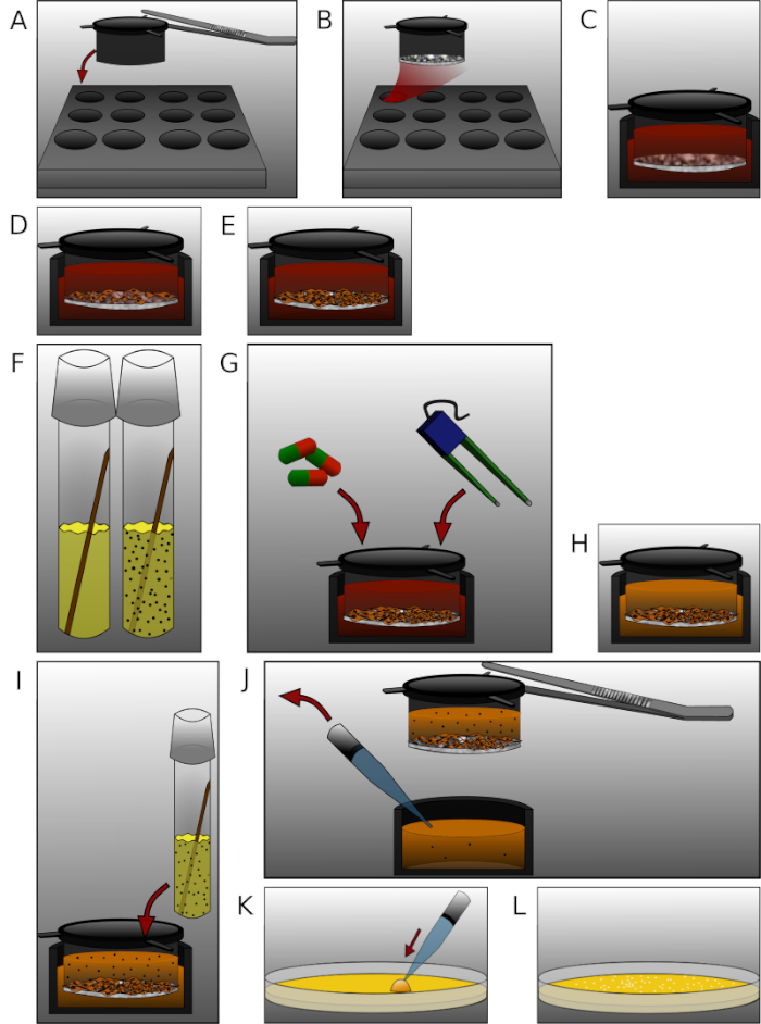
图2:实验方案中各个步骤的详细展示。(A)用灭菌镊子将插入式培养小室放入12孔板中。(B)每孔插入式培养小室中加入90 µL纤连蛋白与IV型胶原蛋白混合液,孵育24 h。(C)用预热的培养基将膜平衡30 min。(D)每孔插入式培养小室接种2 × 10⁵个人脑微血管内皮细胞。(E)将培养板置于适当条件下培养一定时间。(F)检测前一天,将一株E. coli菌落接种至LB液体培养基中,孵育24 h。(G)处理细胞或测量跨内皮电阻(TEER)。(H)将完全培养基更换为无抗生素培养基。(I)每孔上室加入450 µL细菌悬液(OD600 = 0.5),孵育6 h。(J)用镊子移除插入式培养小室,从每孔下室取50 µL培养基样品。(K)将样品涂布于琼脂平板上,孵育24 h。(L)计数菌落并分析数据。请点击此处查看该图的放大版本。
结果
按照实验方案,接种细胞并构建血脑屏障(BBB)模型。接种后第14天,用活性醛类物质乙二醛处理细胞。本实验旨在探讨年龄与糖尿病在术后谵妄(POD)27中的相关性,以及老年患者脑膜炎高发的原因48。由于年龄增长和糖尿病状态下晚期糖基化终末产物(AGEs)水平均升高49,因此有必要进一步研究糖基化在病原微生物穿越血脑屏障致病机制中的作用。糖基化是指蛋白质中的游离氨基与还原性糖或其他羰基化合物的羰基之间发生的非酶促反应。葡萄糖是已知的羰基供体之一,但还存在反应性更强的羰基化合物。这些化合物首先形成不稳定的席夫碱(Schiff's base),随后重排为更稳定且更具反应活性的二羰基化合物,如乙二醛。AGEs作为最终产物,可在蛋白质之间形成交联50,并通过与晚期糖基化终末产物受体(RAGE)相互作用,破坏细胞结构并改变细胞功能51。
细胞用 0.05 和 0.15 mM 乙二醛(GO)溶液处理 1 小时,未处理细胞作为对照。通过免疫印迹及抗-AGE 抗体检测糖基化产物图3)。获得的细菌菌落被计数,并表示为菌落数的绝对值(图4A)或相对于对照组归一化的菌落数量(图4B未处理细胞孔中收集的培养基仅形成极少数菌落。该结果表明,未处理细胞能够构建屏障,可作为对照。经乙二醛处理的样本菌落数量增加,说明乙二醛对THBMECs细胞及其细胞屏障密度具有影响,因为处理组与未处理组细胞间的菌落数量存在显著差异。乙二醛处理后细菌跨屏障数量增加,这可能解释了糖尿病与血脑屏障(BBB)破坏相关疾病之间存在关联的原因。
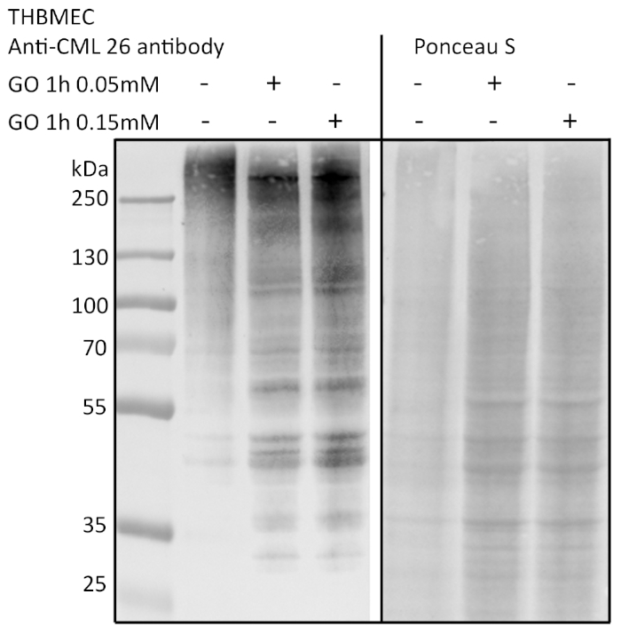
图3:通过免疫印迹检测蛋白质糖基化。 THBMEC细胞用不同浓度的GO处理1小时。提取总蛋白并进行SDS-PAGE分离。使用抗AGE抗体(CML-26)通过免疫印迹检测蛋白质的糖基化水平。请点击此处查看该图的放大版本。
在另一组实验中,通过葡萄糖诱导 THBMECs 的蛋白质糖基化。将灭菌的葡萄糖加入 DMEM/F-12 培养基中,使葡萄糖浓度从正常葡萄糖培养基(NG)的 17.5 mM 升高至高葡萄糖培养基(HG)的 42.5 mM。THBMECs 在两个不同的细胞培养瓶中培养:一个使用正常葡萄糖(NG)培养基,另一个使用高葡萄糖(HG)培养基。这两种不同的培养基也用于在 12 孔板的滤膜上构建血脑屏障(BBB)。在 NG 培养基中培养的细胞作为对照组。获得的菌落数量以菌落绝对数量(图 4C)或相对于对照组归一化后的相对数量表示(图 4D)。结果表明,细菌穿过人源血脑屏障的过程未受到显著影响,由此得出结论:NG 与 HG 之间的差异不足以破坏血脑屏障的完整性。设计不同实验条件旨在验证该模型以及模拟血脑屏障的细胞完整性。
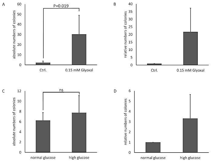
图 4:在 BBB 模型中使用 THBMEC 计数的细菌菌落的绝对数量与相对数量。 THBMEC 经 0.15 mM GO 处理 1 小时,未处理细胞作为对照。每个上室加入 450 µL 的 E. coli 菌悬液(OD600 = 0.5)。6 小时后,将下室培养基接种于琼脂平板上。(A)该图显示计数菌落数量的平均值 ± 标准误(SEM)。(B)该图显示以未处理细胞为对照归一化的菌落数量,数据表示为 ± SEM(n = 4)。在(C)和(D)中,THBMEC 在 NG 和 HG 培养基中培养。每个上室加入 450 µL 的 E. coli 菌悬液(OD600 = 0.5)。6 小时后,将下室培养基接种于琼脂平板上。(C)该图显示计数菌落数量的平均值 ± 标准误(SEM)。(D)该图显示以未处理细胞为对照归一化的菌落数量,数据表示为 ± SEM(n = 3)。请点击此处查看该图的放大版本。
讨论
对微生物穿越血脑屏障致病机制的认识有限,制约了术后谵妄(POD)或脑膜炎治疗手段的进一步发展。这些疾病的高死亡率和高发病率迫切需要改进患者治疗方案,深入研究其潜在机制,并建立一个可靠的化合物筛选平台。这些多因素参与的事件可通过人脑微血管内皮细胞(BMECs)进行研究。已有多种物种BMECs的成功分离方法被报道,但这些方法普遍存在细胞分子特征丢失的问题52,53。本方案中描述的THBMECs在极早期传代阶段即进行转染,此时细胞表现出特异性的脑内皮细胞特征并得以维持43。这一点至关重要,因为目前尚未完全揭示相关通路中的所有作用环节,而该模型似乎能够模拟传统BMECs的功能特性。我们所提出的模型可直接反映BMECs本身的变化以及微生物穿越血脑屏障的过程。
THBMEC 细胞的处理简便,所需的技术设备在大多数生命科学实验室中均具备。当 THBMEC 形成紧密的单层后,本模型即可立即开始进行各项检测操作。由于可将新检测方法与传统检测方法(如跨内皮电阻(TEER)测量或示踪剂标记)相结合,因此该模型的应用范围广泛54。此外,还可加入星形胶质细胞或周细胞,构建共培养或三联培养模型。本模型也可用于检测药物对微生物穿越过程的影响,即在上室接种细菌前,先用化合物处理 THBMEC。事实上,目前已可购得适用于 96 孔板的带滤膜插件,从而实现操作流程的自动化。这有助于建立高通量药物筛选系统,加快针对上述疾病的药物研发进程,并在药物开发过程中减少对血脑屏障的不良影响。
在本方法中,将细菌加入上室后孵育时间是一个关键步骤。在实验方案中必须以小时为单位设定时间线,因为大肠杆菌(E. coli)的代时仅为20分钟55。否则,使用不同的时间点可能导致结果产生误导。此外,在细菌暴露期间,若培养板操作不当,上室与下室之间可能存在交叉污染的风险。在此过程中对12孔板进行任何操作均可能污染下室中的培养基。
E. coli 是细菌性脑膜炎中一种众所周知且非常常见的致病菌。进一步的研究应检测其他与脑膜炎相关的细菌,例如 Neisseria meningitidis56 或 Streptococcus pneumoniae57。这些细菌似乎通过不同的机制穿越血脑屏障,其机制仍需进一步阐明,以更好地治疗患者。在老年患者中,术后谵妄(POD)的发生率升高26,同时伴随的合并症数量也增加。已知不同疾病之间存在相互作用,尤其是糖尿病等全身性疾病。在我们的模型中,可以模拟这些病理条件,或在加入细菌前对细胞进行预处理。
该模型的局限性在于THBMECs与细菌之间的直接接触,因此需要进一步研究以探究可能的接触机制,并确定所涉及的通路和蛋白质。然而,可以取出插入物并收集细胞以进行后续分析。与干细胞模型相比,该模型的TEER值较低38,39,40。我们通过使用一种在6小时后不会穿过未经处理细胞BBB的细菌浓度证实了这一点。
总之,该方法为分析细菌穿越血脑屏障提供了可靠的平台,并具有扩展用于高通量药物筛选的潜力。
披露
作者无任何利益冲突需要披露。
致谢
作者感谢Maryam Hussain博士在本方法早期工作中所做的贡献,感谢PD Dr. Kerstin Danker(柏林夏里特医学院)课题组提供THBMECs,以及Juliane Weber对稿件的审阅。本研究得到了RTK 2155(ProMoAge)项目的资助。
材料
| 姓名 | 公司 | 目录编号 | 评论 |
|---|---|---|---|
| 琼脂 - 琼脂 | Carl Roth | 6494.3 | 生物科学级 |
| 高压灭菌器 | Systec | VX-150 | |
| 细菌 E.coli 菌株 GM2163 | Fermentas Life Sciences, 立陶宛 | ||
| 光度计 | Eppendorf | 6131 | |
| THBMEC 细胞 | M. F. Stins 课题组 | ||
| 细胞培养瓶 | Greiner Bio-One | 658175 | |
| Universal 320 通用离心机 | Hettrichlab | 1401 | |
| IV型胶原蛋白 | SIGMA Aldrich | C6745 | 来源于人细胞培养 |
| Countess 自动细胞计数仪,含细胞计数板载玻片和 0.4% 台盼蓝染色液 | Invitrogen | C10311 | |
| 培养管 | Greiner Bio-One | 191180 | |
| 比色皿 | BRAND | 759015 | |
| 二水合磷酸氢二钠 | MERCK | 1065800500 | |
| DMEM/F-12 | GIBCO/ Thermo Sc. | 11330032 | HEPES |
| 15 ml Falcon 管 | Greiner Bio-One | 188271 | |
| 50 ml Falcon 管 | Greiner Bio-One | 227261 | |
| 胎牛血清 | GIBCO/ Thermo Sc. | 10270 | 优质胎牛血清 - 巴西来源 |
| 纤连蛋白 | SIGMA Aldrich | F0556 | 人成纤维细胞来源溶液 |
| Heracell 150 CO2 培养箱 | Heraeus | 50116047 | |
| New Brunswick I 26 摇床培养箱 | Eppendorf | M1324-0000 | |
| 接种环 | Dr. Ilona Schubert - Laborfachhandel | 641000 | |
| LB 肉汤基础培养基 | GIBCO/ Thermo Sc. | 12780029 | |
| L-谷氨酰胺 | GIBCO/ Thermo Sc. | 25030-081 | |
| 微生物培养箱 B 6200 | Heraeus | 51015192 | |
| 生物安全柜 AURA 2000 M.A.C. II 级 | BIOAIR | 12469 | |
| 倒置显微镜 | Zeiss | TELAVAL 31 | |
| 2 ml 微量离心管 | Sarstedt | 72,695,400 | |
| 1.5 ml 微量离心管 | Sarstedt | 72,706,400 | |
| 青霉素/链霉素 | GIBCO/ Thermo Sc. | 15140122 | |
| 培养皿 | Dr. Ilona Schubert - Laborfachhandel | 464-800 | |
| 氯化钾 | Roth | HN02.3 | |
| 磷酸二氢钾 | Roth | P018.2 | |
| 氯化钠 | Roth | 9265.2 | |
| ThinCerts + 多孔板 | Greiner Bio-One | 665631 | 12 孔,孔径 3.0 µm |
| 胰蛋白酶 - EDTA | GIBCO/ Thermo Sc. | 15400054 | |
| Laboport 真空泵 | KNF | N 86 KT.18 |
参考文献
- Goldmann, E. E. Vitalfärbung am Zentralnervensystem: Beitrag z. Physio-Pathologie d Plexus chorioideus ud Hirnhäute. , (1913).
- Reese, T. S., Karnovsky, M. J. Fine structural localization of a blood-brain barrier to exogenous peroxidase. Journal of Cell Biology. 34 (1), 207-217 (1967).
- Risau, W., Dingler, A., Albrecht, U., Dehouck, M. P., Cecchelli, R. Blood-brain barrier pericytes are the main source of gamma-glutamyltranspeptidase activity in brain capillaries. Journal of Neurochemistry. 58 (2), 667-672 (1992).
- Brightman, M. W., Reese, T. S. Junctions between intimately apposed cell membranes in the vertebrate brain. Journal of Cell Biology. 40 (3), 648-677 (1969).
- Coomber, B. L., Stewart, P. A. Morphometric analysis of CNS microvascular endothelium. Microvascular Research. 30 (1), 99-115 (1985).
- Tilling, T., Korte, D., Hoheisel, D., Galla, H. J. Basement membrane proteins influence brain capillary endothelial barrier function in vitro. Journal of Neurochemistry. 71 (3), 1151-1157 (1998).
- Morris, A. W., et al. Vascular basement membranes as pathways for the passage of fluid into and out of the brain. Acta Neuropathologica. 131 (5), 725-736 (2016).
- Betz, A. L., Goldstein, G. W. Polarity of the blood-brain barrier: neutral amino acid transport into isolated brain capillaries. Science. 202 (4364), 225-227 (1978).
- Butt, A. M., Jones, H. C., Abbott, N. J. Electrical resistance across the blood-brain barrier in anaesthetized rats: a developmental study. Journal of Physiology. 429, 47-62 (1990).
- Calabria, A. R., Weidenfeller, C., Jones, A. R., de Vries, H. E., Shusta, E. V. Puromycin-purified rat brain microvascular endothelial cell cultures exhibit improved barrier properties in response to glucocorticoid induction. Journal of Neurochemistry. 97 (4), 922-933 (2006).
- O'Carroll, S. J., et al. Pro-inflammatory TNFalpha and IL-1beta differentially regulate the inflammatory phenotype of brain microvascular endothelial cells. Journal of Neuroinflammation. 12 (131), (2015).
- Simi, A., Tsakiri, N., Wang, P., Rothwell, N. J. Interleukin-1 and inflammatory neurodegeneration. Biochemical Society Transactions. 35 (Pt 5), 1122-1126 (2007).
- Janzer, R. C., Raff, M. C. Astrocytes induce blood-brain barrier properties in endothelial cells. Nature. 325 (6101), 253-257 (1987).
- Tao-Cheng, J. H., Nagy, Z., Brightman, M. W. Tight junctions of brain endothelium in vitro are enhanced by astroglia. Journal of Neuroscience. 7 (10), 3293-3299 (1987).
- Utsumi, H., et al. Expression of GFRalpha-1, receptor for GDNF, in rat brain capillary during postnatal development of the BBB. American Journal of Physiology and Cell Physiology. 279 (2), (2000).
- Tran, N. D., Correale, J., Schreiber, S. S., Fisher, M. Transforming growth factor-beta mediates astrocyte-specific regulation of brain endothelial anticoagulant factors. Stroke. 30 (8), 1671-1678 (1999).
- Balabanov, R., Washington, R., Wagnerova, J., Dore-Duffy, P. CNS microvascular pericytes express macrophage-like function, cell surface integrin alpha M, and macrophage marker ED-2. Microvascular Research. 52 (2), 127-142 (1996).
- Ramsauer, M., Krause, D., Dermietzel, R. Angiogenesis of the blood-brain barrier in vitro and the function of cerebral pericytes. Faseb Journal. 16 (10), 1274-1276 (2002).
- Lindahl, P., Johansson, B. R., Leveen, P., Betsholtz, C. Pericyte loss and microaneurysm formation in PDGF-B-deficient mice. Science. 277 (5323), 242-245 (1997).
- Young, G. B., Bolton, C. F., Archibald, Y. M., Austin, T. W., Wells, G. A. The electroencephalogram in sepsis-associated encephalopathy. Journal of Clinical Neurophysiology. 9 (1), 145-152 (1992).
- Carlyle Clawson, C., Francis Hartmann, J., Vernier, R. L. Electron microscopy of the effect of gram-negative endotoxin on the blood-brain barrier. Journal of Comparative Neurology. 127 (2), 183-197 (1966).
- Jeppsson, B., et al. Blood-brain barrier derangement in sepsis: cause of septic encephalopathy? The American Journal of Surgery. 141 (1), 136-142 (1981).
- Tighe, D., Moss, R., Bennett, D. Cell surface adrenergic receptor stimulation modifies the endothelial response to SIRS. Systemic Inflammatory Response Syndrome. New Horizons (Baltimore, Md). 4 (4), 426-442 (1996).
- Kim, K. S., et al. The K1 capsule is the critical determinant in the development of Escherichia coli meningitis in the rat. Journal of Clinical Investigation. 90 (3), 897-905 (1992).
- Kim, K. S., Wass, C. A., Cross, A. S. Blood-brain barrier permeability during the development of experimental bacterial meningitis in the rat. Experimental Neurology. 145 (1), 253-257 (1997).
- Arshi, A., et al. Predictors and Sequelae of Postoperative Delirium in Geriatric Hip Fracture Patients. Geriatric Orthopaedic Surgery and Rehabililation. 9, 2151459318814823(2018).
- Smulter, N., Lingehall, H. C., Gustafson, Y., Olofsson, B., Engstrom, K. G. Delirium after cardiac surgery: incidence and risk factors. Interactive CardioVascular and Thoracic Surgery. 17 (5), (2013).
- Kratz, T., Heinrich, M., Schlauss, E., Diefenbacher, A. Preventing postoperative delirium. Deutsches Arzteblatt International. 112 (17), 289-296 (2015).
- Uchida, Y., et al. Quantitative targeted absolute proteomics of human blood-brain barrier transporters and receptors. Journal of Neurochemistry. 117 (2), 333-345 (2011).
- Warren, M. S., et al. Comparative gene expression profiles of ABC transporters in brain microvessel endothelial cells and brain in five species including human. Pharmacology Research. 59 (6), 404-413 (2009).
- Omidi, Y., et al. Evaluation of the immortalised mouse brain capillary endothelial cell line, b.End3, as an in vitro blood-brain barrier model for drug uptake and transport studies. Brain Research. 990 (1-2), 95-112 (2003).
- Steiner, O., Coisne, C., Engelhardt, B., Lyck, R. Comparison of immortalized bEnd5 and primary mouse brain microvascular endothelial cells as in vitro blood-brain barrier models for the study of T cell extravasation. Journal of Cerebral Blood Flow and Metabolism. 31 (1), 315-327 (2011).
- Burek, M., Salvador, E., Forster, C. Y. Generation of an immortalized murine brain microvascular endothelial cell line as an in vitro blood brain barrier model. Journal of Visualized Experiments. (66), e4022(2012).
- Ohtsuki, S., et al. Quantitative targeted absolute proteomic analysis of transporters, receptors and junction proteins for validation of human cerebral microvascular endothelial cell line hCMEC/D3 as a human blood-brain barrier model. Molecular Pharmacolgy. 10 (1), 289-296 (2013).
- Urich, E., Lazic, S. E., Molnos, J., Wells, I., Freskgard, P. O. Transcriptional profiling of human brain endothelial cells reveals key properties crucial for predictive in vitro blood-brain barrier models. PLoS One. 7 (5), e38149(2012).
- Lopez-Ramirez, M. A., et al. Cytokine-induced changes in the gene expression profile of a human cerebral microvascular endothelial cell-line, hCMEC/D3. Fluid Barrier CNS. 10 (27), (2013).
- Cucullo, L., et al. Immortalized human brain endothelial cells and flow-based vascular modeling: a marriage of convenience for rational neurovascular studies. Journal of Cerebral Blood Flow Metabolism. 28 (2), 312-328 (2008).
- Lippmann, E. S., et al. Derivation of blood-brain barrier endothelial cells from human pluripotent stem cells. Nature Biotechnology. 30 (8), 783-791 (2012).
- Boyer-Di Ponio, J., et al. Instruction of circulating endothelial progenitors in vitro towards specialized blood-brain barrier and arterial phenotypes. PLoS One. 9 (1), e84179(2014).
- Cecchelli, R., et al. A stable and reproducible human blood-brain barrier model derived from hematopoietic stem cells. PLoS One. 9 (6), e99733(2014).
- Lippmann, E. S., Al-Ahmad, A., Azarin, S. M., Palecek, S. P., Shusta, E. V. A retinoic acid-enhanced, multicellular human blood-brain barrier model derived from stem cell sources. Science Reports. 4, 4160(2014).
- Lim, R. G., et al. Huntington's Disease iPSC-Derived Brain Microvascular Endothelial Cells Reveal WNT-Mediated Angiogenic and Blood-Brain Barrier Deficits. Cell Reports. 19 (7), 1365-1377 (2017).
- Stins, M. F., Badger, J., Sik Kim, K. Bacterial invasion and transcytosis in transfected human brain microvascular endothelial cells. Microbiological Pathogens. 30 (1), 19-28 (2001).
- Gaschignard, J., et al. Neonatal Bacterial Meningitis: 444 Cases in 7 Years. Pediatric Infectious Disease Journal. 30 (3), 212-217 (2011).
- Hussain, M. The Effect of Glycation on the Permeability of an in vitro Blood-brain Barrier Model. , University Halle-Wittenberg. (2015).
- Weber, V. The effect of glycation on the permeability of human blood-brain barrier. , University Halle-Wittenberg. (2019).
- Hussain, M., et al. Novel insights in the dysfunction of human blood-brain barrier after glycation. Mechanism of Ageing Development. 155, 48-54 (2016).
- Choi, C. Bacterial meningitis in aging adults. Clinical Infectious Disease. 33 (8), 1380-1385 (2001).
- Furth, A. J. Glycated proteins in diabetes. British Journal of Biomedical Science. 54 (3), 192-200 (1997).
- Sajithlal, G. B., Chithra, P., Chandrakasan, G. Advanced glycation end products induce crosslinking of collagen in vitro. Biochimica and Biophysica Acta. 1407 (3), 215-224 (1998).
- Ray, R., Juranek, J. K., Rai, V. RAGE axis in neuroinflammation, neurodegeneration and its emerging role in the pathogenesis of amyotrophic lateral sclerosis. Neuroscience and Biobehavior Reviews. 62, 48-55 (2016).
- DeBault, L. E., Cancilla, P. A. Gamma-Glutamyl transpeptidase in isolated brain endothelial cells: induction by glial cells in vitro. Science. 207 (4431), 653-655 (1980).
- Diglio, C. A., Grammas, P., Giacomelli, F., Wiener, J. Primary culture of rat cerebral microvascular endothelial cells. Isolation, growth, and characterization. Laboratory Investigation. 46 (6), 554-563 (1982).
- Buchert, M., Turksen, K., Hollande, F. Methods to examine tight junction physiology in cancer stem cells: TEER, paracellular permeability, and dilution potential measurements. Stem Cell Reviews. 8 (3), 1030-1034 (2012).
- Gibson, B., Wilson, D. J., Feil, E., Eyre-Walker, A. The distribution of bacterial doubling times in the wild. Proceedings of the Royal Society B: Biological Sciences. 285 (1880), (2018).
- Pron, B., et al. Interaction of Neisseria maningitidis with the components of the blood-brain barrier correlates with an increased expression of PilC. Journal of Infectious Diseases. 176 (5), 1285-1292 (1997).
- Iovino, F., Orihuela, C. J., Moorlag, H. E., Molema, G., Bijlsma, J. J. Interactions between blood-borne Streptococcus pneumoniae and the blood-brain barrier preceding meningitis. PLoS One. 8 (7), e68408(2013).
重印与许可
