本方案详细介绍了经导管二尖瓣修复术中如何进行实时超声心动图引导。针对手术的每个阶段,均描述了基本切面及必要的测量方法。
需要JoVE订阅才能观看此内容。 请登录或开始免费试用
本文内容
摘要
摘要
经皮导管二尖瓣缘对缘修复术对于外科手术风险较高的重度有症状二尖瓣反流患者而言,是一种安全且成熟的治疗方式。除X线透视外,超声心动图引导是目前的金标准,应采用标准化技术进行操作。
本文介绍了我们可重复的超声心动图操作逐步指南,包括成像切面、测量方法,并强调了操作过程中可能出现的潜在困难。
本文为该手术每个步骤提供了详细且按时间顺序排列的超声心动图切面,特别比较了二维与三维成像之间的选择偏好。如有需要,文中还描述了脉冲波、连续波及彩色多普勒测量方法。此外,由于目前尚无关于经皮缘对缘修复术中二尖瓣反流定量评估的官方指南,本文也包含了在夹合二尖瓣瓣叶后以及器械释放后进行超声心动图定量评估的建议。同时,本文还阐述了术中为预防和处理可能并发症所需的重要超声心动图切面。
经导管二尖瓣修复术中的超声心动图引导是必需的。采用系统化方法可改善介入医师与影像医师之间的协作,对于安全、有效地实施手术至关重要。
引言
二尖瓣反流(MR)是欧洲第二常见的瓣膜手术指征1。若不加以治疗,可能导致严重的心力衰竭和生活质量下降2,3,4。经皮二尖瓣修复术(PMVR)是一种基于导管的技术,通过连接A2和P2瓣叶,模拟Alfieri缝合手术方法进行二尖瓣修复5。对于外科手术风险较高的患者,该技术为治疗重度MR提供了一种微创治疗方案。来自多个注册研究和临床试验的数据表明,MitraClip手术(一种经导管二尖瓣修复疗法)是一种有效且安全的方法6,7,8,9。2019年,一种类似装置——PASCAL经导管瓣膜修复系统——进入市场,其在治疗重度MR患者方面已显示出可行性和可接受的安全性10。PMVR手术的持续时间和成功率依赖于操作者的个体技能和经验11。与其他经皮技术(如仅需透视引导的经导管主动脉瓣置换术[TAVR])不同,PMVR需要超声心动图引导12,13。
本文逐步介绍了经导管二尖瓣修复术(PMVR)期间的超声心动图操作方法,包括测量步骤、术中二尖瓣反流(MR)程度定量评估的建议,以及预防围术期并发症所需的重要切面。
访问受限。请登录或开始试用以查看此内容。
方案
本方案遵循本机构人类研究伦理委员会的指南。
1. 干预前评估
- 在进行房间隔穿刺前,排除心包积液。若存在少量心包积液,应在以右心室(RV)为重点的四腔心(4Ch)切面、经食管中段右心室流入-流出道切面以及长轴(LAX)切面上,测量舒张末期最大无回声间隙。
- 使用脉冲波多普勒(PW)评估左上肺静脉(LUPV)的肺静脉血流模式,并排除左心耳(LAA)内血栓形成。显示以左心耳为重点的短轴(SAX)切面,随后在40–60°范围内扫查,并逆时针旋转探头以显示左上肺静脉。在90–110°范围内扫查,评估右上肺静脉(RUPV)的血流(图1 和 补充图1)。
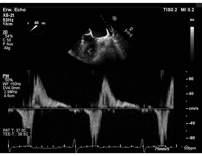
图1:改良短轴切面:左上肺静脉的脉冲多普勒血流 请点击此处查看该图的放大版本。
- 确保在手术前后评估期间血流动力学状态保持一致。
注意:由于二尖瓣反流(MR)是一种动态性瓣膜疾病,在全身麻醉下反流程度可能显得较轻。此时应咨询操作者,并增加后负荷和/或前负荷。 - 找到最佳的交界间切面(50–70°),在三个节段分别获取带和不带彩色多普勒的垂直切面(X-plane),测量后叶二尖瓣(PML)长度。然后再次检查瓣叶形态(图2 和 补充图2)。

图 2:二尖瓣的二维双平面彩色多普勒图像:内侧反流束 请点击此处查看此图的放大版本。
- 在长轴切面(120–140°)使用连续波多普勒(CW)评估二尖瓣跨瓣压差。
注:平均压差(MPG)> 5 mmHg 是经导管二尖瓣修复术(PMVR)的相对禁忌证。 - 采集带有彩色多普勒的三维数据集,或带有彩色多普勒的宽扇区缩放图像,并测量三维收缩颈宽度(3D-VCA)(图3)。
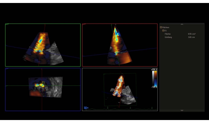
图3:三维数据集的多平面重建结合彩色多普勒:三维缩流颈面积 请点击此处查看该图的放大版本。
- 不使用彩色,利用三维容积测量二尖瓣面积(MVA)补充图3).
注意:一个区域 < 4 cm2 是相对禁忌症,一个区域 < 3 cm2 进行该操作的绝对禁忌证。否则,在经胃底短轴切面中评估MVA。 - 显示二尖瓣的三维 en-face 外科心房视角(主动脉瓣位于12点钟位置)。
注意:瓣膜的节段中,节段1称为“外侧”,节段3称为“内侧”。在正面手术视野中,各节段的排列顺序与交界视图中的顺序相反。执行 180° 顺时针旋转至三维 en-face 外科视野(主动脉瓣位于6点钟位置),将使两个视野中的节段顺序保持一致图4 和 图5).
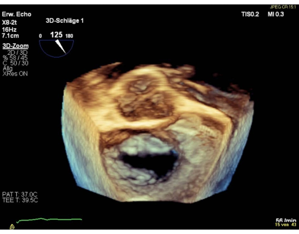
图4:宽扇区放大图像:三维正面外科心房视图(主动脉瓣位于12点钟位置)请点击此处查看该图的放大版本。
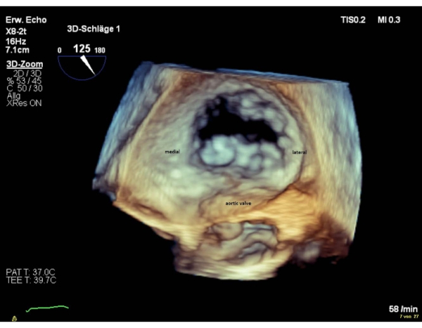
图5:宽扇区放大图像:三维心房面观(主动脉瓣位于6点钟位置)请点击此处查看此图的放大版本。
- 最后,使用 X-plane 获取上下腔静脉观(90–110°),以显示主动脉瓣(AV),用于经房间隔穿刺。
2. 策略
- 在将可操控引导导管(SGC)和夹合器输送系统(CDS)插入左心房之前,应与操作者讨论相关策略。
- 如果孔口满足条件,则评估单装置策略 < 1 厘米宽,并在反流束正上方放置夹子,如果开口为圆形。
- 若存在较大的椭圆形反流口或多股反流束,需评估植入≥2个夹合装置的可行性。装置植入应从反流口内侧开始,因为相较于从外侧开始,先在内侧植入第一个装置通常更有利于后续第二个装置的定位。补充图4).
3. 房间隔穿刺
- 显示结合短轴切面的上下腔静脉观,确保可见主动脉瓣,以避免损伤主动脉。
- 确保穿刺部位略偏上方且偏后方(图6)。

图6:二维双平面视图:经房间隔穿刺 请点击此处查看该图的放大版本。
- 对于退行性二尖瓣反流(如二尖瓣脱垂),选择 4-5 cm 的穿刺高度;对于功能性二尖瓣反流,选择 > 3.5 cm 的穿刺高度。应避免经卵圆孔途径,因其位置过于靠前。
- 当穿间隔针导致房间隔出现帐篷样改变时,在四腔心视图(4Ch)的中期收缩阶段测量穿刺高度(补充图5)。
注意:对于心房较大的患者,若穿刺部位过于靠后,则在四腔心视图中无法观察到帐篷征象。此时应后弯探头并将其更深地插入食管。 - 完成穿间隔穿刺后,始终需在四腔心视图中排除心包积液。
- 展示以左心耳(LAA)和肺静脉为重点的短轴视图(SAX),以观察硬导丝进入左上肺静脉(LUPV)的过程。
4. 将SGC引入LA
- 在短轴(SAX)切面下,结合连续二维超声心动图和透视引导,观察导丝引导导管(SGC)在扩张器作用下的 tenting 和前移,以避免损伤左心房壁。
注意:SGC 的尖端由一个不透射线且超声回声明亮的双环标记定义(补充图6)。 - 向操作者展示短轴(SAX)切面和双腔静脉(bicaval)切面(90–120°),以将 SGC 定向至左心室(LV)方向。
5. 将CDS推进至左心房
- 采集包含房间隔、左侧嵴和二尖瓣的三维容积图像,并确保可见左侧嵴,因为冠状窦开口(CDS)突出较为常见(图7)。

图7:宽扇区放大图像:左心房内包含房间隔、左侧嵴和二尖瓣的SGC 请点击此处查看此图的放大版本。
- 否则应选择短轴切面(SAX)和长轴切面(LAX,X平面),以确保CDS未与嵴部及左心房壁接触。
注意:操作者可要求成像人员显示房间隔,并将SGC回撤数毫米以越过嵴部。若无法在三维图像中观察到双环结构,应切换至二维模式,并在短轴切面中显示SGC。
- 检查CDS是否垂直于对合线,以确保正确的路径。
- 以约60°显示二维的交界视图,以展示内侧-外侧平面,并以120-140°显示长轴视图,以识别二尖瓣的前-后平面(补充图7)。
- 或者,在三维端面视图中优化CDS的内侧、外侧、前侧和后侧位置(补充图8)。
6. 二尖瓣上方和下方装置的方向定位
- 采用三维 en-face 视图,显示瓣叶对合线与瓣臂呈垂直定位。
- 若图像质量不佳,可结合使用交界间视图与长轴视图(图8 和 图9)。
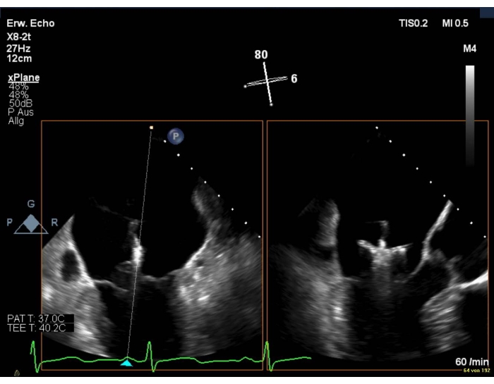
图8:二尖瓣的二维双平面视图:器械在二尖瓣上的定位 请点击此处查看此图的放大版本。

图9:宽扇区放大图像:装置在二尖瓣上的定位 请点击此处查看该图的放大版本。
注意:夹子臂仅在 LAX 视图中可见。
- 调整前后连合切面角度,以观察位于内侧和外侧装置的完整双臂长度。对于位于内侧的装置,以约 30–45° 角度扫查;对于位于外侧的装置,以约 70–90° 角度扫查。
- 选择联合使用交界连合切面与长轴切面,以观察CDS向左心室的推进情况。
- 确保CDS置于二尖瓣下方仅数毫米处。
- 通过三维 en-face 视图确认夹子臂仍处于预定位置,因为在穿过瓣膜时夹子发生旋转较为常见。
注意:若夹子臂位置已发生改变,需进行顺时针或逆时针旋转以实现对称抓合。操作过程中应谨慎,尽量减少腱索及腱下结构的缠绕。 - 若需对器械进行大幅度重新定向,应使用交界连合切面结合X-plane视图,观察夹子翻转并将其回收至左心房的过程。
7. 二尖瓣瓣叶的抓取及夹闭器置入前后二尖瓣反流的评估
- 在交界间切面结合长轴切面(X-plane)或仅在长轴切面中记录瓣叶的夹合情况(补充图9)。
- 请麻醉医师执行屏气操作,以减少通气过程中的心脏移动,便于顺利夹合瓣叶。
- 持续观察瓣叶附着点,避免瓣叶或腱索发生卷曲。
注意:夹合卷曲的瓣叶或腱索可能导致部分瓣叶撕脱和/或加重二尖瓣反流。 - 在释放夹合器前,仔细评估反流程度的减轻情况。操作者与超声成像医师均需共同分析这一关键步骤。
- 将经食管超声(TEE)探头向夹合器内侧和外侧旋转,或使用X-plane模式结合彩色多普勒,以检测靠近夹合器的偏心性反流束(补充图10)。
注意:由于夹合器产生的声影伪影,可能导致对二尖瓣反流程度的低估。可将探头更深地插入食管,或显示经胃短轴切面,以无伪影的方式观察残余反流束。 - 评估肺静脉的脉冲多普勒(PW)血流频谱。
注意:若先前存在的收缩期血流逆向转变为收缩期主导的血流模式,则提示反流可能已显著减轻。 - 测量二尖瓣跨瓣平均压差(MPG)。
注意:若压差 > 5 mmHg,则为夹合器释放的相对禁忌证(补充图11 和 补充图12)。 - 采用二尖瓣的三维 en-face 视图或经胃短轴切面(SAX)显示双孔形态(图10)。
- 将经食管超声(TEE)探头向夹合器内侧和外侧旋转,或使用X-plane模式结合彩色多普勒,以检测靠近夹合器的偏心性反流束(补充图10)。
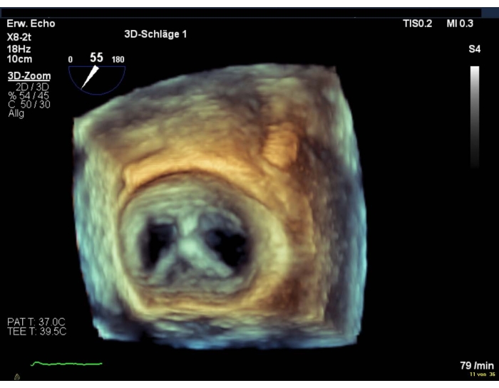
图10:宽扇区放大图像:器械置入后二尖瓣的双孔开口 请点击此处查看该图的高清版本。
- 最后,如果结果满意,在二维图像中检查瓣叶的植入情况。
- 释放CDS上的夹子后,重复最后五个步骤。
注意:由于系统对二尖瓣(MV)的张力,释放装置后残余的反流射流可能会加重。 - 在X-plane的LAX切面中显示输送导管尖端从SGC中撤出的过程,并确保倒刺避免与左心房(LA)接触(补充图13)。
8. 最终磁共振评估
- 若存在残余反流束,结合彩色多普勒显示联合间切面,并辅以垂直X平面成像。
- 在三维容积图像中计算三维瓣口面积(3D-VCA)。注意:通常各瓣口并不位于同一平面,此时应在相应平面上分别测量每个瓣口的平面面积(补充图14)。
- 再次评估肺静脉血流及二尖瓣跨瓣平均压差。
注意:在经导管二尖瓣修复术中,持续监测左心房压力可能是一项有用的辅助手段。 - 最后,显示二尖瓣的三维端面观。
9. 植入其他设备
- 确保二尖瓣反流(MR)的减轻程度足够。
注意:如果效果不理想,应评估是否需植入额外装置。 - 确保额外装置不会与已植入并伸入左心室的装置发生接触。
注意:必须使用透视成像(fluoroscopy)来显示夹子之间的实际距离。 - 显示三维正面观,以观察连合线,因为在植入第一个夹子后,连合线位置可能已发生偏移。
- 重复第7.4点所述的五个步骤,以评估在使用额外夹子夹合瓣叶后的二尖瓣反流情况。
访问受限。请登录或开始试用以查看此内容。
结果
经皮缘对缘瓣膜修复术为不适合手术且有症状的重度二尖瓣反流(MR)患者提供了外科瓣膜修复或置换之外的替代治疗方案。MitraClip 的首次临床应用是在 Endovascular Valve Edge-to-Edge Repair Study I(EVEREST I)14 试验中进行研究的。许多其他试验已证实该手术的有效性,可改善症状,并具有较低的院内死亡率和不良事件发生率(ACCESS EU、TRAMI、EVEREST II)15,16,17。经皮缘对缘修复术已被纳入欧洲关于原发性和继发性 MR 干预治疗的指南18。两项随机对照试验 MITRA-FR 和 COAPT 将 MitraClip 手术与最优药物治疗进行了比较,验证了该干预措施在继发性 MR 患者中的可行性。尽管 MITRA-FR 试验在 12 个月时未显示 MitraClip 组在复合终点(全因死...
访问受限。请登录或开始试用以查看此内容。
讨论
经食管超声引导下的二尖瓣修复术(PMVR)是一种安全的方法。尽管超声心动图可能导致并发症,但通常很少引起显著损害。然而,在进行经食管超声心动图检查后,仍有可能出现食管损伤。此类事件的发生率随着手术持续时间的缩短而降低34。相反,经导管二尖瓣缘对缘修复术相关的多种并发症已有报道35,36。其中,大出血是最常见的严重并发症,且经导管瓣膜手术后输血与死亡率显著相关37。心包填塞较为罕见,但可能在手术多个步骤中发生。在房间隔穿刺、将夹合装置输送导管(SGC)插入左心房以及夹合器释放后回收SGC导管头端时保持谨慎,可降低这种危及生命并发症的发生概率。夹合器特异性并发症(如栓塞和夹合器部分脱落)的发生率较低,通过在夹合瓣叶过程中及之后进行准确的超声心动图评估,可进一步减少此类风险。
尽管在进行超声心动图检查时已格外谨慎,二尖瓣反流(MR)仍可能加重。在内侧和外侧位置追加放置夹子可能会...
访问受限。请登录或开始试用以查看此内容。
披露
作者无任何利益冲突需要披露。
致谢
感谢 Dorothea Scheurlen 女士提供技术视频支持。
访问受限。请登录或开始试用以查看此内容。
材料
| 姓名 | 公司 | 目录编号 | 评论 |
|---|---|---|---|
| EPIQ 7 超声系统 | Philips | US218B0542 | 心脏超声设备 |
| X8-2t xMATRIX 3D-TEE 探头 | Philips | B34YYK | 经食管超声探头 |
| 6F 25 cm 套管 | Merit Medical | B60N25AQ | 套管 |
| 16 F 扩张器 | Abbott | 405544 | 扩张器 |
| BRK-1 房间隔穿刺针 71 cm | St. Jude Medical | ABVA407201 | 房间隔穿刺针 |
| Swartz 90° 导引鞘 | St. Jude Medical | 407356 | 房间隔穿刺用可调弯导引鞘 |
| Amplatz 超硬导丝 | Kook Medical | 46509 | 导丝 |
| 可操控引导导管 | Abbott | SGC0302 | 可操控引导导管 |
| MitraClip NTR 输送系统 | Abbott | CDS0602-NTR | 夹合器输送系统 |
| MitraClip NTR 组件 | Abbott | MSK0602-NTR | 器械 |
参考文献
- Iung, B., et al. A prospective survey of patients with valvular heart disease in Europe: The Euro Heart Survey on Valvular Heart Disease. European Heart Journal. 24 (13), 1231-1243 (2003).
- Hauptman, P. J., Rector, T. S., Wentworth, D., Kubo, S. Quality of life in advanced heart failure: role of mitral regurgitation. American Heart Journal. 151 (1), 213-218 (2006).
- Lewis, E. F. Assessing the impact of heart failure therapeutics on quality of life and functional capacity. Current Treatment Options in Cardiovascular Medicine. 15 (4), 425-436 (2013).
- Vaishnava, P., Lewis, E. F. Assessment of quality of life in severe heart failure. Current Heart Failure Reports. 4 (3), 170-177 (2007).
- Feldman, T., et al. Percutaneous repair or surgery for mitral regurgitation. New England Journal of Medicine. 364 (15), 1395-1406 (2011).
- Feldman, T., et al. Percutaneous mitral repair with the MitraClip system: safety and midterm durability in the initial EVEREST (Endovascular Valve Edge-to-Edge REpair Study) cohort. Journal of the American College of Cardiology. 54 (8), 686-694 (2009).
- Whitlow, P. L., et al. Acute and 12-month results with catheter-based mitral valve leaflet repair: the EVEREST II (Endovascular Valve Edge-to-Edge Repair) High Risk Study. Journal of the American College of Cardiology. 59 (2), 130-139 (2012).
- Baldus, S., et al. MitraClip therapy in daily clinical practice: initial results from the German transcatheter mitral valve interventions (TRAMI) registry. European Journal of Heart Failure. 14 (9), 1050-1055 (2012).
- Stone, G. W., et al. Transcatheter Mitral-Valve Repair in Patients with Heart Failure. New England Journal of Medicine. 379 (24), 2307-2318 (2018).
- Lim, D. S., et al. Transcatheter Valve Repair for Patients With Mitral Regurgitation: 30-Day Results of the CLASP Study. JACC: Cardiovascular Interventions. 12 (14), 1369-1378 (2019).
- Schillinger, W., et al. Impact of the learning curve on outcomes after percutaneous mitral valve repair with MitraClip and lessons learned after the first 75 consecutive patients. European Journal of Heart Failure. 13 (12), 1331-1339 (2011).
- Katz, W. E., Conrad Smith, A. J., Crock, F. W., Cavalcante, J. L. Echocardiographic evaluation and guidance for MitraClip procedure. Cardiovascular Diagnosis and Therapy. 7 (6), 616-632 (2017).
- Altiok, E., et al. Optimized guidance of percutaneous edge-to edge repair of the mitral valve using real-time 3-D transesophageal echocardiography. Clinical Research in Cardiology. 100 (8), 675-681 (2011).
- Feldman, T., et al. Percutaneous mitral valve repair using the edge-to-edge technique: six-month results of the EVEREST Phase I Clinical Trial. Journal of the American College of Cardiology. 46 (11), 2134-2140 (2005).
- Maisano, F., et al. Percutaneous mitral valve interventions in the real world: early and 1-year results from the ACCESS-EU, a prospective, multicenter, nonrandomized post-approval study of the MitraClip therapy in Europe. Journal of the American College of Cardiology. 62 (12), 1052-1061 (2013).
- Kalbacher, D., et al. Long-term outcome, survival and predictors of mortality after MitraClip therapy: Results from the German Transcatheter Mitral Valve Interventions (TRAMI) registry. International Journal of Cardiology. 277, 35-41 (2019).
- Glower, D. D., et al. Percutaneous mitral valve repair for mitral regurgitation in high-risk patients: results of the EVEREST II study. Journal of the American College of Cardiology. 64 (2), 172-181 (2014).
- Baumgartner, H., et al. 2017 ESC/EACTS Guidelines for the management of valvular heart disease. European Heart Journal. 38 (36), 2739-2791 (2017).
- Obadia, J. F., et al. MITRA-FR Investigators. Percutaneous Repair or Medical Treatment for Secondary Mitral Regurgitation. New England Journal of Medicine. 379 (24), 2297-2306 (2018).
- Stone, G. W., et al. COAPT Investigators. Transcatheter Mitral-Valve Repair in Patients with Heart Failure. New England Journal of Medicine. 379 (24), 2307-2318 (2018).
- Vahanian, A., et al. Joint Task Force on the Management of Valvular Heart Disease of the European Society of Cardiology (ESC); European Association for Cardio-Thoracic Surgery (EACTS), Guidelines on the management of valvular heart disease (version 2012). European Heart Journal. 33 (19), 2451-2496 (2012).
- American College of Cardiology/American Heart Association Task Force on Practice Guidelines; Society of Cardiovascular Anesthesiologists; Society for Cardiovascular Angiography and Interventions. ACC/AHA 2006 guidelines for the management of patients with valvular heart disease: a report of the American College of Cardiology/American Heart Association Task Force on Practice Guidelines. Circulation. 114 (5), 84(2006).
- Praz, F., et al. Compassionate use of the PASCAL transcatheter mitral valve repair system for patients with severe mitral regurgitation: a multicentre, prospective, observational, first-in-man study. Lancet. 390 (10096), 773-780 (2017).
- McCarthy, K. P., Ring, L., Rana, B. S. Anatomy of the mitral valve: understanding the mitral valve complex in mitral regurgitation. European Journal of Echocardiography. 11 (10), 3-9 (2010).
- Zoghbi, W. A., et al. Recommendations for Noninvasive Evaluation of Native Valvular Regurgitation: A Report from the American Society of Echocardiography Developed in Collaboration with the Society for Cardiovascular Magnetic Resonance. Journal of the American Society of Echocardiography. 30 (4), 303-371 (2017).
- Zoghbi, W. A., et al. Guidelines for the Evaluation of Valvular Regurgitation Percutaneous Valve Repair or Replacement: A Report from the American Society of Echocardiography Developed in Collaboration with the Society for Cardiovascular Angiography and Interventions, Japanese Society of Echocardiography, and Society for Cardiovascular Magnetic Resonance. Journal of the American Society of Echocardiography. 32 (4), 431-475 (2019).
- Abudiab, M. M., Chao, C. J., Liu, S., Naqvi, T. Z. Quantitation of valve regurgitation severity by three-dimensional vena contracta area is superior to flow convergence method of quantitation on transesophageal echocardiography. Echocardiography. 34 (7), 992-1001 (2017).
- Dietl, A., et al. 3D vena contracta area after MitraClip procedure: precise quantification of residual mitral regurgitation and identification of prognostic information. Cardiovasc Ultrasound. 16 (1), 1(2018).
- Ikenaga, H., et al. Usefulness of Intraprocedural Pulmonary Venous Flow for Predicting Recurrent Mitral Regurgitation and Clinical Outcomes After Percutaneous Mitral Valve Repair With the MitraClip. JACC: Cardiovascular Interventions. 12 (2), 140-150 (2019).
- Tang, G. H. L., et al. Continuous invasive hemodynamic monitoring using steerable guide catheter to optimize mitraclip transcatheter mitral valve repair: A multicenter, proof-of-concept study. Journal of Interventional Cardiology. 31 (6), 907-915 (2018).
- Maor, E., et al. Acute Changes in Left Atrial Pressure After MitraClip Are Associated With Improvement in 6-Minute Walk Distance. Circulation: Cardiovascular Interventions. 10 (4), 004856(2017).
- Kuwata, S., et al. Continuous Direct Left Atrial Pressure: Intraprocedural Measurement Predicts Clinical Response Following MitraClip Therapy. JACC: Cardiovascular Interventions. 12 (2), 127-136 (2019).
- Corrigan, F. E., et al. Pulmonary Venous Waveforms Predict Rehospitalization and Mortality After Percutaneous Mitral Valve Repair. JACC Cardiovasc Imaging. 12 (10), 1905-1913 (2019).
- Ruf, T. F., et al. ELMSTREET (Esophageal Lesions during MitraClip uSing TRansEsophageal Echocardiography Trial. EuroIntervention. 13 (12), 1444-1451 (2017).
- Eggebrecht, H., et al. Risk and outcomes of complications during and after MitraClip implantation: Experience in 828 patients from the German TRAnscatheter mitral valve interventions (TRAMI) registry. Catheterization and Cardiovascular Interventions. 86 (4), 728-735 (2015).
- von Bardeleben, R. S., et al. Incidence and in-hospital safety outcomes of patients undergoing percutaneous mitral valve edge-to-edge repair using MitraClip: five-year German national patient sample including 13,575 implants. EuroIntervention. 14 (17), 1725-1732 (2019).
- Généreux, P., et al. Incidence, predictors, and prognostic impact of late bleeding complications after transcatheter aortic valve replacement. Journal of the American College of Cardiology. 64 (24), 2605-2615 (2014).
- Benito-González, T., Estévez-Loureiro, R., Gualis, J. Chordal Rupture Following MitraClip Implantation Resulting in Massive Mitral Regurgitation. Journal of Invasive Cardiology. 27 (10), 224-225 (2015).
- Kaneko, H., Neuss, M., Weissenborn, J., Butter, C. Impact of residual mitral regurgitation after MitraClip implantation. International Journal of Cardiology. 227, 813-819 (2017).
- Buzzatti, N., et al. What is a "good" result after transcatheter mitral repair? Impact of 2+ residual mitral regurgitation. Journal of Thoracic and Cardiovascular Surgery. 151 (1), 88-96 (2016).
- Feldman, T., Guerrero, M. Assessing the Balance Between Less Mitral Regurgitation and More Residual Transmitral Pressure Gradient After MitraClip. JACC: Cardiovascular Interventions. 10 (9), 940-941 (2017).
- Neuss, M., et al. Elevated Mitral Valve Pressure Gradient After MitraClip Implantation Deteriorates Long-Term Outcome in Patients With Severe Mitral Regurgitation and Severe Failure. JACC: Cardiovascular Interventions. 10 (9), 931-939 (2017).
- Katz, W. E., Conrad Smith, A. J., Crock, F. W., Cavalcante, J. L. Echocardiographic evaluation and guidance for MitraClip procedure. Cardiovascular Diagnosis and Therapy. 7 (6), 616-632 (2017).
- Labrousse, L., et al. Guidance of the MitraClip procedure by 2D and 3D imaging. Archives of Cardiovascular Diseases. 111 (6-7), 432-440 (2018).
- Khalique, O. K., Hahn, R. T. Percutaneous Mitral Valve Repair: Multi-Modality Cardiac Imaging for Patient Selection and Intra-Procedural Guidance. Frontiers in Cardiovascular Medicine. 6, 142(2019).
- Bushari, L. I., et al. Percutaneous Transcatheter Edge-to-Edge MitraClip Technique: A Practical "Step-by-Step" 3-Dimensional Transesophageal Echocardiography Guide. Mayo Clinic Proceedings. 94 (1), 89-102 (2019).
- Sherif, M. A., et al. MitraClip step by step; how to simplify the procedure. Netherlands Heart Journal. 25 (2), 125(2017).
- Guarracino, F., et al. Transesophageal echocardiography during MitraClip procedure. Anesthesia & Analgesia. 118 (6), 1188-1196 (2014).
- Avenatti, E., et al. Diagnostic Value of 3-Dimensional Vena Contracta Area for the Quantification of Residual Mitral Regurgitation After MitraClip Procedure. JACC: Cardiovascular Interventions. 12 (6), 582-591 (2019).
- Altiok, E., et al. Analysis of procedural effects of percutaneous edge-to-edge mitral valve repair by 2D and 3D echocardiography. Circulation: Cardiovascular Imaging. 5 (6), 748-755 (2012).
访问受限。请登录或开始试用以查看此内容。
重印与许可
