本文描述了一种使用环钻和钝性高尔夫球棒状刮匙在小鼠角膜中央制造上皮擦伤伤口的实验方案。该角膜伤口愈合模型具有高度可重复性,目前正被用于评估在疾病背景下受损的角膜伤口愈合情况。
需要JoVE订阅才能观看此内容。 请登录或开始免费试用
本文内容
摘要
摘要
角膜对视觉至关重要,约占眼球屈光力的三分之二。角膜在视觉功能中发挥关键作用,其透明性尤为关键。然而,由于角膜位于眼球表面,极易受到多种损伤,这些损伤可导致角膜透明性丧失,最终引发失明。针对这些损伤,有效的角膜伤口愈合对于维持角膜稳态、保持角膜透明性和屈光能力至关重要。当角膜伤口愈合受损时,角膜易发生感染、溃疡和瘢痕形成。鉴于角膜伤口愈合在维持角膜透明性和视力方面具有根本重要性,深入了解正常角膜伤口愈合过程是理解与感染和疾病相关的角膜愈合障碍的前提。为此,小鼠角膜损伤模型已被证明有助于进一步阐明在正常生理条件下角膜伤口愈合的机制。本文描述了一种使用环钻和钝性高尔夫棒状刮匙在小鼠角膜中央制造上皮擦伤的实验方案。在此模型中,将直径为2 mm的圆形环钻置于角膜中央,用于界定伤口区域。谨慎使用高尔夫棒状刮匙去除上皮组织,形成一个圆形伤口,同时不损伤角膜上皮基底膜。随后发生的炎症反应表现为一系列特征明确的细胞和分子事件级联,这些事件对有效伤口愈合至关重要。这一简单的角膜伤口愈合模型具有高度可重复性,已有大量文献报道,目前正被用于评估在疾病背景下受损的角膜伤口愈合情况。
引言
角膜是眼球前部透明的三分之一部分。角膜具有多种功能,包括保护眼球内部结构,以及形成一道结构屏障,防止眼部感染1。更重要的是,角膜对视觉至关重要,提供了眼球约三分之二的屈光力2,3。角膜在视觉中发挥关键作用的关键在于其透明性。然而,由于角膜位于眼球表面,日常容易遭受多种损伤,可能导致其屏障功能受损、透明度丧失,最终导致失明。角膜透明性的丧失是全球范围内视力障碍的主要原因之一4,5。角膜擦伤是急诊科(ER)就诊的常见原因,占所有与眼部相关急诊病例的一半6。据估计,美国每年有超过100万人遭受与眼部相关的损伤7。针对这些损伤,有效的角膜伤口愈合对于维持角膜稳态及其透明性和屈光功能至关重要。当角膜伤口愈合受损时,角膜易发生感染、溃疡和瘢痕形成8,9。此外,屈光手术日益普及也给角膜带来了独特的创伤性挑战10。鉴于角膜伤口愈合在维持角膜透明性和视力方面具有根本重要性,深入了解正常的角膜伤口愈合过程是理解与感染和疾病相关的角膜愈合障碍的前提。
为此,已开发出多种角膜伤口愈合的动物模型11,12,13,14,15。小鼠角膜伤口愈合模型已被证明有助于加深我们对正常生理条件下角膜伤口愈合机制的理解。在角膜伤口愈合研究中已采用多种不同类型的角膜伤口,每种模型均适用于探究伤口愈合过程的不同方面。角膜伤口愈合研究中最常见的伤口模型为机械性伤口模型和化学性伤口模型。化学性角膜伤口通常涉及在角膜上造成碱烧伤,适用于研究角膜溃疡、混浊和新生血管形成13。机械性角膜伤口则包括清创(擦伤)伤口和角膜切除伤口14,15,16。完整或破损的角膜上皮基底膜分别定义了清创伤口和角膜切除伤口。在清创伤口中,上皮基底膜保持完整;而在角膜切除伤口中,基底膜被破坏,穿透通常达到角膜前基质层。清创伤口最适用于研究角膜损伤后的再上皮化、上皮细胞增殖、免疫反应以及神经再生。而角膜切除伤口则最适用于研究角膜瘢痕形成14,15。
本文描述了一种使用环钻和钝性高尔夫球棒形刮匙在小鼠角膜中央制造上皮擦伤伤口的实验方案。这种简单的角膜伤口愈合模型具有高度可重复性,已有大量文献报道,目前正被用于评估在疾病背景下受损的角膜伤口愈合能力17。
访问受限。请登录或开始试用以查看此内容。
方案
所有动物实验方案均经休斯顿大学和贝勒医学院机构动物照护与使用委员会批准。在小鼠的饲养与使用过程中,遵循了视力与眼科研究协会(ARVO)关于动物在视力与眼科研究中使用声明中所列的指导原则。
1. 准备
- 荧光素溶液的制备
- 通过将10 mg荧光素钠盐溶解于1 mL无菌生理盐水或无菌1×磷酸盐缓冲液(PBS)中,配制1%的荧光素溶液。
注意:为避免微生物污染,应在使用当天或使用前一天配制荧光素钠溶液。若提前一天配制,需将荧光素溶液避光保存于4 °C。用铝箔包裹试管以防止光漂白。 - 将溶液分装成适合单次实验使用的等份。每只小鼠每个时间点使用1–1.5 µL荧光素溶液。使用以下公式计算单次实验所需分装体积:

注意:例如,若单次实验中研究6只小鼠,且在5个时间点监测伤口大小,则该实验所需的分装体积为:

- 通过将10 mg荧光素钠盐溶解于1 mL无菌生理盐水或无菌1×磷酸盐缓冲液(PBS)中,配制1%的荧光素溶液。
- 小鼠麻醉用氯胺酮/赛拉嗪混合液的配制。
- 配制10 mL混合液时,将2.0 mL氯胺酮(100 mg/mL)与1.0 mL赛拉嗪(20 mg/mL)混合,再加入7.0 mL无菌PBS。可在手术当天或前一天配制氯胺酮/赛拉嗪混合液。
注意:除非特别说明,所有溶液均应在室温下使用。
- 配制10 mL混合液时,将2.0 mL氯胺酮(100 mg/mL)与1.0 mL赛拉嗪(20 mg/mL)混合,再加入7.0 mL无菌PBS。可在手术当天或前一天配制氯胺酮/赛拉嗪混合液。
2. 麻醉
- 称重小鼠(8-12周龄C57BL/6野生型小鼠),以确定所需麻醉剂的合适剂量。使用双手固定法固定和操作小鼠,以便注射麻醉剂18。腹腔内注射(i.p.)氯胺酮/赛拉嗪混合液,终浓度为每千克体重80 mg氯胺酮和8 mg赛拉嗪。
- 在对小鼠进行角膜损伤操作前,需等待其完全麻醉。通过轻捏脚趾后观察足底反射来评估麻醉深度。当达到充分麻醉时,轻捏脚趾不应引起小鼠移动。
注意:由于小鼠在麻醉期间会迅速丧失体温,因此提供热源以防止低体温至关重要。在麻醉及恢复期间,应将小鼠置于加热装置(加热垫)上。
3. 角膜伤口的制备
- 仅损伤右眼或左眼。在不同小鼠之间操作时,应保持所选眼睛(左眼或右眼)的一致性。
注意:由于角膜擦伤会损伤角膜神经并降低视力,双侧眼损伤可能导致显著的不适和视觉障碍。由于镇痛药物可能抑制炎症反应,在研究角膜炎症机制的某些实验中使用镇痛剂可能会引入混杂因素。本机构动物护理与使用委员会(IACUC)已批准在角膜损伤实验中避免使用镇痛药物。 - 创建角膜上皮损伤
- 在解剖显微镜下,使用直径为2 mm的角膜环钻(见图1)标记角膜中央区域,用拇指和食指拉开眼睑以保持眼睛充分睁开。轻轻旋转环钻,在角膜上皮表面留下压痕。
注意:使用环钻时需避免施加过大压力,否则可能导致角膜穿孔。同时应注意将环钻置于中央位置,可利用瞳孔作为解剖标志来确定角膜中心。 - 在解剖显微镜下,将钝性高尔夫球棒状刮刀(见图1)以约45°角置于环钻标记区域内的角膜表面。在标记区域内,小心且连续地用刮刀刮除上皮组织,以完成上皮清创。
注意:清创过程中切勿施加过大力量,以免导致角膜穿孔。小鼠眼睛在操作过程中可能变干,增加清创难度。此时可在眼球表面滴加无菌PBS,以维持角膜的最佳湿润状态。
- 在解剖显微镜下,使用直径为2 mm的角膜环钻(见图1)标记角膜中央区域,用拇指和食指拉开眼睑以保持眼睛充分睁开。轻轻旋转环钻,在角膜上皮表面留下压痕。

图 1:2 mm 钻孔器和钝性高尔夫棒。 钻孔器用于在角膜中央标记一个圆形区域,高尔夫棒用于去除标记区域内的上皮组织。 请点击此处查看该图的放大版本。
4. 伤口闭合与再上皮化的监测
- 用移液器将 1-1.5 µL 的 1% 荧光素溶液滴加至损伤表面,并使用配备蓝光光源的数码显微镜对角膜成像。
- 在加入荧光素溶液后的第一分钟内获取图像,以避免溶液扩散至周围上皮组织,导致伤口面积被高估。在特定时间点(即 0 h、12 h、18 h、24 h 和 30 h)对受损角膜进行成像。
注意:在所有时间点,成像均在麻醉条件下进行;造伤时使用氯胺酮/赛拉嗪麻醉,后续时间点则使用异氟烷麻醉。在伤口愈合监测期间,小鼠需单独饲养于不同笼中。若将小鼠同笼饲养,它们倾向于舔舐同窝小鼠的眼睛,该行为已被证明会影响多种组织的伤口愈合19,20。 - 分析所采集的图像,使用图像分析软件勾画并测量伤口面积。各时间点的伤口面积以 0 h 时原始伤口面积的百分比表示。
5. 免疫荧光成像与分析
- 在损伤后的预定时间点,采用经IACUC批准的方法(本实验中为过量二氧化碳安乐死,随后行颈椎脱位)处死小鼠。
- 用虹膜弯剪轻轻按压外眦部,使眼球突出,将剪刀伸至眼球后方,牢固夹住视神经并剪断,从而完整摘除眼球。
- 将每个眼球置于含1 mL 1×PBS和2%多聚甲醛的溶液中,室温固定1小时,随后用1 mL 1×PBS洗涤3次,每次5分钟。
- 在解剖显微镜下,使用手术刀在距角膜缘约500 µm的巩膜处做切口,并切开眼球壁。用镊子轻轻将虹膜组织从角膜上分离,然后小心修剪去除巩膜组织,注意保留角膜缘结构完整。
- 从角膜周边向中心做四个部分径向切口,每个切口长约1 mm,但不贯穿中心,以便角膜展平。
- 将角膜置于含1 mL 1×PBS、2%牛血清白蛋白(BSA)和0.01% Triton X-100的溶液中,室温透化和封闭15分钟,随后在含2% BSA的1×PBS中继续封闭45分钟。
- 将角膜在4 °C下与直接标记的特异性荧光染料偶联抗体混合液(溶于含2% BSA的1×PBS中)孵育过夜。
注:抗体用于标记特定细胞和组织。例如,内皮细胞、中性粒细胞和血小板分别使用抗CD3121,22、抗Ly-6G23,24和抗CD4125,26抗体进行标记。在抗体混合液中加入4',6-二脒基-2-苯基吲哚(DAPI)以显示细胞核。 - 孵育结束后,用1×PBS洗涤角膜3次,每次15分钟。
- 将角膜置于载玻片上的抗荧光淬灭封片液滴中,加盖盖玻片,使用荧光显微镜以所需放大倍数(4×至100×物镜)进行成像。按照图2A所示区域,采集角膜不同部位的全层图像。
注:图2A所示的显微分析模式用于评估炎症反应和细胞分裂的区域性变化。DAPI和Ly-6G染色联合使用可识别血管外中性粒细胞。DAPI染色下,球形中性粒细胞的细胞核呈明显的马蹄形或环形(图2B)。
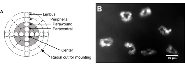
图 2:中央刮伤后角膜成像策略及中性粒细胞浸润情况。 (A)角膜全层铺片的示意图,显示跨越角膜直径的九个显微视野。灰色区域代表原始伤口区域。每个区域的宽度为 500 µm。(B)注意经 DAPI 染色后中性粒细胞核呈现明显的马蹄形或环形结构。请点击此处查看该图的放大版本。
访问受限。请登录或开始试用以查看此内容。
结果
图3 显示了使用钝头高尔夫球杆尖端工具制造的角膜伤口的透射电子显微照片,证明了上皮基底膜在损伤后确实保持完整。
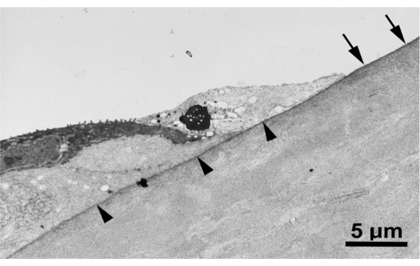
图3:角膜擦伤后上皮基底膜保持完整。 使用钝性高尔夫球杆头制造的角膜伤口的透射电子显微镜图像。箭头指示位于伤口周围残留上皮下方的基底膜,而箭头则指示脱失区域中的基底膜。该图像显示了从未擦伤区域到擦伤区域角膜基底膜的连续性,表明清创术后基底膜仍保持完整。
访问受限。请登录或开始试用以查看此内容。
讨论
本方法学论文的目的是描述一种使用环钻和钝性高尔夫球棒状刮匙在小鼠角膜中央制造上皮擦伤创面的实验方案。该小鼠模型已被用于研究角膜炎症及其在伤口愈合过程中的作用。此类模型可用于在正常生理条件以及病理状态下研究角膜伤口愈合的机制17,28,29,41,42。已有研究证明,该模型可用于病理条件下的角膜伤口愈合研究,例如在饮食诱导肥胖小鼠模型中观察到的角膜伤口愈合受损现象17。该模型的一个显著优势在于,它能够产生精确(2 mm)且可重复的上皮损伤范围,并准确定位于角膜中央,同时不破坏基底膜。这种角膜损伤模型操作简便、快速。除了钝性高尔夫球棒状刮匙29,39
访问受限。请登录或开始试用以查看此内容。
披露
作者无任何利益冲突需要披露。
致谢
基金支持:由美国国立卫生研究院(NIH)EY018239(A.R.B.、C.W.S. 和 R.E.R.)、P30EY007551(A.R.B.)以及Sigma Xi研究援助基金(P.K.A.)资助。本内容仅由作者负责,不代表美国国立卫生研究院或Sigma Xi的官方观点。
访问受限。请登录或开始试用以查看此内容。
材料
| 姓名 | 公司 | 目录编号 | 评论 |
|---|---|---|---|
| 抗CD31抗体 | BD Bioscience, Pharmingen | 550274 | |
| 抗CD41抗体 | BD Bioscience, Pharmingen | 553847 | |
| 抗Ly6G抗体 | BD Bioscience, Pharmingen | 551459 | |
| 牛血清白蛋白(BSA) | ThermoFisher Scientific | B14 | |
| C57BL/6小鼠 | Jackson Laboratories | 664 | |
| DAPI | Sigma Aldrich | D8417 | |
| DeltaVision宽场去卷积荧光显微镜 | GE Life Sciences | ||
| 解剖显微镜 | Leica Microsystems | ||
| 电子台秤(称重仪) | Fisher Scientific | ||
| 乙醇 | ThermoFisher Scientific | T038181000CS | |
| 高尔夫球杆形刮匙 | Stephens Instruments | S2-1135 | |
| 虹膜弯剪 | Fisher Scientific | 31212 | |
| 异氟烷 | Patterson Veterinary | 07-893-1389 | |
| 氯胺酮 | Patterson Veterinary | 07-890-8598 | |
| 磷酸盐缓冲液(PBS) | ThermoFisher Scientific | AM9624 | |
| 荧光素钠盐 | Sigma Aldrich | 46970 | |
| 手术刀片(手术刀刀片) | Fine Science Tools | 10022-00 | |
| 环钻 | Integra Miltex | 33-31 | |
| 吐温X-100 | Fisher Scientific | 50-295-34 | |
| 镊子 | Fine Science Tools | 11923-13 | |
| 赛拉嗪 | Patterson Veterinary | 07-808-1947 |
参考文献
- DelMonte, D. W., Kim, T. Anatomy and physiology of the cornea. Journal of Cataract and Refractive Surgery. 37 (3), 588-598 (2011).
- Meek, K. M., Knupp, C. Corneal structure and transparency. Progress in Retinal and Eye Research. 49, 1-16 (2015).
- Sridhar, M. S. Anatomy of cornea and ocular surface. Indian Journal of Ophthalmology. 66 (2), 190-194 (2018).
- Flaxman, S. R., et al. Global causes of blindness and distance vision impairment 1990-2020: a systematic review and meta-analysis. The Lancet Global Health. 5 (12), 1221-1234 (2017).
- Robaei, D., Watson, S. Corneal blindness: A global problem. Clinical & Experimental Ophthalmology. 42 (3), 213-214 (2014).
- McGwin, G., Owsley, C. Incidence of emergency department-treated eye injury in the United States. Archives of Ophthalmology. 123 (5), 662-666 (2005).
- Ljubimov, A. V., Saghizadeh, M. Progress in corneal wound healing. Progress in Retinal and Eye Research. 49, 17-45 (2015).
- Wilson, S. L., Haj, A. J. E., Yang, Y. Control of scar tissue formation in the cornea: Strategies in clinical and corneal tissue engineering. Journal of Functional Biomaterials. 3 (3), 642(2012).
- Vaidyanathan, U., et al. Persistent corneal epithelial defects: A review article. Medical Hypothesis, Discovery and Innovation in Ophthalmology. 8 (3), 163-176 (2019).
- Netto, M., et al. Wound healing in the cornea: a review of refractive surgery complications and new prospects for therapy. Cornea. 24 (5), 509-522 (2005).
- Friedenwald, J. S., Buschke, W. Some factors concerned in the mitotic and wound-healing activities of the corneal epithelium. Transactions of the American Ophthalmological Society. 42, 371-383 (1944).
- Xu, K., Yu, F. -S. X. Impaired epithelial wound healing and EGFR signaling pathways in the corneas of diabetic rats. Investigative Ophthalmology & Visual Science. 52 (6), 3301-3308 (2011).
- Bai, J. Q., Qin, H. F., Zhao, S. H. Research on mouse model of grade II corneal alkali burn. International Journal of Ophthalmology. 9 (4), 487-490 (2016).
- Blanco-Mezquita, J. T., Hutcheon, A. E. K., Stepp, M. A., Zieske, J. D. αVβ6 integrin promotes corneal wound healing. Investigative Ophthalmology and Visual Science. 52 (11), 8505-8513 (2011).
- Blanco-Mezquita, J. T., Hutcheon, A. E. K., Zieske, J. D. Role of thrombospondin-1 in repair of penetrating corneal wounds. Investigative Ophthalmology and Visual Science. 54 (9), 6262-6268 (2013).
- Stepp, M. A., et al. Wounding the cornea to learn how it heals. Experimental Eye Research. 121, 178-193 (2014).
- Hargrave, A., et al. Corneal dysfunction precedes the onset of hyperglycemia in a mouse model of diet-induced obesity. PLoS ONE. 15, 0238750(2020).
- Machholz, E., Mulder, G., Ruiz, C., Corning, B. F., Pritchett-Corning, K. R. Manual restraint and common compound administration routes in mice and rats. Journal of Visualized Experiments: JoVE. (67), e2771(2012).
- Bodner, L., Dayan, D. Effect of parotid submandibular and sublingual saliva on wound healing in rats. Comparative Biochemistry and Physiology. Part A, Physiology. 100 (4), 887-890 (1991).
- Abbasian, B., Azizi, S., Esmaeili, A. Effects of rat's licking behavior on cutaneous wound healing. Iranian Journal of Basic Medical Sciences. 13 (1), 242-247 (2010).
- DeLisser, H. M., et al. Involvement of endothelial PECAM-1/CD31 in angiogenesis. The American Journal of Pathology. 151 (3), 671-677 (1997).
- Piali, L., et al. CD31/PECAM-1 is a ligand for alpha v beta 3 integrin involved in adhesion of leukocytes to endothelium. The Journal of Cell Biology. 130 (2), 451-460 (1995).
- Fleming, T. J., Fleming, M. L., Malek, T. R. Selective expression of Ly-6G on myeloid lineage cells in mouse bone marrow. RB6-8C5 mAb to granulocyte-differentiation antigen (Gr-1) detects members of the Ly-6 family. The Journal of Immunology. 151 (5), 2399-2408 (1993).
- Fleming, T. J., Malek, T. R. Multiple glycosylphosphatidylinositol-anchored Ly-6 molecules and transmembrane Ly-6E mediate inhibition of IL-2 production. The Journal of Immunology. 153 (5), 1955-1962 (1994).
- Phillips, D. R., Charo, I. F., Scarborough, R. M. GPIIb-IIIa: the responsive integrin. Cell. 65 (3), 359-362 (1991).
- Nieswandt, B., et al. Acute systemic reaction and lung alterations induced by an antiplatelet integrin gpIIb/IIIa antibody in mice. Blood. 94 (2), 684-693 (1999).
- Li, Z., Burns, A. R., Rumbaut, R. E., Smith, C. W. γδ T cells are necessary for platelet and neutrophil accumulation in limbal vessels and efficient epithelial repair after corneal abrasion. American Journal of Pathology. 171 (3), 838-845 (2007).
- Liu, Q., Smith, C. W., Zhang, W., Burns, A. R., Li, Z. NK cells modulate the inflammatory response to corneal epithelial abrasion and thereby support wound healing. American Journal of Pathology. 181 (2), 452-462 (2012).
- Gao, Y., et al. NK cells are necessary for recovery of corneal CD11c+ dendritic cells after epithelial abrasion injury. Journal of Leukocyte Biology. 94 (2), 343-351 (2013).
- Xiao, C., et al. Acute tobacco smoke exposure exacerbates the inflammatory response to corneal wounds in mice via the sympathetic nervous system. Communications Biology. 2, 33(2019).
- Wang, H., et al. Epothilone B speeds corneal nerve regrowth and functional recovery through microtubule stabilization and increased nerve beading. Scientific Reports. 8 (1), 2647(2018).
- Li, Z., Burns, A. R., Smith, C. W. Lymphocyte function-associated Antigen-1-dependent inhibition of corneal wound healing. Cell Injury. 169, 1590-1600 (2006).
- Wu, M., et al. The neuroregenerative effects of topical decorin on the injured mouse cornea. Journal of Neuroinflammation. 17 (1), 1-14 (2020).
- Rodrigues, M., Kosaric, N., Bonham, C. A., Gurtner, G. C. Wound healing: A cellular perspective. Physiological Reviews. 99 (1), 665-706 (2019).
- Rennard, S. I. Inflammation and repair processes in chronic obstructive pulmonary disease. American Journal of Respiratory and Critical Care Medicine. 160 (5), Pt 2 12-16 (1999).
- Landén, N. X., Li, D., Ståhle, M. Transition from inflammation to proliferation: a critical step during wound healing. Cellular and Molecular Life Sciences. 73 (20), 3861-3885 (2016).
- Li, Z., Rumbaut, R. E., Burns, A. R., Smith, C. W. Platelet response to corneal abrasion is necessary for acute inflammation and efficient re-epithelialteation. Investigative Ophthalmology and Visual Science. 47, 4794-4802 (2006).
- Lam, F. W., Burns, A. R., Smith, C. W., Rumbaut, R. E. Platelets enhance neutrophil transendothelial migration via P-selectin glycoprotein ligand-1. American Journal of Physiology - Heart and Circulatory Physiology. 300 (2), 468-475 (2011).
- La Cruz, A. D., et al. Platelet and erythrocyte extravasation across inflamed corneal venules depend on CD18, neutrophils, and mast cell degranulation. International Journal of Molecular Sciences. 22 (14), 7360(2021).
- Li, Z., Burns, A. R., Smith, C. W. Two waves of neutrophil emigration in response to corneal epithelial abrasion: Distinct adhesion molecule requirements. Investigative Ophthalmology and Visual Science. 47 (5), 1947-1955 (2006).
- Li, Z., Burns, A. R., Han, L., Rumbaut, R. E., Smith, C. W. IL-17 and VEGF Are Necessary for Efficient Corneal Nerve Regeneration. The American Journal of Pathology. 178 (3), 1106-1116 (2011).
- Xue, Y., et al. Modulation of circadian rhythms affects corneal epithelium renewal and repair in mice. Investigative Ophthalmology and Visual Science. 58 (3), 1865-1874 (2017).
- Zhang, W., Magadi, S., Li, Z., Smith, C. W., Burns, A. R. IL-20 promotes epithelial healing of the injured mouse cornea. Experimental Eye Research. 154, 22-29 (2017).
- Li, Z., Burns, A. R., Miller, S. B., Smith, C. W. CCL20, γδ T cells, and IL-22 in corneal epithelial healing. FASEB Journal. 25 (8), 2659-2668 (2011).
- Li, Z., Burns, A. R., Han, L., Rumbaut, R. E., Smith, C. W. IL-17 and VEGF are necessary for efficient corneal nerve regeneration. American Journal of Pathology. 178 (3), 1106-1116 (2011).
- Reins, R. Y., Hanlon, S. D., Magadi, S., McDermott, A. M. Effects of topically applied Vitamin D during corneal wound healing. PLoS ONE. 11 (4), 0152889(2016).
- Gagen, D., et al. ICAM-1 mediates surface contact between neutrophils and keratocytes following corneal epithelial abrasion in the mouse. Experimental Eye Research. 91 (5), 676-684 (2010).
- Li, Z., Rivera, C. A., Burns, A. R., Smith, C. W. Hindlimb unloading depresses corneal epithelial wound healing in mice. Journal of Applied Physiology. 97 (2), 641-647 (2004).
- Byeseda, S. E., et al. ICAM-1 is necessary for epithelial recruitment of γδ T cells and efficient corneal wound healing. American Journal of Pathology. 175 (2), 571-579 (2009).
- Li, Z., Burns, A. R., Rumbaut, R. E., Smith, C. W. γδ T cells are necessary for platelet and neutrophil accumulation in limbal vessels and efficient epithelial repair after corneal abrasion. American Journal of Pathology. 171 (3), 838-845 (2007).
- Petrescu, M. S., et al. Neutrophil interactions with keratocytes during corneal epithelial wound healing: A role for CD18 integrins. Investigative Ophthalmology and Visual Science. 48 (11), 5023-5029 (2007).
- Pal-Ghosh, S., Pajoohesh-Ganji, A., Tadvalkar, G., Stepp, M. A. Removal of the basement membrane enhances corneal wound healing. Experimental Eye Research. 93 (6), 927-936 (2011).
- Pal-Ghosh, S., et al. Cytokine deposition alters leukocyte morphology and initial recruitment of monocytes and γδT cells after corneal injury. Investigative Ophthalmology & Visual Science. 55 (4), 2757-2765 (2014).
- Pal-Ghosh, S., Tadvalkar, G., Jurjus, R. A., Zieske, J. D., Stepp, M. A. BALB/c and C57BL6 mouse strains vary in their ability to heal corneal epithelial debridement wounds. Experimental Eye Research. 87 (5), 478-486 (2008).
- Kato, T., Chang, J. H., Azar, D. T. Expression of type XVIII collagen during healing of corneal incisions and keratectomy wounds. Investigative Ophthalmology and Visual Science. 44 (1), 78-85 (2003).
- Kure, T., et al. Corneal neovascularization after excimer keratectomy wounds in matrilysin-deficient mice. Investigative Ophthalmology and Visual Science. 44 (1), 137-144 (2003).
- Lin, A., et al. Bacterial keratitis preferred practice pattern. Ophthalmology. 126 (1), 1-55 (2019).
- Cable, E. J., Onishi, K. G., Prendergast, B. J. Circadian rhythms accelerate wound healing in female Siberian hamsters. Physiology and Behavior. 171, 165-174 (2017).
- Lyons, A. B., Moy, L., Moy, R., Tung, R. Circadian rhythm and the skin: A review of the literature. Journal of Clinical and Aesthetic Dermatology. 12 (9), 42-45 (2019).
- Westman, J., Grinstein, S., Marques, P. E. Phagocytosis of Necrotic Debris at Sites of Injury and Inflammation. Frontiers in Immunology. 10, 3030(2020).
- Gaudry, M., et al. Intracellular pool of vascular endothelial growth factor in human neutrophils. Blood. 90 (10), 4153-4161 (1997).
- Pan, Z., et al. Vascular endothelial growth factor promotes anatomical and functional recovery of injured peripheral nerves in the avascular cornea. FASEB Journal. 7, 2756-2767 (2013).
- Di, G., et al. VEGF-B promotes recovery of corneal innervations and trophic functions in diabetic mice. Scientific Reports. 7 (1), 1-13 (2017).
- Thomas, M. R., Storey, R. F. The role of platelets in inflammation. Thrombosis and Haemostasis. 114 (3), 449-458 (2015).
- Margraf, A., Zarbock, A. Platelets in inflammation and resolution. The Journal of Immunology. 203 (9), 2357-2367 (2019).
访问受限。请登录或开始试用以查看此内容。
重印与许可
