本文介绍了一些成本低廉且操作简便的方法,可在细胞培养实验室中常规建立多种3D皮肤模型。研究人员可根据自身需求自行构建模型,而无需依赖 commercially available(商业化的)模型。
方法文章
从二维(2D)单层培养到三维(3D)多细胞共培养:构建适用于多种应用的皮肤模型
* These authors contributed equally
本文内容
摘要
摘要
由于皮肤具有复杂的结构和重要的功能,因此成为化妆品、制药和医疗行业一个引人关注的研究模型。在欧盟,已全面禁止对化妆品及其成分进行动物试验。对于药品和制药领域,此类试验的可能性也受到持续限制。根据“3R”原则,对人工构建的模型进行单个化合物乃至完整配方的测试正变得越来越普遍。其中成本最低且应用最广泛的是二维模型,它由单层细胞构成,但无法反映组织中细胞之间的真实相互作用。尽管市面上可获得的三维模型能更真实地模拟组织结构,但其大规模应用仍受限,原因在于价格昂贵、等待时间较长,且现有模型通常仅限于少数常规类型。
为了将已开展的研究提升到更高水平,我们优化了多种3D皮肤模型制备的实验流程。所述方法成本低廉且制备简便,可适用于众多实验室以及具有不同细胞培养经验的研究人员。
引言
皮肤是一个具有多细胞相互作用的连续结构,反映了这一复杂器官的正常功能与稳态。皮肤由形态学上不同的层次构成:内层为真皮,外层为表皮。在表皮的最上方,我们还可进一步区分出角质层(由扁平的死亡细胞——角质细胞组成),其对外界环境提供最强的保护作用。皮肤的一些最重要被动与主动功能包括:抵御外界因素对身体的侵害、参与免疫学过程、分泌、吸收、体温调节以及感觉功能1,2,3。由于皮肤被认为是人体最大的器官之一,不可避免地会接触到各种病原体、过敏原、化学物质以及紫外线(UV)辐射。因此,皮肤由多种具有特定功能的细胞构成。表皮中存在的主要细胞类型包括角质形成细胞(几乎占所有细胞的90%,在表皮深层具有结构和免疫功能,随后经历角质化过程,最终转化为表皮最上层的角质细胞)、黑色素细胞(仅占表皮细胞群体的3%–7%,可产生具有紫外线防护作用的黑色素)以及朗格汉斯细胞(属于免疫系统)。在真皮中,主要细胞为成纤维细胞(可产生生长因子和蛋白质)、树突状细胞以及肥大细胞(后两者均属于免疫系统细胞)4,5,6。此外,皮肤还含有多种细胞外蛋白(如I型和IV型胶原蛋白、纤连蛋白和层粘连蛋白;图1)以及蛋白纤维(胶原蛋白和弹性蛋白),这些成分不仅维持皮肤的特定结构,还促进细胞结合、细胞黏附及其他相互作用7。

图 1:皮肤结构示意图。 该图标注了存在于皮肤各层中的四种基本细胞类型,并标明了细胞外基质中的不同蛋白质。本图使用 Microsoft PowerPoint 制作。请点击此处查看此图的放大版本。
化妆品和药品的安全性是一个非常重要的问题,保护消费者和患者的健康是首要任务8直到最近,这一目标被认为可通过包括动物实验在内的大量测试来确保。然而,这些实验通常需要采用极端手段,导致用于研究目的的动物(通常是小鼠、大鼠和猪)承受疼痛与痛苦。1959年,《人道实验技术原则》(3R原则)被提出:(1-替代)以非动物方法替代动物实验, 体外, 在计算机中 或 离体 模型,(2 - 减量)减少用于研究的动物数量,以及(3 - 优化)改善仍需用于研究的动物的福利,同时改进所开发的替代方法9此外,在欧洲联盟(EU)内,动物化妆品测试受到法律监管。自2004年9月11日起,禁止销售经动物测试的化妆品的禁令正式生效。2009年3月11日,欧盟进一步禁止对化妆品原料进行动物测试,禁止销售使用新经动物测试的原料制成的化妆品;然而,针对重复剂量毒性、生殖毒性及毒物动力学等复杂的人体健康问题,仍允许对产品进行动物测试。自2013年3月11日起,欧盟全面禁止销售任何成品或其成分曾进行动物测试的化妆品。10因此,目前在美容学领域,研究在三个层面上进行: 体外 (细胞),离体 (真实组织),以及 体内 (志愿者)11在药品研发中,动物实验的需求仍然存在,但已大幅减少并受到严格管控。12.
作为动物实验的替代方法以及对新型活性成分有效性进行初步评估的方法, 体外 皮肤细胞培养被用于此类研究。在无菌实验室条件下分离不同类型的皮肤细胞并进行培养,可评估活性物质的安全性和毒性。皮肤细胞系也被广泛认可为研究模型,因为这些细胞由认证公司提供,使得不同实验室之间的实验结果具有可比性。这些测试通常在简单的人体皮肤细胞单层培养的二维模型上进行。一些更先进的模型包括共培养体系(如角质形成细胞与成纤维细胞、角质形成细胞与黑素细胞的共培养),以及三维模型,后者包含无支架培养(如球体培养)和基于支架的表皮、真皮乃至全层皮肤替代物的模拟结构。13值得一提的是,除最后一类(皮肤等效物)外,其余模型均未商业化供应,如有需要,科研人员需自行制备。
尽管如今已有大量此类模型被保存下来并 routinely 出售(表1),为了验证大多数研究结果,还需不断开发新的模型。因此,新构建的模型应能更好地模拟人体内发生的真实相互作用。当采用多种不同类型细胞的混合物构建此类模型时,可再现组织的多细胞特性 体内 可以实现。结果,建立了器官型培养(图2).
| 名称 | 描述 | |||||
| 正常皮肤 | EpiSkin | 重建人表皮 - 胶原膜上的角质形成细胞 | ||||
| SkinEthic RHE | 重建人表皮 - 聚碳酸酯膜上的角质形成细胞 | |||||
| SkinEthic RHE-LC | 含朗格汉斯细胞的人表皮模型 - 聚碳酸酯膜上的角质形成细胞和朗格汉斯细胞 | |||||
| SkinEthic RHPE | 重建人色素表皮 - 聚碳酸酯膜上的角质形成细胞和黑色素细胞 | |||||
| T-Skin | 重建人全层皮肤模型 - 生长在聚碳酸酯膜上的成纤维细胞层及其表面的角质形成细胞 | |||||
| Phenion FT Skin model | 水凝胶中的角质形成细胞和成纤维细胞 | |||||
| 病变皮肤 | 黑色素瘤全层皮肤模型 | 正常人源性角质形成细胞和成纤维细胞,以及人恶性黑色素瘤细胞系 A375 | ||||
| 银屑病组织模型 | 正常人角质形成细胞和成纤维细胞 | |||||
表1:用于各类研究的最常用商用皮肤等效物。
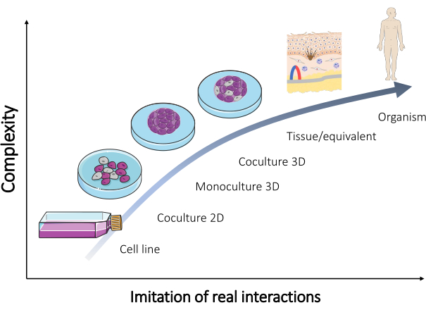
图2:不同复杂性 体外 模型 不同复杂性之间的关系 体外 利用模型重建生物体以及直接在人体内发生的实际相互作用。该图改编自 Servier 公司的“微生物学与细胞培养”系列(Servier Medical Art,https://smart.servier.com/)。 请点击此处以查看此图的放大版本。
商业等效模型最重要的局限性之一是仅提供非常通用的研究模型,且所含细胞类型很少(通常为1-2种,极少达到3种)。然而,皮肤中实际存在的细胞种类远多于此,这些细胞之间的相互作用可能显著影响各种成分的耐受性14。某些免疫组分的缺失会降低其在多种研究中的应用价值,包括免疫治疗研究。这是一个严重问题,因为黑色素瘤是一种危及生命的皮肤癌,其特点为早期发生转移以及对治疗方案频繁产生耐药性15。为了改进人工皮肤模型,研究人员正尝试建立免疫细胞与细胞系及类器官的共培养体系16,这被认为是所研究模型的重大进步。例如,肥大细胞参与皮肤中的多种生理过程(如伤口愈合、组织重塑)和病理过程(如炎症、血管生成及肿瘤进展)17。因此,模型中是否存在肥大细胞会显著改变模型对研究化合物的反应。最后,目前仍缺乏大量与皮肤相关的基础信息,这些信息只能通过基础研究来揭示。因此,构建和优化不同类型的人工皮肤模型(表2)是一项极为重要的工作。本文介绍了多种构建先进皮肤模型的方法,包括球状体和皮肤等效物。
| 体外 皮肤模型 | 尝试重建组织中发生的相互作用 | 所用细胞示例 |
| 2D 或 3D 细胞培养 | 表皮 | 角质形成细胞 |
| 黑色素细胞 | ||
| 角质形成细胞 + 黑色素细胞 | ||
| 真皮 | 成纤维细胞 | |
| 肥大细胞 | ||
| 成纤维细胞 + 肥大细胞 | ||
| 皮肤 | 角质形成细胞 + 成纤维细胞 | |
| 角质形成细胞 + 肥大细胞 | ||
| 黑色素细胞 + 成纤维细胞 | ||
| 黑色素细胞 + 肥大细胞 | ||
| 角质形成细胞 + 成纤维细胞 + 黑色素细胞 | ||
| 角质形成细胞 + 成纤维细胞 + 肥大细胞 |
表2:在二维和三维培养中重建皮肤组织的细胞类型混合示例。
方案
本研究遵循赫尔辛基宣言的指导原则,并已获得华沙医科大学伦理委员会(KB/7/2022)的批准。所有参与研究的受试者均签署了知情同意书。
注意:高级皮肤模型制备所描述的操作步骤可使用市售的原代皮肤细胞与细胞系,或从患者中分离的原代细胞进行。商业来源的细胞通常附带相关文件资料,其在大多数国家的研究使用无需额外审批。然而,部分国家对此有强制性规定,因此必须查阅当地伦理委员会的相关法规。若研究中需使用从患者分离的原代细胞,则必须首先获得当地伦理委员会的批准,并严格按照其制定的严格指南开展实验。此外,必须从所有皮肤组织捐献者处获取书面知情同意书。本文未涉及原代皮肤细胞的分离方法,但可参考以下文献中的示例性分离流程:Kosten 等人(角质形成细胞)18,Ścieżyńska 等人(黑素细胞)19,Kröger 等人(成纤维细胞与肥大细胞)20。大多数正常皮肤细胞及细胞系属于生物安全等级 BSL1 类别,不会造成任何危害。然而,所使用的实验室设备必须符合在受控条件下进行人和动物细胞培养的相关标准。
1. 皮肤细胞培养
注意:皮肤细胞培养必须在专用的培养瓶中进行,根据细胞类型选择用于贴壁细胞或悬浮细胞的培养瓶,并在 37 °C、含 5% 二氧化碳的培养箱中培养。与细胞培养及用于研究相关的所有操作均需在无菌条件下进行,且必须在经紫外线 C(UVC)照射 15–30 分钟后的超净工作台内操作。获取细胞悬液(用于构建二维和三维模型)需根据细胞类型采用相应的操作步骤(对于贴壁细胞,如角质形成细胞、成纤维细胞和黑色素细胞,参见步骤 1.1;对于非贴壁细胞,如肥大细胞,参见步骤 1.2)(图 3)。针对不同规格的培养瓶,本方法中所用各试剂(如培养基、磷酸盐缓冲液或胰蛋白酶溶液)的体积详见 Table 3。与细胞类型相关的参数(如试剂浓度与组成、离心方法等)列于 Table 4。接种时所用的细胞密度见 Table 5。以上所有表格均位于本节末尾。
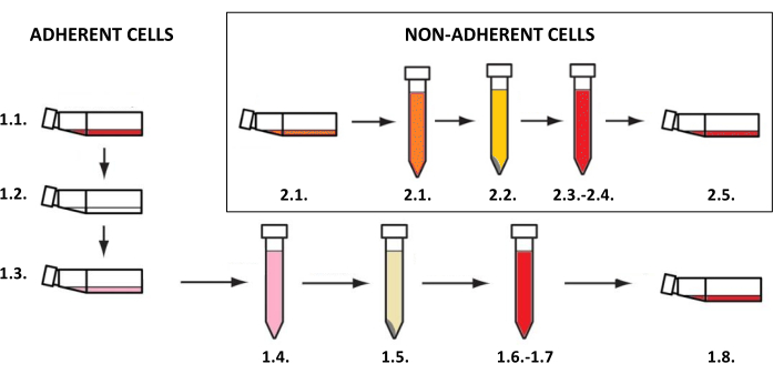
图3:贴壁细胞与非贴壁细胞的培养。贴壁细胞与非贴壁细胞培养的一般分步流程(数字对应步骤1.1和1.2的描述)。该图使用MS PowerPoint绘制。请点击此处查看此图的放大版本。
- 获得贴壁细胞的悬浮液
- 从培养瓶中移除培养基。
- 用磷酸盐缓冲液(PBS,表 3)轻轻洗涤细胞。
- 加入含乙二胺四乙酸的胰蛋白酶溶液(胰蛋白酶-EDTA 溶液,表 3)。将培养瓶置于 37 °C 培养,并在光学显微镜下观察细胞从表面脱落的情况。
- 将脱落的细胞悬浮于至少两倍体积的完全培养基或胰蛋白酶中和液中以灭活胰蛋白酶(比例为 2:1)(体积参见表 3,试剂参见 表 4)。将培养瓶中的内容物定量转移至 15 mL 离心管中。
- 取少量细胞悬液(20 µL)至 1.5 mL 离心管中,使用手动或自动细胞计数板进行细胞计数。
- 离心(参数见 表 4),弃去大部分上清液,将细胞沉淀重悬于少量残留液体中。然后,加入足够的新鲜培养基以恢复至离心前的体积(体积见 表 3),若细胞密度适合接种;否则重新计算所需的新鲜培养基体积。
注意:某些细胞(如黑素细胞)对离心非常敏感,因此应避免在短时间内重复离心。 - 根据实验需求(皮肤细胞的二维/三维单培养或共培养),制备所需密度的细胞悬液(细胞数/mL,细胞密度见 表 5)。
注意:若需继续培养细胞,将 5,000–8,000 个细胞/mL 接种至新培养瓶中,并加入新鲜培养基(体积见 表 3)。
- 获得非贴壁细胞的悬浮液
- 将含有细胞悬液的培养基从培养瓶中移出,并定量转移至 15 mL 离心管中。
- 取少量细胞悬液(20 µL)至新的 1.5 mL 离心管中,使用手动或自动细胞计数板进行细胞计数。
- 离心(参数见 表 4),弃去大部分上清液,将细胞重悬于少量残留液体中。然后,加入新鲜培养基以恢复至离心前的体积(见 表 3),若细胞密度适合接种;否则重新计算所需的新鲜培养基体积。
- 根据实验需求(皮肤细胞的二维/三维单培养或共培养),制备所需密度的细胞悬液(每毫升细胞数,建议值见 表 5)。
注意:若需继续培养细胞,将 5,000–8,000 个细胞/mL 接种至新培养瓶中,并加入新鲜培养基。
| 25 cm2 培养瓶 | 75 cm2 培养瓶 | |
| 培养基 [mL] | 4–5 | 8–12 |
| PBS [mL] | 5 | 10 |
| 胰蛋白酶–EDTA [mL] | 0.5–1 | 1–2 |
| 中和培养基 [mL] | 1–2 | 2–4 |
表3:细胞培养及细胞悬液制备过程中所用试剂的体积。
| 皮肤细胞单层培养 | 胰蛋白酶 | 胰蛋白酶失活剂 | 离心 | 二维单层培养的培养基类型 | |
| 角质形成细胞 | 0.25% | 含胰蛋白酶中和剂 | 300 × g,5 分钟,室温 | Keratinocyte Growth Medium 2 | |
| 成纤维细胞 | 0.25% | 用培养基 | 300 × g,5 分钟,室温 | DMEM, 10% FBS | |
| 黑素细胞 | 0.025% | 含胰蛋白酶中和剂 | 300 × g,3 分钟,室温 | Medium 254, PMA-Free Human Melanocyte Growth Supplement-2 | |
| 肥大细胞 | 无需 | 无需 | 300 × g,3 分钟,室温 | IMDM, 10% FBS, 1% 非必需氨基酸, 226 µM α-巯基甘油 | |
表4:胰蛋白酶消化、离心参数以及培养基类型取决于细胞种类。
| 模型类型 | 细胞密度 [cell/mL] | ||
| 2D | 单层培养 | 成纤维细胞 | 2 x 10⁵ |
| 肥大细胞 | |||
| 角质形成细胞 | |||
| 黑素细胞 | |||
| 3D | 球体(悬滴法) | 成纤维细胞 | 5 x 10⁵ |
| 肥大细胞 | |||
| 角质形成细胞 | |||
| 黑素细胞 | |||
| 混合细胞 | |||
| 球体(限制性贴壁法) | 成纤维细胞 | 2 x 10⁵ | |
| 肥大细胞 | |||
| 角质形成细胞 | |||
| 黑素细胞 | |||
| 混合细胞 | |||
| 等效组织 | 成纤维细胞 | 1 x 10⁵ | |
| 肥大细胞 | 1 x 10⁴ | ||
| 角质形成细胞 | 8 x 10⁵ | ||
| 黑素细胞 | 5 x 10⁴ | ||
表5:不同类型皮肤模型的细胞接种密度。
2. 皮肤细胞球的制备
注意:步骤 2.1(图 4)中描述了使用悬滴法形成球体的方法,而步骤 2.2(图 5)则展示了通过限制细胞黏附来形成球体的方法。然而,由于球体非常微小且容易不稳定,采用这些方法进行操作时需要耐心、细致和缓慢的动作。
- 悬滴法
- 使用适当的细胞密度以获得所需球体大小(推荐细胞密度为 5 x 10⁵ 个细胞/mL,表 5)。
- 用移液器将 20 µL 细胞悬液滴加到培养皿或微量孔板的盖子上(图 4A)。
- 用培养皿/孔板的底部盖住,并轻轻翻转(悬滴将自动形成在盖子上,图 4B)。
- 在培养皿/孔板的孔中加入无菌水/PBS 溶液,以防止液滴中的培养基蒸发。
- 将液滴在 37 °C 下孵育 48–72 小时。
注意:重力使细胞下沉,而缺乏可附着的表面可防止细胞贴壁,从而促进细胞聚集。然而,某些细胞类型可能需要更长的孵育时间。 - 在进行下一步操作前,向新孔板的孔中加入完全培养基(100 µL)(或使用已从孔中移除水/PBS 的旧孔板)。
注意:在下一步操作前,取 200 µL 吸头,剪去每根吸头末端的 1/5,并在使用前进行灭菌。 - 使用末端剪切过的无菌移液吸头将细胞球体转移至多孔板的孔中。取 200 µL 吸头,剪去每根吸头末端的 1/5,并在使用前灭菌(图 4C)。
注意:此步骤可能较困难,因为在翻转培养皿/孔板时液体流动可能会破坏球体。 - 将转移后的球体在多孔板中于 37 °C 下继续孵育 1 天,然后再进行后续实验(例如化合物添加、细胞毒性检测、将球体引入类器官系统等)(图 4D)。
- 限制细胞贴壁法
- 在接种细胞前,用表面活性剂溶液(例如 Pluronic F-127、聚乙二醇、聚乙烯醇)21 预处理 U 型底孔板的孔。在 PBS 中配制 1% 的表面活性剂溶液,每孔加入 100 µL。将孔板在 37 °C 下孵育 24 小时(图 5A-C)。如需长期保存,可继续储存,但需通过补充 PBS 缓冲液维持液面高度。
- 将细胞悬液按每孔 50 µL 的体积和所需细胞密度进行制备(推荐接种密度为 2 x 10⁵/mL,表 5)。
- 在接种细胞前,从孔中移除表面活性剂溶液,以避免细胞膜因裂解而受损(图 5D)。
- 将细胞悬液加入孔板中,在 37 °C 下孵育 24 小时以形成细胞聚集体(图 5E)。大约 1–3 天后,球体将形成(图 5F),并可用于后续实验。
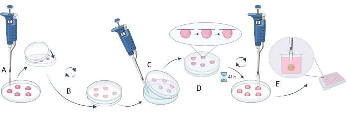
图4:悬滴法。(A)将细胞悬液滴加至培养皿盖内表面,并用培养皿底部盖住皿盖;(B)旋转培养皿使液滴悬于皿盖内侧形成悬滴;(C)向培养皿底部加入水或PBS(以减少液体蒸发);(D)将含悬滴的培养皿进行孵育,以形成细胞球体;(E)收集已形成球体的液滴,并将转移后的球体置于多孔板中进行稳定培养。该图由Biorender.com制作。请点击此处查看此图的放大版本。
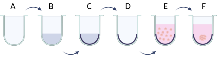
图 5:通过限制性细胞黏附法逐步制备细胞球。 (A)U型底孔板;(B、C)通过表面活性剂溶液实现限制性细胞黏附;(D)从孔板中移除溶液;(E)接种细胞;(F)细胞聚集并形成细胞球。该图由 Biorender.com 制作。 请点击此处查看此图的放大版本。
3. 全层皮肤替代物的制备
注意:全层(表皮和真皮)皮肤等效物的构建可分为三个步骤(图6):首先制备含典型真皮细胞(如成纤维细胞和肥大细胞,图6A)的人工真皮层;随后在人工表皮中接种细胞(主要为角质形成细胞和黑色素细胞,图6B);最后角质形成细胞垂直生长并可能发生角化过程(形成角质层,图6C)。全层皮肤等效物的制备详见步骤3.1(3.1.1–3.1.10)。若需要构建较简单的皮肤等效物(例如仅表皮型),可将选定的细胞类型(如角质形成细胞)直接接种于 commercially available collagen 或 polycarbonate membranes 上,并进行培养,也可诱导其发生角化过程(可直接进入步骤3.1.9–3.1.10)。
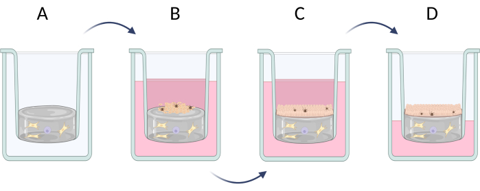
图6:在插入式培养器中逐步制备全层皮肤等效物。(A)含真皮细胞的假真皮层制备,(B)表皮细胞接种,(C)在培养基中进一步培养皮肤等效物,(D)气-液界面培养促进复层上皮的形成。该图由Biorender.com制作。请点击此处查看此图的放大版本。
- 在24孔板中制备全层皮肤等效物
- 将装有水、PBS(10×)、1 M NaOH 和 I 型胶原蛋白溶液的试管置于冰上。
- 确定适宜数量的真皮细胞(如成纤维细胞和肥大细胞,比例为10:1)接种于水凝胶中。将适量真皮细胞转移至1.5 mL离心管中500 µL 成纤维细胞的 500 µL 肥大细胞的数量,依据细胞密度 表5) 并离心细胞(300 × g) g,3 分钟,室温。
- 移去上清液,并将细胞在混合液中轻轻重悬 695 µL 水/100 µL PBS(10倍浓度)/ 5 µL NaOH。
注意:如果总体积溶液1 mL不足,可使用 表6 重新计算每种试剂的体积。 - 添加 200 µL 将胶原溶液加入混合物中,并用移液器轻轻混匀。
注意:需小心操作,因为混合物的质地会较为浓稠。 - 添加 200 µL 将制备好的混合物加入24孔板的插入式培养小室中。用于无角质层模型的实验, 添加 500 µL 向24孔板的每个孔中加入。
- 将培养板在室温(RT)孵育10分钟,然后转移至培养箱中孵育30分钟。
注意:在进行任何其他操作前,须检查水凝胶是否已聚合。 - 在将细胞接种到水凝胶表面之前,用 PBS 缓冲液冲洗水凝胶表面(500 µL/well).
- 确定接种在水凝胶顶部的表皮细胞(如角质形成细胞和黑色素细胞,比例为15:1)的合适数量。制备细胞混合液于 500 µL 含10% FBS的DMEM培养基(添加 250 µL 角质形成细胞的 250 µL 黑色素细胞,细胞密度在 表 4)并轻柔地加入孔板中。
注意:在某些情况下,可先接种黑素细胞,使其在水凝胶上充分铺展,24 小时后更换培养基并接种角质形成细胞。在此情况下,添加 250 µL 细胞悬液 250 µL 培养基的 - 将培养板置于培养箱中孵育 37 °C 培养2-5天,具体时间取决于细胞生长速度,每48小时更换一次培养基(FBS浓度从10%逐步降至1%),并在光学显微镜下监测细胞状态。
- 如果需要在水凝胶表面的角质形成细胞形成单层后诱导角化过程,则使用不含FBS、额外添加钙离子和L-抗坏血酸的培养基继续培养2-7周(浓度为 1.5 µM CaCl2 和 50 µg/mL L-抗坏血酸
注意:孵育时间取决于所使用的角质形成细胞及其分化速度。
| 试剂 | 计算试剂体积的公式 | 示例计算 | |
| (最终体积 Vtotal = 1 mL) | |||
| I型胶原溶液 | 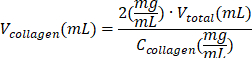 | (Ccollagen = 10 mg/mL) | |
| 0.2 mL = 200 μL | |||
| PBS (10x) |  | 0.1 mL = 100 μL | |
| 1 M NaOH |  | 0.005 mL = 5 μL | |
| 无菌 H2O |  | 0.695 mL = 695 μL | |
表6:制备2 mg/mL I型胶原蛋白水凝胶所需试剂体积的计算。
4. 通过细胞染色方法鉴定三维皮肤模型中的细胞类型
注意:为了确认所构建的皮肤模型包含预期的细胞类型,建议进行细胞染色。在对特定模型开展任何靶向实验之前,这一步骤至关重要22。对于三维皮肤模型,必须先将模型包埋于石蜡中,并使用切片机将人工组织切成薄片以制备显微镜载玻片(步骤 4.1),然后进行细胞染色(图 7)。
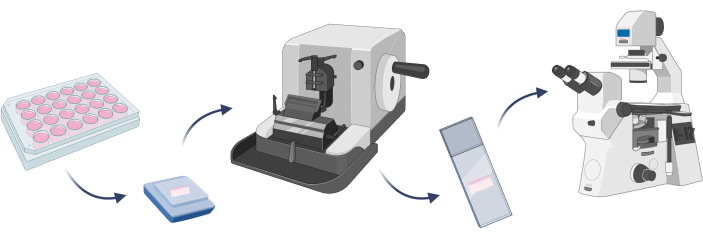
图7:三维皮肤模型包埋、细胞染色及显微观察的基本步骤。 该图由Biorender.com制作。 请点击此处查看此图的放大版本。
- 3D皮肤模型的包埋
- 在室温下用PBS洗涤皮肤等效物两次,每次5分钟,并使用PBS中的3.7%多聚甲醛溶液固定(30分钟,室温)。重复用PBS洗涤步骤。
- 包埋前,通过将皮肤等效物依次孵育于浓度递增的乙醇溶液中进行脱水:50%(15分钟)、70%(15分钟)、96%(2次,每次30分钟)和99.8%(2次,每次30分钟)。
- 将固定并脱水后的皮肤等效物放入盛有石蜡的模具中。
注:将等效物以适当的方向放置。 - 用包埋盒盖住模具,并在顶部添加更多石蜡。在室温下使其凝固最多30分钟。
- 将石蜡包埋的皮肤等效物在-80 °C下冷冻至少1小时。
- 打开切片机,插入石蜡包埋的皮肤等效物,切取5 µm厚的切片。将切下的类组织切片置于载玻片上,并在37 °C下干燥至少8小时。
- 将载玻片浸入二甲苯中(2次,每次10分钟),然后用浓度递减的乙醇溶液再水化:99.8%(5分钟)、96%(5分钟)、70%(5分钟)和50%(5分钟)。
- 从乙醇溶液中取出载玻片,用蒸馏水冲洗两次(每次5分钟)。
注:传统的细胞染色可通过应用特异性染料(苏木精、伊红23)或使用特异性靶向生物标志物的抗体进行(包括成纤维细胞的胶原蛋白1A2、角质形成细胞的角蛋白14、黑素细胞的melan-A或酪氨酸酶24)。常规的苏木精-伊红染色可按照不同公司提供的方案进行(步骤4.2)。另一方面,若需进行免疫荧光或免疫组织化学染色,则操作流程不同且耗时更长(步骤4.3和4.4)。为避免非特异性反应,应使用来自不同物种的一抗,随后使用相应的二抗。
- 3D皮肤模型的苏木精-伊红染色
- 在室温下用苏木精溶液对载玻片染色3分钟。
- 用酸性乙醇溶液洗涤载玻片1分钟。
注:通过将2 mL 35%-38%盐酸与98 mL 99.8%乙醇混合制备酸性乙醇溶液。 - 接着,用0.1%碳酸氢钠溶液洗涤载玻片,以获得明显的淡蓝紫色。
注:将100 mg碳酸氢钠溶解于100 mL超纯水中,即可配制0.1%碳酸氢钠溶液。 - 用95%乙醇洗涤载玻片1分钟。
- 在室温下用伊红溶液对载玻片染色1分钟。
- 用95%乙醇洗涤载玻片1分钟,再用99.8%乙醇洗涤2分钟。
- 用二甲苯洗涤载玻片,每次2分钟,共两次。
- 用加拿大树胶封片,并在载玻片上加盖盖玻片。样品即可用于显微镜观察。
- 3D皮肤模型的免疫荧光染色
- 用PBS冲洗载玻片(5分钟)。
- 制备封闭液(含0.1% Triton X-100和0.1% Tween 20的PBS缓冲液中加入3%牛血清白蛋白[BSA]或脱脂奶粉),将载玻片在其中孵育1小时(室温)。
- 用PBS缓冲液洗涤载玻片两次(每次5分钟)。
- 根据生产商推荐条件(表7),用PBS缓冲液稀释一抗,并在室温下孵育1–2小时,或在4 °C下过夜。
- 用PBS缓冲液洗涤载玻片两次(每次5分钟)。
- 根据生产商推荐条件(表7),用PBS缓冲液稀释二抗,并在室温下孵育1小时。
- 用PBS洗涤载玻片两次(每次5分钟)。
- 制备用于细胞核染色的染料溶液(如Hoechst 33342或DAPI,表7),并在室温下孵育最多15分钟。
- 用PBS洗涤载玻片(5分钟)。
- 用加拿大树胶封片,加盖盖玻片,并使用荧光显微镜观察细胞染色结果。
- 3D皮肤模型的免疫组织化学染色
- 执行步骤4.3.1–4.3.5。
- 额外使用与二抗偶联酶相匹配的缓冲液进行一次洗涤步骤。
- 根据生产商推荐条件,用与偶联酶相匹配的缓冲液稀释二抗,并在室温下孵育1小时。
- 用PBS洗涤载玻片两次(每次5分钟)。
- 配制适用于所用酶的合适底物溶液,并根据生产商推荐条件孵育载玻片。
- 用缓冲液洗涤载玻片(5分钟),然后用加拿大树胶封片。
- 使用明场显微镜观察细胞染色结果。
| 检测的细胞类型/细胞类器官 | 染色剂 | 稀释倍数 / 浓度 | ||
| 肥大细胞 | 亲和素−磺基罗丹明 101 | 1 μg/mL | ||
| 成纤维细胞 | 兔源 Col1A2 抗体 | 1:50 | ||
| FITC 标记的山羊抗兔二抗 | 1:250 | |||
| 角质形成细胞 | 鼠源细胞角蛋白 14 抗体 | 1:50 | ||
| FITC 标记的山羊抗鼠二抗 | 1:250 | |||
| 黑色素细胞 | 鼠源 Melan-A 抗体 | 1:50 | ||
| FITC 标记的山羊抗鼠二抗 | 1:250 | |||
| 细胞核 | Hoechst 33342 | 1 μg/mL | ||
| DAPI | 1 μg/mL | |||
表7:细胞染色所用试剂的浓度与稀释比例。
结果
在实验室开始构建皮肤模型之前,必须确定所用细胞的类型(原代细胞/细胞系)以及为这些细胞选择合适的培养基。大多数细胞库均推荐并可提供适用于各类细胞培养的培养基。对于共培养模型,需选择一种能够满足培养中所有细胞类型需求的培养基。表818,19,20,25,26,27 汇总了常用于原代皮肤细胞培养及常见皮肤细胞系的一些典型培养基。用于原代细胞培养的培养基通常价格较高,且成分复杂;而细胞系通常可适应成分简单的培养基。某些细胞类型(主要是成纤维细胞和肥大细胞)能够产生并分泌促进其他细胞(如角质形成细胞和黑素细胞)生长的因子28,29。若模型中包含此类细胞,则无需额外补充培养基成分。
| 细胞类型 | 细胞名称 | 培养基 | 参考文献 | ||||
| 角质形成细胞 | HaCaT 细胞系 | DMEM, 10% FBS, 100 U/mL 青霉素, 100 μg/mL 链霉素 | 根据供应商说明 | ||||
| 原代正常人表皮角质形成细胞 (NHEK) | 角质形成细胞生长培养基2(基础培养基 + 补充剂混合物) | 根据供应商说明 | |||||
| 原代人表皮角质形成细胞;正常成人 (HEKa) | 真皮细胞基础培养基, 0.4% 牛垂体提取物, 0.5 ng/mL 重组人转化生长因子-α, 6 mM L-谷氨酰胺, 100 ng/mL 氢化可的松半琥珀酸酯, 5 mg/mL 重组人胰岛素, 1 mM 肾上腺素, 5 mg/mL 去唾液酸转铁蛋白, 100 U/mL 青霉素(如需要), 100 μg/mL 链霉素(如需要) | 根据供应商说明 | |||||
| 原代角质形成细胞 | DMEM/F-12 (3:1), 1% Ultroser G, 1 µM 氢化可的松, 1 µM 异丙肾上腺素, 0.1 µM 胰岛素, 1 ng/mL 角质形成细胞生长因子, 1% 青霉素-链霉素 | 18 | |||||
| 黑素细胞 | 原代黑素细胞 | Medium 254, 无PMA人黑素细胞生长补充剂-2, 1% 抗生素溶液 | 19 | ||||
| 原代黑素细胞 | RPMI-1640, 10% FBS, 14.7 μg/mL 酚红溶液, 1% L-谷氨酰胺, 1% 青霉素/链霉素 | 27 | |||||
| HEMa-LP 细胞系 | Medium 254, 5 µg/mL 重组人胰岛素, 50 µg/mL 抗坏血酸, 6 mM L-谷氨酰胺, 1 µM 肾上腺素, 1.5 mM 氯化钙, 100 U/mL 青霉素(如需要), 100 μg/mL 链霉素(如需要) | 根据供应商说明 | |||||
| 原代正常人表皮黑素细胞 (NHEM) | 黑素细胞生长培养基(基础培养基 + 补充剂混合物) | 根据供应商说明 | |||||
| 成纤维细胞 | 原代人Tenon成纤维细胞 (HTFs) | EMEM, 5% FBS, 5 ng/mL 重组人碱性成纤维细胞生长因子, 5 μg/mL 重组人胰岛素, 50 μg/mL 抗坏血酸, 7 mM L-谷氨酰胺, 100 U/mL 青霉素, 100 μg/mL 链霉素, 0.25 μg/mL 两性霉素B | 28 | ||||
| 原代HTFs,以及 | DMEM, 10% FBS, 100 U/mL 青霉素, 100 μg/mL 链霉素, 0.25 μg/mL 两性霉素B | 20.28 | |||||
| 原代人皮肤成纤维细胞 | |||||||
| HFF-1 细胞系 | DMEM, 15% FBS, 100 U/mL 青霉素, 100 μg/mL 链霉素 | 根据供应商说明 | |||||
| BJ 细胞系 | EMEM, 10% FBS, 100 U/mL 青霉素, 100 μg/mL 链霉素 | 根据供应商说明 | |||||
| 肥大细胞 | 原代人皮肤肥大细胞 (hsMCs) | IMDM, 10% FBS, 1% 非必需氨基酸, 226 µM α-巯基甘油, 100 U/mL 青霉素, 100 μg/mL 链霉素 | 20 | ||||
| LAD2 细胞系 | StemPro-34, 2.5% StemPro-34 营养补充剂, 2 mM L-谷氨酰胺, 100 ng/mL 重组人干细胞因子, 100 U/mL 青霉素, 100 μg/mL 链霉素 | 29 | |||||
| HMC-1.1 和 1.2 细胞系 | IMDM, 10% FBS, 2 mM L-谷氨酰胺, 25 mM HEPES, 100 U/mL 青霉素, 100 μg/mL 链霉素 | 29 | |||||
表8:培养原代皮肤细胞和细胞系最常用培养基的概述。
说明:Dulbecco's 最低必需培养基(DMEM)、Eagle's 最低必需培养基(EMEM)、胎牛血清(FBS)、Ham's F-12 营养混合物(F12)、Iscove's 改良 Dulbecco's 培养基(IMDM)、重组人源(rh)、Roswell Park Memorial Institute 培养基(RPMI)。
本文使用角质形成细胞、黑色素细胞、成纤维细胞和肥大细胞等原代细胞构建了皮肤模型。这些原代细胞的培养条件比细胞系略为严格,用于单细胞培养时推荐使用的培养基分别为:添加补充剂的角质形成细胞生长培养基2(用于角质形成细胞)、添加补充剂的Medium 254(用于黑色素细胞)、添加补充剂的DMEM培养基(用于成纤维细胞)以及添加补充剂的IMDM培养基(用于肥大细胞)。在上述培养基中,各细胞均表现出与其细胞类型相对应的典型形态(代表性图像见图8)。在两种或多种细胞共培养的情况下,选择一种能够支持所有培养细胞类型生长的培养基至关重要。经过数次测试后,最终选定含10%胎牛血清和1%抗生素混合液的DMEM培养基,用于构建更为复杂的三维球体模型和等效皮肤模型。
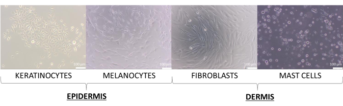
图8:二维单层培养中观察到的不同形态的皮肤细胞。 比例尺:100 µm。 请点击此处查看该图的放大版本。
球体(通常称为球状体)是细胞与组织工程研究人员开发的最简单的三维模型之一,尽管由皮肤细胞构建的球体并不常见。在此模型中,既可以构建单细胞培养球体,也可以构建多细胞共培养球体。文献中已报道了多种球体构建方法(例如悬滴法、限制细胞贴壁法、磁力悬浮法、旋转法、微流控技术等)30。由于制备简便、成本低廉,且所需材料和设备易于获取,前两种方法特别适合三维(3D)细胞模型初学者,其操作方案见上文(步骤2.1和2.2)。根据文献31,32,这些方法中最重要的参数包括细胞数量、细胞悬液体积以及培养时间。
球体可由不同数量的细胞构建,但每种细胞类型以及细胞共培养体系的接种细胞密度均需单独优化。角质形成细胞和成纤维细胞单层培养的优化过程表明,接种 1 x 10⁴ 每孔细胞数对两种细胞类型均获得了最佳结果。所呈现的类球体在 图9 使用U型底培养板通过限制性细胞黏附法(步骤2.2)制备。建议至少制备4个孔的技术重复样本(最佳为6个孔)。由1 x 10组成的类球体4 细胞在孔中可见,因而更易于操作。因此,即使不移除类球体,也能在实验过程中从孔中更换旧培养基。经过上述操作后,类球体的形态大多保持不变且可重复。在操作过程中,较大的类球体稳定性较差。还需指出的是,不同类型的细胞可形成不同颜色的类球体(例如., 角质形成细胞形成的球体颜色较深,而成纤维细胞形成的球体则明显更浅。
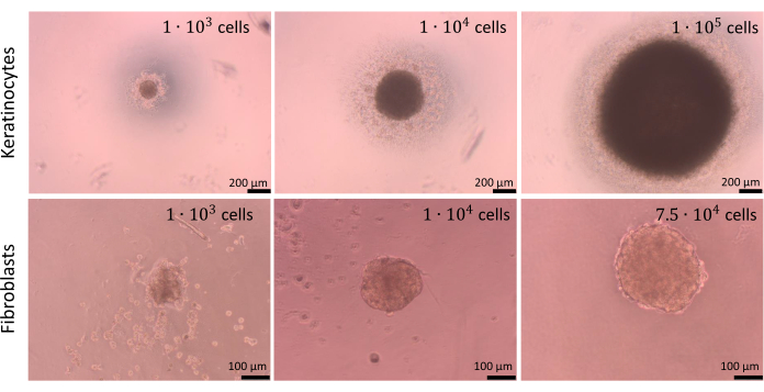
图9:球体的形成。 使用限制性细胞黏附方法,由不同类型和数量的皮肤细胞形成的球体。比例尺(上图):200 µm。比例尺(下图):100 µm。 请点击此处查看该图的放大版本。
如悬滴法(步骤2.1)中所述,在某一阶段需要将球形结构从培养皿盖转移至孔中。此操作过程可能对球形结构造成损伤,因此本步骤要求操作具有高度精确性。所形成的球形结构若未被小心处理,容易失去正常形态(图10)。第一张图像(图10A)显示了一个各向均等、形态良好的球体。第二和第三张图像(图10B、C)中观察到球体出现轻微的形态改变,但细胞聚集体仍保持圆形。最后三张图像(图10D–F)展示了球体损伤的不同阶段。为获得可重复的球形结构形态与结构,需要积累操作经验。对于初次尝试构建球体的研究人员,建议采用限制细胞黏附的方法(步骤2.2),该方法可减少操作者操作差异的影响,从而获得更具可比性的结果。
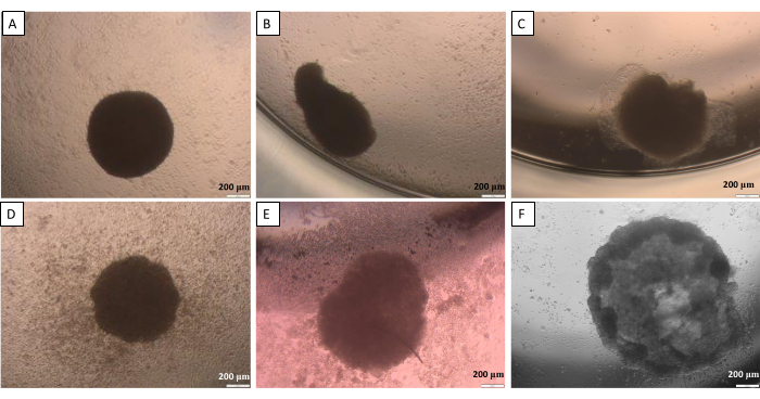
图10:悬滴法中将类球体从盖子转移到孔板时可能出现的困难 (A) 一个良好的球体,(B,C) 轻微受损的球状体,(D-F) 严重受损的球状体。图像拍摄于转移后 24 小时。比例尺: 200 µm. 请点击此处以查看此图的放大版本。
等效皮肤模型相较于球体是更为先进的三维人工皮肤模型。在构建皮肤模型的过程中,需考虑多个方面,包括模型的层数(仅表皮、仅真皮、含表皮和角质层的真皮)、所使用的细胞类型、应用的材料、理想的模型尺寸,以及后续将用于何种类型的研究等14。皮肤等效模型可置于特制的插入式培养插件中,这些插件可放入标准的多孔板中,孔板尺寸可根据需要选择(如96孔、48孔、24孔等)。尽管使用插件更便于在不同孔之间转移以及更换培养基时操作,且能避免损伤等效模型,但其成本较高。如果模型无需包含角质层,则更经济的方案是在多孔板的孔内直接制备等效模型。
人工真皮层通常采用基于支架的模型构建,使用天然(如明胶、胶原蛋白、纤维蛋白、透明质酸、壳聚糖-海藻酸盐等)或合成(如聚乙二醇二丙烯酸酯和聚乳酸)水凝胶33。为了模拟真实皮肤的真皮层,该层主要由水组成,并含有一定量的细胞外基质(ECM)成分(包括胶原蛋白或纤连蛋白),以介导细胞黏附、细胞间相互作用及其他细胞活动34。在本研究中,选用Ⅰ型胶原蛋白,因其易于制备成水凝胶形式,且其结构柔韧,便于后续潜在的研究操作(例如将凝胶从一个培养皿转移至另一个培养皿)。来源于大鼠尾部的Ⅰ型胶原蛋白溶液通常通过将粉末溶于20 mM乙酸中制备。为实现胶原蛋白的聚合,需将pH值调节至6.5–7.5的适宜范围,可通过精确加入氢氧化钠实现。为方便操作,一些公司提供了特定的计算方法,有助于确定制备此类水凝胶所需的精确体积(表6)。尽管文献中报道的水凝胶中胶原蛋白浓度各不相同(例如0.5–2 mg/mL35;5–30 mg/mL36;低和高胶原含量37),但在本描述的模型中,选用2 mg/mL的浓度,因其形成的水凝胶仍具有足够的柔韧性,同时结构足够致密,必要时可完整地从孔板中取出。
为了制备一种较为逼真的全层皮肤,应以尽可能接近人体内实际比例的细胞比例进行接种。在表皮中,根据身体部位的不同,一个黑素细胞与其相关的一群角质形成细胞之间的比例约为1:36,这一结构被称为表皮黑素单元(Epidermal Melanin Unit, EMU)38。因此,在人工表皮中采用的比例为1个黑素细胞对应15个角质形成细胞(表5)。为了构建人工真皮层,使用了I型胶原蛋白水凝胶,并将成纤维细胞与肥大细胞以1个肥大细胞比10个成纤维细胞的比例包埋其中。通过倒置光学显微镜观察时,可实时监测所构建的每层等效皮肤结构,只需调节观察样本的焦平面深度即可(示例图像见图11)。
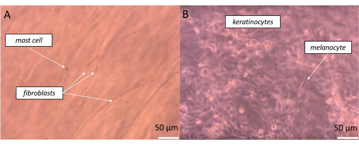
图 11:在构建的全层皮肤等效物特定层次中对不同类型细胞的实时观察。 (A)假真皮层和(B)假表皮层,通过明场显微镜观察。比例尺:50 µm。 请点击此处查看该图的放大版本。
通过染色检测等效物,可以更准确地观察并确认获得预期的模型结构。固定的等效物需先包埋于石蜡中,随后用切片机进行切片。含有薄层人工组织的载玻片可进一步用不同染料染色,包括苏木精和伊红(医学实验室常规进行的基础染色)。通过该操作,可区分等效物中的人工真皮与表皮,并识别单个皮肤细胞(图12)。在 图12 中,不仅展示了特定类型的细胞,还可观察到角质形成细胞处于细胞分裂过程的不同阶段(如末期和中期)。对于肥大细胞,其细胞内特异性颗粒清晰可辨。这些图像初步证实所构建的皮肤等效物具有活性(细胞在其中生长),且细胞可在所建立的模型中正常发挥功能。然而,对于三维表皮及全层皮肤模型,尤其重要的是需检测所获得结构的质量与功能性。为评估角质层的通透性,应进行跨上皮电阻(TEER)测量或荧光黄(Lucifer-Yellow)染色39,40。此外,在结构合理的人工皮肤中,应存在特定的标志物,包括分化标志物(如Filaggrin、Involucrin、Loricrin、Keratin 10、Keratin 5,以及包含神经酰胺的脂质类别)、真皮-表皮连接标志物(如IV型胶原、Laminin V、Alpha6Beta4-整合素、BP抗原)41、表皮层紧密连接标志物(如claudin-1、occludin、闭合环蛋白(ZO)-1)42,以及基底层增殖标志物(Ki67)41。

图 12:皮肤细胞形态与功能。 全层皮肤等效物经苏木精和伊红染色后,观察皮肤细胞的形态与功能(细胞分裂的观察)。比例尺:100 µm(上图),50 µm(中图左),100 µm(中图右),50 µm(下图)。 请点击此处查看此图的放大版本。
确认生物标志物存在的最常用方法是进行特异性染色,例如免疫组织化学或免疫荧光染色。可使用不同的抗体和荧光染料,在显微镜下可视化模型中的特定细胞。培养细胞染色的示例结果如图13所示。为观察角质形成细胞,使用了抗细胞角蛋白14的抗体;对于黑色素细胞,则使用了抗黑色素A特异性抗体。使用胶原蛋白1A2抗体对成纤维细胞进行染色,而荧光素硫代罗丹明101与亲和素偶联后用于检测肥大细胞中存在的肝素。

图13:细胞荧光染色结果。 (A)角质形成细胞中的细胞角蛋白14。比例尺:50 µm。(B)黑色素细胞中的Melan-A。比例尺:50 µm。(C)成纤维细胞中的胶原蛋白1A2。比例尺:100 µm。(D)肥大细胞中的肝素。比例尺:10 µm。请点击此处查看该图的高清版本。
讨论
本文介绍了一种可用于制备自主先进人工皮肤模型的方法学。当计划开展的研究需要特定定义的模型,而这些模型在市场上可能无法获得或价格极为昂贵时,该方法是一种理想的解决方案。如前所述,目前市场上已有若干种商用皮肤等效物(例如., EpiSkin,EpiDerm FT)。然而,其成本(€100-€每件400)以及较短的交付时间(数天至数周)可能促使研究人员尝试自行制备此类模型。所提出的操作流程即使对于经验较少的科研人员而言也易于实施,同时能够获得高度复杂的皮肤模型。值得强调的是,模型的细胞组成完全由研究人员自主决定。此外,该模型可在此基础上进一步开发与优化,从而开辟全新的研究视角。相比之下,商业化的模型则需购买不同的等效产品以作替代。
尽管3D细胞培养模型可能包含多种细胞类型,且易于操作和获取,但它们仍然是人工模型,无法完全重现组织的复杂性和功能性(例如免疫功能、血管化)。因此,在大多数研究中,需要使用多种模型来验证所获得的结果。部分此类模型的优缺点及其局限性汇总于表9中。另一方面,商用模型可确保较高的质量标准,实现实验的可重复性以及不同实验室间数据的可比性。若要将一种新化合物应用于研究,通常有必要购买相应的商用等效模型。但在准备阶段,使用自制的3D皮肤模型(多细胞类型球体或等效结构)有助于减少在商用等效模型上所需进行的实验数量。制备和使用所述模型的目的并非规避使用经认证的研究模型的必要性,而是为了促进研究并降低相关费用。
| 对比的模型组合 | 优势 | 劣势 | ||||
| 细胞培养 vs. 动物 | 最大程度减少动物痛苦 | 难以获取受试因素对整个机体影响的完整信息 | ||||
| 实验高度标准化——结果更易重复 | 单一模型不足以反映体内发生的全部过程 | |||||
| 不会对整个生物体产生副作用 | - | |||||
| 对实验条件的控制更佳 | - | |||||
| 可实现自动化操作(例如,生物打印) | - | |||||
| 成本较低 | - | |||||
| 所需样本量小 | - | |||||
| 产生的废弃物量有限 | - | |||||
| 3D 培养 vs. 2D 培养 | 更接近完整生物体的结构与功能 | 培养过程耗时较长 | ||||
| 可构建具有功能性的组织 | 成本较高 | |||||
| 可根据研究需求定制模型 | 几乎无法自发形成 3D 结构 | |||||
| - | 缺乏标准化的检测方法来量化不同化合物的作用效果 | |||||
| - | 市场上可获得的不同 3D 培养模型有限 | |||||
| 细胞系 vs. 原代细胞 | 经过认证并获批的模型 | 可用的细胞系种类有限 | ||||
| 实验高度标准化——结果更易重复 | 难以从同一供体获取多种类型的细胞 | |||||
| 生命周期更长 | 可能具有与天然细胞不同的特性 | |||||
| 增殖速度相对较快 | 细胞功能常出现异常 | |||||
| 对多种操作(如冻存、离心)耐受性较强 | - | |||||
表9:不同研究模型应用的比较——优势与劣势
已有若干文章描述了如何制备3D皮肤模型(除了综述类文章对市售模型进行总结14,43,44外,通常这些文章仅聚焦于获得球体45或类似结构46的单一方法)。
本文描述了两种用于皮肤细胞成球的方法。悬滴法应用广泛,但在某些情况下其重复性和稳定性可能不足。该方法多数步骤需要特定操作,例如由于液滴转移过程中水分蒸发较快,需快速操作。同时建议动作轻柔,缺乏此类操作技巧可能导致细胞聚集体受损31,32。因此,另一种更简便的成球方法聚焦于限制细胞黏附。当缺乏适宜的细胞附着表面时,细胞间的相互作用会增强,从而促使细胞聚集体形成。该方法的重复性更高,且无需转移细胞球。通过这些方法,已确定形成一个细胞球所需的最优皮肤细胞数量为 1 x 10⁴ 个细胞/球。
接下来,描述了皮肤等效物制备的相关步骤。其在研究中的外观和功能表现可能在很大程度上取决于构成它们的组分,包括细胞(表2)、支架材料和培养基。用于制备人工皮肤的3D支架可分为合成水凝胶和来源于天然物质的水凝胶。根据所用材料及其形成水凝胶的特性,可能需要额外补充培养基。合成水凝胶需要将生物活性分子(如蛋白质、酶和生长因子)引入其网络结构中,以介导特定的细胞功能47。文献中报道的实现生长因子在水凝胶中可控释放的主要方法包括直接负载、静电相互作用、共价结合以及使用载体48。由细胞外基质(ECM)蛋白和聚合物等天然来源形成的水凝胶可在3D支架内形成液体通道,加速营养物质的分布,因此无需额外补充培养基。研究表明,小分子(如细胞因子和生长因子)和大分子(包括糖胺聚糖和蛋白聚糖)可通过扩散在ECM中进行转运47。然而,氧气、营养物质及其他生物活性分子的分子扩散可能受到ECM水凝胶自身特性的阻碍。较低的扩散效率与水凝胶厚度增加有关,同时也与胶原蛋白浓度过高相关37。本研究中,为构建皮肤等效物,采用了低浓度胶原蛋白(2 mg/mL),表明分子在水凝胶中的扩散应良好且迅速。因此,在此阶段未对培养基或水凝胶本身进行额外补充。为模拟真皮层,将肥大细胞与成纤维细胞(1:10)包埋于胶原蛋白水凝胶中。随后,将黑素细胞与角质形成细胞(1:15)接种至水凝胶表面,并将整个等效结构置于培养基中进行培养。需要指出的是,基础培养基包含多种氨基酸、无机酸和维生素,并额外添加了血清(含有多种成分:细胞生长和贴附因子、脂类、激素、营养物质和能量来源、载体蛋白、结合与转运蛋白等)。为获得合适的表皮结构,应在特定时间向培养基中添加不同的补充成分。启动表皮分化的最重要刺激因子是钙离子,因其可激活细胞内信号通路。抗坏血酸可刺激与钙介导通路相似的信号路径,同时还能增强抗坏血酸的转运并防止亲水性抗氧化剂的耗竭41。此外,当向培养基中添加其他成分(如咖啡因、氢化可的松、三碘甲腺原氨酸、腺嘌呤和霍乱毒素)时,细胞分化效果得到进一步改善41,44。重要的是,制备的模型必须始终通过检测确认特定类型的细胞存在于相应的层次中。通过H&E染色,在所构建的等效物结构中确认了四种皮肤细胞均存在。
最常见的问题是处理所获得模型时的操作精细程度和依赖经验判断。一些困难可能与细胞球体的形成以及水凝胶的制备有关。在细胞培养过程中,还可能发生其他多种问题,包括微生物污染、细胞增殖速率低、模型中所用原代细胞的老化、由原代细胞与细胞系构建的2D和3D模型的最大培养时间等。在表10中,汇总了一些实用建议,用以指导遇到上述任一问题时应采取的措施。
| 细胞培养中的常见问题 | 建议 | |||
| 微生物污染 | 如果某个细胞培养瓶/培养皿中发生微生物污染,应尽快移除受污染的培养物(以避免污染其他培养瓶/培养皿中的细胞)。重新冻存新的一管细胞。 如果污染反复出现,可尝试扩大所用抗生素的抗菌谱并提高其浓度。 | |||
| 细胞增殖速率低 | 某些细胞具有较长的倍增时间。为促进其增殖,可在基础培养基中添加多种细胞特异性的生长因子。此外,提高基础培养基中胎牛血清(FBS)或L-谷氨酰胺的浓度也可能有助于促进细胞生长。 | |||
| 模型中所用原代细胞的老化 | 经过数次传代后,原代细胞会进入衰老状态并停止分裂。为解决模型中的这一问题,建议尽可能使用早期代次的细胞来构建模型。 | |||
| 由原代细胞与细胞系构建的2D和3D模型的最大培养时间 | 模型的培养时间很大程度上取决于所用细胞的类型。使用原代细胞时,由于其寿命较短,培养时间将更短。 | |||
| 细胞球体形成困难 | 某些细胞可能需要更长时间才能形成球体。如果数天后仍未形成球体,可收集样本中的细胞,并使用台盼蓝染色等方法检测其活力。 | |||
| 球体稳定性问题 | 如果球体不稳定且在操作过程中易被破坏,可尝试使用较少数量的细胞来形成球体。务必始终轻柔地转移正在生长球体的培养皿。 | |||
| 水凝胶制备困难 | 请检查各组分(水、PBS [10x]、NaOH、I型胶原蛋白)的比例是否正确。胶原蛋白储存液通常非常黏稠,因此务必缓慢移液。气泡会影响水凝胶的形态,因此采用反向移液法可能有助于解决此问题。 | |||
表10:细胞培养问题排查
所建立的模型在制备完成后可用于多个领域,包括(1)用于药物和化妆品的新型生物活性化合物的细胞毒性和遗传毒性实验49,(2)不同因子刺激的实验50(3)基础研究,增进我们对皮肤细胞及其生物学功能、与其他细胞及环境相互作用的了解51,52(4)针对特定疾病实体的研究,可将特定类型的细胞引入所构建的模型中(如癌细胞、特定基因突变的细胞, 等等。14,53以及更多应用。毋庸置疑,这些模型的应用符合3Rs原则,有助于在产品测试和科学研究中更伦理地使用动物,且不违反化妆品产品动物测试禁令法规。
披露
作者声明无利益冲突。
致谢
作者感谢华沙理工大学通过“卓越计划——研究型大学”项目提供的两项资助:POB BIB BIOTECHMED-2 启动基金(编号:1820/2/ZO1/POB4/2021)以及校长资助的学生科研小组项目(SKIN-ART,编号:1820/116/Z16/2021)。此外,作者还感谢 Joanna Cieśla 教授以及药物与化妆品生物技术系 和华沙理工大学化学学院生物技术科学俱乐部“Herbion”所提供的支持。特别感谢 Michał Stepulak 博士提供了 Pluronic F-127 化合物。
材料
| 姓名 | 公司 | 目录编号 | 评论 |
|---|---|---|---|
| 用于贴壁细胞培养的24孔板 | Biologix Europe GmbH | 07-6024 | - |
| 35%–38% HCL | Chempur | 115752837 | - |
| 60 mm 细胞培养培养皿 | 巢 | 705001 | - |
| 亲和素−磺基罗丹明101 | Sigma Aldrich | A2348-5MG | - |
| 倒置明场显微镜 | 奥林巴斯 | CKX41 | - |
| 氯化钙 | Avantor | 874870116 | - |
| T75 贴壁细胞培养瓶 | Genoplast | G77080033 | - |
| 15 mL 离心管 | GoogLab Scientific | G66010522 | - |
| CO2 培养箱 | Heal Force | Galaxy 170R | - |
| 兔源 Col1A2 抗体 | Novus | NBP2-92790 | - |
| Corning(R) Transwell(R) 聚碳酸酯 | Corning | CLS3422-48EA | - |
| 小鼠源细胞角蛋白14抗体 | Novus | NBP1-79069 | - |
| 组织学用DPX封片剂 | Sigma Aldrich | 06522-100ML | - |
| 杜尔贝科改良伊格尔培养基(DMEM) | VWR Chemicals | L0102-500 | - |
| 伊红Y | Kolchem | - | 0.5 % 水溶液 |
| Eppendorf管 1.5 mL | Sarstedt | 72.690.001 | - |
| 2 mL 离心管 | Sarstedt | 72.691 | - |
| 无水乙醇 99.8% | Avantor | 396480111 | 用超纯水稀释至所需浓度 |
| 胎牛血清 | Gibco | 10270106 | - |
| 荧光倒置显微镜 | Olympus | IX71 | - |
| FITC标记的山羊抗小鼠二抗 | Sigma Aldrich | F0257-1mL | |
| FITC标记的山羊抗兔二抗 | Novus | NB7159 | - |
| Harris苏木精 | 科尔切姆 | - | 1 mg/mL,溶于95%乙醇 |
| Hoechst 33342 | ThermoFisher | H3570 | - |
| 层流室 | Heal Force | HFSafe-1200 | - |
| 小鼠来源的Melan-A抗体 | Santa Cruz Biotechnology | sc-20032 | - |
| 切片机 | 微米 | HM355S | - |
| NaOH | Avantor | 810981997 | - |
| 石蜡颗粒 | Sigma Aldrich | 1.07164 | - |
| 多聚甲醛 | Sigma Aldrich | 1581227 | - |
| 青霉素/链霉素溶液 | Sigma Aldrich | P4333 | - |
| 移液器吸头,1000 µL | Sarstedt | 70.305 | - |
| 移液器吸头,20 µL | Sarstedt | 70.3021 | - |
| 移液器吸头,200 µL | Sarstedt | 70.303 | - |
| Pluronic F-127 | BASF | 50401036 | - |
| 10 mL 血清移液管 | GoogLab Scientific | G33270011 | - |
| 25 mL 血清移液管 | GoogLab Scientific | G33280011 | - |
| 5 mL 血清移液管 | GoogLab Scientific | G33260011 | - |
| 碳酸氢钠 | Sigma Aldrich | S5761 | - |
| 碳酸氢钠 | Chempur | 118105307 | |
| 0.25% 胰蛋白酶-EDTA 溶液,含酚红 | Sigma Aldrich | 25200072 | - |
| I型胶原蛋白 | IBIDI | 50201 | - |
| U型底96孔板 | Sarstedt | 83.3925500 | - |
| 二甲苯 | Sigma Aldrich | 534056 | - |
参考文献
- Farage, M. A., Miller, K. W., Elsner, P., Maibach, H. I. Characteristics of the aging skin. Advances in Wound Care. 2 (1), 5-10 (2013).
- Zhu, H., Alikhan, A., Maibach, H. I. Biology of Stratum Corneum: Tape Stripping and Protein Quantification. Textbook of Aging Skin. Farage, M. A., Miller, K. W., Maibach, H. I. , Springer Berlin Heidelberg. Berlin, Heidelberg. (2015).
- Boer, M., Duchnik, E., Maleszka, R., Marchlewicz, M. Structural and biophysical characteristics of human skin in maintaining proper epidermal barrier function. Postepy Dermatogogii I Alergologii. 33 (1), 1-5 (2016).
- De Falco, M., Pisano, M. M., De Luca, A. Embryology and Anatomy of the Skin. In Skin Cancer: A Practical Approach. Current Clinical Pathology. Baldi, A., Pasquali, P., Spugnini, E. P. , Springer. New York. (2014).
- Dehdashtian, A., Stringer, T. P., Warren, A. J., Mu, E. W., Amirlak, B., Shahabi, L. Anatomy and Physiology of the Skin. Melanoma: A Modern Multidisciplinary Approach. Riker, A. I. , Springer International Publishing: Cham. 15-26 (2018).
- Graham, H. K., Eckersley, A., Ozols, M., Mellody, K. T., Sherratt, M. J. Human Skin: Composition, Structure and Visualisation Methods. Skin Biophysics; Studies in Mechanobiology, Tissue Engineering, and Biomaterials. Limbert, G. 22, Springer International Publishing: Cham. 1-18 (2019).
- Piasek, A. M., Musolf, P., Sobiepanek, A. Aptamer-based advances in skin cancer research. Current Medicinal Chemistry. 30 (8), 953-973 (2023).
- Bergfeld, W. F., Belsito, D. V., Marks, J. G., Andersen, F. A. Safety of ingredients used in cosmetics. Journal of the American Academy of Dermatology. 52 (1), 125-132 (2005).
- Hubrecht, R. C., Carter, E. The 3Rs and humane experimental technique: Implementing change. Animals. 9 (10), 754(2019).
- Sreedhar, D., Manjula, N., Ajay, P., Shilpa, P., Ligade, V. Ban of cosmetic testing on animals: A brief overview. International Journal of Current Research and Review. 12 (14), 113-116 (2020).
- Silva, R. J., Tamburic, S. A state-of-the-art review on the alternatives to animal testing for the safety assessment of cosmetics. Cosmetics. 9 (5), 90(2022).
- Xu, Y., Shrestha, N., Préat, V., Beloqui, A. An overview of in vitro, ex vivo and in vivo models for studying the transport of drugs across intestinal barriers. Advanced Drug Delivery Reviews. 175, 113795(2021).
- Boelsma, E., Ponec, M. Basics (Guidelines) on Cell Culture Testing for Topical/Dermatological Drugs/Products and Cosmetics With Regard to Efficacy and Safety of the Preparations. In Dermatopharmacology of Topical Preparations. Gabard, B., Surber, C., Elsner, P., Treffel, P. , Springer Berlin Heidelberg. Berlin, Heidelberg. 37-57 (2000).
- Suhail, S., Sardashti, N., Jaiswal, D., Rudraiah, S., Misra, M., Kumbar, S. G. Engineered skin tissue equivalents for product evaluation and therapeutic applications. Biotechnology Journal. 14 (7), 1900022(2019).
- Sobiepanek, A., et al. Novel diagnostic and prognostic factors for the advanced melanoma based on the glycosylation-related changes studied by biophysical profiling methods. Biosensors and Bioelectronics. 203, 114046(2022).
- Yang, H., Sun, L., Liu, M., Mao, Y. Patient-derived organoids: A promising model for personalized cancer treatment. Gastroenterology Report. 6 (4), 243-245 (2018).
- Baran, J., et al. Mast cells as a target-A comprehensive review of recent therapeutic approaches. Cells. 12 (8), 1187(2023).
- Kosten, I. J., Buskermolen, J. K., Spiekstra, S. W., de Gruijl, T. D., Gibbs, S. Gingiva equivalents secrete negligible amounts of key chemokines involved in langerhans cell migration compared to skin equivalents. Journal of Immunology Research. 2015, 627125(2015).
- cieżyńska, A., et al. A novel and effective method for human primary skin melanocytes and metastatic melanoma cell isolation. Cancers. 13 (24), 6244(2021).
- Kröger, M., et al. In vivo non-invasive staining-free visualization of dermal mast cells in healthy, allergy and mastocytosis humans using two-photon fluorescence lifetime imaging. Scientific Reports. 10 (1), 14930(2020).
- Liu, D., Chen, S., Win Naing, M. A review of manufacturing capabilities of cell spheroid generation technologies and future development. Biotechnology and Bioengineering. 118 (2), 542-554 (2021).
- Rossi, A., Appelt-Menzel, A., Kurdyn, S., Walles, H., Groeber, F. Generation of a three-dimensional full thickness skin equivalent and automated wounding. Journal of Visualized Experiments: JoVE. (96), e52576(2015).
- Kim, K., Kim, J., Kim, H., Sung, G. Y. Effect of α-lipoic acid on the development of human skin equivalents using a pumpless skin-on-a-chip model. International Journal of Molecular Sciences. 22 (4), 2160(2021).
- Curto, E. V., Lambert, G. W., Davis, R. L., Wilborn, T. W., Dooley, T. P. Biomarkers of human skin cells identified using DermArray DNA arrays and new bioinformatics methods. Biochemical and Biophysical Research Communications. 291 (4), 1052-1064 (2002).
- Godwin, L. S., et al. Isolation, culture, and transfection of melanocytes. Current Protocols in Cell Biology. 63, 1-20 (2014).
- Przekora, A., Zarnowski, T., Ginalska, G. A simple and effective protocol for fast isolation of human tenon's fibroblasts from a single trabeculectomy biopsy - a comparison of cell behaviour in different culture media. Cellular & Molecular Biology Letters. 22, 5(2017).
- Rådinger, M., Jensen, B. M., Kuehn, H. S., Kirshenbaum, A., Gilfillan, A. M. Generation, Isolation, and Maintenance of Human Mast Cells and Mast Cell Lines Derived from Peripheral Blood or Cord Blood. Current Protocols in Immunology. Coligan, J. E., Bierer, B. E., Margulies, D. H., Shevach, E. M., Strober, W. , John Wiley & Sons, Inc. Hoboken, NJ. (2010).
- Artuc, M., Muscha Steckelings, U., Henz, B. M. Mast cell-fibroblast interactions: Human mast cells as source and inducers of fibroblast and epithelial growth factors. Journal of Investigative Dermatology. 118 (3), 391-395 (2002).
- Panos, R. J., Rubin, J. S., Csaky, K. G., Aaronson, S. A., Mason, R. J. Keratinocyte growth factor and hepatocyte growth factor/scatter factor are heparin-binding growth factors for alveolar type ii cells in fibroblast-conditioned medium. Journal of Clinical Investigation. 92 (2), 969-977 (1993).
- Lv, D., Hu, Z., Lu, L., Lu, H., Xu, X. Threedimensional cell culture: A powerful tool in tumor research and drug discovery (Review). Oncology Letters. 14 (6), 6999-7010 (2017).
- Amaral, R. L. F., Miranda, M., Marcato, P. D., Swiech, K. Comparative analysis of 3D bladder tumor spheroids obtained by forced floating and hanging drop methods for drug screening. Frontiers in Physiology. 8, 605(2017).
- Gao, B., Jing, C., Ng, K., Pingguan-Murphy, B., Yang, Q. Fabrication of three-dimensional islet models by the geometry-controlled hanging-drop method. Acta Mechanica Sinica. 35 (2), 329-337 (2019).
- Zhang, C., et al. 3D culture technologies of cancer stem cells: Promising ex vivo tumor models. Journal of Tissue Engineering. 11, (2020).
- Sobiepanek, A., Paone, A., Cutruzzolà, F., Kobiela, T. Biophysical characterization of melanoma cell phenotype markers during metastatic progression. European Biophysics Journal: EBJ. 50 (3-4), 523-542 (2021).
- Jin, G. -Z., Kim, H. -W. Effects of Type I collagen concentration in hydrogel on the growth and phenotypic expression of rat chondrocytes. Tissue Engineering and Regenerative. 14 (4), 383-391 (2017).
- Osidak, E. O., et al. Concentrated collagen hydrogels: A new approach for developing artificial tissues. Materialia. 20, 101217(2021).
- Antoine, E. E., Vlachos, P. P., Rylander, M. N. Review of collagen I hydrogels for bioengineered tissue microenvironments: Characterization of mechanics, structure, and transport. Tissue Engineering Part B: Reviews. 20 (6), 683-696 (2014).
- Jimbow, K., Salopek, T. G., Dixon, W. T., Searles, G. E., Yamada, K. The epidermal melanin unit in the pathophysiology of malignant melanoma. The American Journal of Dermatopathology. 13 (2), 179-188 (1991).
- Van Den Bogaard, E., et al. Perspective and consensus opinion: Good practices for using organotypic skin and epidermal equivalents in experimental dermatology research. Journal of Investigative Dermatology. 141 (1), 203-205 (2021).
- Bednarek, R. In vitro methods for measuring the permeability of cell monolayers. Methods and Protocols. 5 (1), 17(2022).
- Idrees, A., et al. Fundamental in vitro 3D human skin equivalent tool development for assessing biological safety and biocompatibility - towards alternative for animal experiments. 4 Open. 4, (2021).
- Park, H. -Y., Kweon, D. -K., Kim, J. -K. Upregulation of tight junction-related proteins by hyaluronic acid in human HaCaT keratinocytes. Bioactive Carbohydrates and Dietary Fibre. 30, 100374(2023).
- Choudhury, S., Das, A. Advances in generation of three-dimensional skin equivalents: Pre-clinical studies to clinical therapies. Cytotherapy. 23 (1), 1-9 (2021).
- Zhang, Z., Michniak-Kohn, B. B. Tissue engineered human skin equivalents. Pharmaceutics. 4 (1), 26-41 (2012).
- Woappi, Y., Altomare, D., Creek, K. E., Pirisi, L. Self-assembling 3D spheroid cultures of human neonatal keratinocytes have enhanced regenerative properties. Stem Cell Research. 49, 102048(2020).
- Ouwehand, K., Spiekstra, S. W., Waaijman, T., Scheper, R. J., de Gruijl, T. D., Gibbs, S. Technical advance: Langerhans cells derived from a human cell line in a full-thickness skin equivalent undergo allergen-induced maturation and migration. Journal of Leukocyte Biology. 90 (5), 1027-1033 (2011).
- Akther, F., Little, P., Li, Z., Nguyen, N. -T., Ta, H. T. Hydrogels as artificial matrices for cell seeding in microfluidic devices. RSC Advances. 10 (71), 43682-43703 (2020).
- Silva, A. K. A., Richard, C., Bessodes, M., Scherman, D., Merten, O. -W. Growth factor delivery approaches in hydrogels. Biomacromolecules. 10 (1), 9-18 (2009).
- Lee, H. -R., et al. Effect of Aronia extract on collagen synthesis in human skin cell and dermal equivalent. Oxidative Medicine and Cellular Longevity. 2022, 4392256(2022).
- Mulder, P. P. G., Raktoe, R. S., Vlig, M., Elgersma, A., Middelkoop, E., Boekema, B. K. H. L. Full skin equivalent models for simulation of burn wound healing, exploring skin regeneration and cytokine response. Journal of Functional Biomaterials. 14 (1), 29(2023).
- Goncalves, K., et al. Investigation into the effect of skin tone modulators and exogenous stress on skin pigmentation utilizing a novel bioengineered skin equivalent. Bioengineering & Translational Medicine. 8 (2), 10415(2023).
- Michel, M., L'Heureux, N., Pouliot, R., Xu, W., Auger, F. A., Germain, L. Characterization of a New Tissue-Engineered Human Skin Equivalent with Hair. In Vitro Cellular & Developmental Biology. Animal. 35 (6), 318-326 (1999).
- Müller, I., Kulms, D. A 3D organotypic melanoma spheroid skin model. Journal of Visualized Experiments: JoVE. (135), e57500(2018).
重印与许可
