本文介绍了一种实验室自建的细胞外囊泡组织因子活性检测方法。目前已采用基于活性的检测方法和基于抗原的检测方法来测定人血浆样本中细胞外囊泡的组织因子含量。其中,基于活性的检测方法比基于抗原的检测方法具有更高的灵敏度和特异性。
需要JoVE订阅才能观看此内容。 请登录或开始免费试用
本文内容
摘要
摘要
组织因子(TF)是凝血因子(F)VII和FVIIa的跨膜受体。TF/FVIIa复合物通过激活FIX和FX来启动凝血级联反应。TF以细胞外囊泡(EV)的形式从细胞释放进入血液循环。TF阳性(+)EV的水平在多种疾病中升高,包括癌症、细菌和病毒感染以及肝硬化,并与血栓形成、弥散性血管内凝血、疾病严重程度和死亡率相关。目前有两种方法可用于检测血浆中的TF+ EV:基于抗原的检测法和基于活性的检测法。研究数据表明,基于活性的检测法比基于抗原的检测法具有更高的灵敏度和特异性。本文介绍了我们实验室自行建立的EVTF活性检测方法,该方法基于两阶段FXa生成实验。将FVIIa、FX和钙离子加入含有TF+ EV的样本中,在有或无抗TF抗体的条件下生成FXa,以区分TF依赖性和非依赖性的FXa生成。通过FXa切割的显色底物来测定FXa水平,同时利用重组再脂化TF建立的标准曲线来确定TF浓度。该实验室自建的EVTF活性检测方法相比商用TF活性检测试剂盒具有更高的灵敏度和特异性。
引言
血液凝固始于凝血因子(F)VII/VIIa 与组织因子(TF)的结合1。TF/FVIIa 复合物可激活 FIX 和 FX,从而启动血液凝固过程1。全长型膜结合 TF 存在两种形式:加密态和活性态。此外,还存在一种选择性剪接形式的 TF(asTF)。细胞膜外小叶中的鞘磷脂和磷脂酰胆碱可使 TF 保持在加密状态2,3,4。当细胞被激活或受损时,磷脂翻转酶将磷脂酰丝氨酸及其他带负电荷的磷脂转移至外小叶1。细胞活化还会导致酸性鞘磷脂酶转位至外小叶,在此处将鞘磷脂降解为神经酰胺5。这两种机制共同促使加密态 TF 转变为活性形式。另有研究提出,蛋白质二硫键异构酶介导加密态 TF 中 Cys186 与 Cys209 之间形成二硫键,从而实现 TF 的去加密化6,7,8。asTF 也存在于血液循环中,但由于缺乏跨膜结构域,因此呈可溶性状态9,10。重要的是,与全长活性 TF 相比,asTF 的促凝血活性极低10,11。
细胞外囊泡(EVs)可由静息状态、活化状态及濒死的宿主细胞以及癌细胞释放12。EVs 表达其亲代细胞来源的蛋白质12。携带活性组织因子(TF)的 EVs 可由活化的单核细胞、内皮细胞和肿瘤细胞释放进入血液循环13,14,15。血浆中 TF 的水平可通过基于活性和基于抗原的检测方法进行测定。基于抗原的检测方法包括 ELISA 和流式细胞术16。基于活性的检测方法有两种:单阶段和双阶段 TF 活性检测。单阶段检测基于血浆凝固试验,即将含 TF 的样本加入血浆后,通过再钙化测定形成凝块所需的时间。双阶段检测通过加入 FVII 或 FVIIa、FX 和钙离子,测定样本中 FXa 的生成量,并使用可被 FXa 裂解的底物来确定 FXa 的水平。
在单阶段和双阶段组织因子(TF)活性检测中,TF浓度通过使用重组TF生成的标准曲线来确定。双阶段检测比单阶段检测具有更高的灵敏度和特异性。许多研究已证实,基于活性的检测方法比基于抗原的检测方法具有更高的灵敏度和特异性17,18,19,20,21。此外,我们实验室自建的活性检测方法比商用活性检测试剂盒具有更高的灵敏度和特异性22。健康个体血浆中外泌体组织因子(EVTF)活性水平极低或无法检测。相反,患有病理状态(如癌症、肝硬化、脓毒症和病毒感染)的个体中可检测到EVTF活性,且其水平与血栓形成、弥散性血管内凝血、疾病严重程度及死亡率相关23,24,25,26,27,28。本文将详细介绍本实验室建立的双阶段EVTF活性检测方法。
访问受限。请登录或开始试用以查看此内容。
方案
该研究已获得北卡罗来纳大学教堂山分校机构审查委员会的批准(方案编号:14-2108)。
1. 从供体采集血液
- 使用21号针头经清洁的静脉穿刺法从肘前静脉采集全血。弃去最初采集的3 mL血液,因为这部分血液可能含有来自血管周围细胞的组织因子(TF)。
- 将2.7 mL或1.8 mL(根据试管规格)血液采集至含有3.2%枸橼酸钠(0.109 mol/L)的真空采血管中。切勿过度或不足填充试管。采血后应立即轻轻颠倒混匀试管,以充分分散枸橼酸钠。
- 若在处理前需运输试管,应避免震荡。应在采血后2小时内完成血浆制备。
2. 阴性对照血浆的制备
注意:样品在最终冷冻前的任何阶段均不应置于冰上。
- 使用健康志愿者的全血制备阴性对照血浆。采集血液后立即按如下步骤制备无血小板血浆:
- 将真空采血管中的血液样本转移至15 mL离心管中。
- 在室温(20–24 °C)下以2,500 × g 离心15分钟,离心时不启用刹车。
- 将上清液(血小板贫乏血浆)转移至新管中,并在相同条件下重复离心一次。
- 将上清液(去血小板血浆)转移至新管中。
- 将样本分装为每份≥100 µL的等分试样,每管底部保留约100 µL。
- 立即在−80 °C下冷冻保存去血小板血浆(图1)。
阳性对照血浆的制备
注意:不同个体对脂多糖的反应存在差异。
- 将血液样本从真空采血管转移至15 mL离心管中。
- 使用健康志愿者的全血,经脂多糖(LPS)刺激后制备阳性对照血浆 大肠杆菌 37 °C下振荡孵育5小时,加入O111:B4(10 µg/mL)。
- 孵育5小时后,按照步骤2.1.2至2.1.6进行操作。
4. 从血浆中分离细胞外囊泡
注意:EV沉淀物可能不可见。解冻时间取决于样品体积,但通常我们将100 µL血浆在37 °C下解冻30分钟。
- 在 37 °C 下解冻血浆样本。
- 在 1.5 mL 离心管中,向每 100 µL 血浆样本加入 1 mL 无钙 HBSA [HBSA-Ca(-)] 缓冲液 [137 mM NaCl、5.38 mM KCl、5.55 mM 葡萄糖、10 mM HEPES、0.1% (w/v) 牛血清白蛋白]。
- 在 4 °C 下以 20,000 × g 离心 15 分钟。
- 小心吸除上清液,保留约 20 µL,避免扰动细胞外囊泡(EV)沉淀。
- 向每管中加入 1 mL HBSA-Ca(-) 缓冲液重悬 EV 沉淀,在沉淀位置反复吹打混匀,并涡旋振荡各管,随后在 4 °C 下再次以 20,000 × g 离心 15 分钟。
- 小心吸除上清液,保留约 20 µL,避免扰动 EV 沉淀。
- 用 80 µL HBSA-Ca(-) 缓冲液重悬 EV 沉淀,并在沉淀位置反复吹打混匀(图 2)。
5. 可选方案:使用超速离心机分离小型细胞外囊泡
- 步骤4.3之后,收集上清液,并在4 °C下以100,000 × g超速离心70分钟。
- 吸走上清液至剩余20 µL,注意不要扰动EV沉淀。
- 向每管中加入1 mL HBSA-Ca(-)缓冲液重悬EV沉淀,充分涡旋混匀后,在4 °C下再次以100,000 × g超速离心70分钟。
- 吸走上清液至剩余20 µL,注意不要扰动EV沉淀。
- 用80 µL HBSA-Ca(-)缓冲液重悬EV沉淀。
6. 细胞外囊泡组织因子活性的测定
注意:在将EV样品加入孔板前,应充分移取并涡旋混匀。乙二胺四乙酸二钠会抑制FXa切割显色底物的能力,因此在步骤6.7中,乙二胺四乙酸二钠不能替代乙二胺四乙酸四钠使用。
- 向96孔板的两个孔中各加入40 µL EV样品。
- 向其中一个EV样品孔中加入11 µL抑制性小鼠抗人组织因子IgG [(36.4 µg/mL,终浓度7.8 µg/mL)],向另一孔中加入11 µL对照小鼠IgG [(36.4 µg/mL,终浓度7.8 µg/mL)]。
- 将96孔板在室温下孵育15分钟。
- 在孵育期间,使用重脂化重组组织因子制备TF标准品(0、0.32、0.63、1.25、2.5、5、10和20 pg/mL),每种浓度设两个复孔,每孔50 µL。
- 通过混合以下组分制备因子混合液:4 mL含钙HBSA [HBSA-Ca(+)] [HBSA-Ca(-) + 10 mM CaCl2,终浓度10 mM]、800 µL溶于HBSA-Ca(+)中的900 nM FX(终浓度:146.4 nM)以及120 µL溶于HBSA-Ca(+)中的200 nM FVIIa(终浓度:4.8 nM)。
- 向每个孔中加入50 µL因子混合液,用封板膜覆盖孔板,于37 °C孵育箱中孵育2小时。
- 孵育2小时后,每孔加入25 µL含乙二胺四乙酸(EDTA)的HBSA缓冲液 [HBSA-Ca(-) + 25 mM EDTA四钠盐,终浓度5 mM] 以终止FXa生成,并在室温下继续孵育5分钟。
- 将FXa可切割的显色底物重悬至4 mM浓度(向一支25 mg的试剂瓶中加入8.7 mL蒸馏水)。
- 向每个孔中加入25 µL显色底物溶液,先用封板膜覆盖孔板,再用铝箔包裹以避光,于37 °C孵育15分钟。
- 孵育15分钟后,用针头去除气泡,或在1,500 × g 离心1分钟以去除气泡。
- 使用酶标仪在405 nm波长处读取吸光度,读数前进行一次混匀。
- 利用重脂化重组组织因子生成的标准曲线,将各孔的FXa生成量转换为组织因子活性。
- 使用公式(1)计算TF依赖性FXa生成量(EVTF活性):
TF依赖性FXa生成量(EVTF活性 [pg/mL])= 总FXa生成量(对照IgG孔) - TF非依赖性FXa生成量(抗TF IgG孔)(图3)(1)
访问受限。请登录或开始试用以查看此内容。
结果
成功的结果应显示阳性对照值 ≥0.5 pg/mL,阴性对照值 <0.5 pg/mL。最好选择LPS应答较强、EVTF活性 >1.0 pg/mL 的样本作为阳性对照。代表性结果展示了从11名健康供体全血血浆中分离出的EVs在有或无LPS激活情况下的EVTF活性(图4)。11名供体中有6名(供体2、4、5、8、10、11)为中至高LPS应答者,而另外5名(供体1、3、6、7、9)为低LPS应答者。
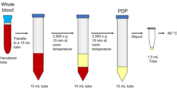
图 1:血小板耗竭血浆的制备。
访问受限。请登录或开始试用以查看此内容。
讨论
本文介绍了我们实验室内部使用的EVTF活性检测方案。该方案包含三个关键步骤。在重悬EV沉淀时,即使沉淀不可见,也必须在沉淀所在位置反复吹打,以确保充分重悬;若EV沉淀未完全重悬,将导致样本EVTF活性值出现假阴性或低估结果。第二,在步骤6.5中必须使用含钙的HBSA-Ca(+)缓冲液,因为缺乏钙离子将无法生成FXa。第三,必须使用EDTA四钠而非EDTA二钠来终止FXa的生成,因为后者会抑制FXa切割显色底物的能力。
我们建议每种试剂按以下方法储存和使用。我们将检测缓冲液和复溶后的底物储存在 4 °C,并重复使用直至用完。如果底物在 2-3 周内未用完,我们会将其丢弃,因为它会变黄。我们将重组 TF、抗体、FVIIa 和 FX 储存在 -80 °C,并建议分装成单次检测用量,以避免冻融循环。
在一项初步研究中,我们将血浆样本在室温下放置9小时,然后在-80 °C下重新冷冻过夜,并于次日进行检测。作为对照,我们将相同的血浆样本解冻后立即重新冷冻。结果发现,室温下放置9小时的样本其EVTF活性较立即重新冷冻的样本降低了16%(Hi...
访问受限。请登录或开始试用以查看此内容。
披露
不存在需要披露的竞争性利益。
致谢
本工作由美国国立卫生研究院心肺血液研究所R35HL155657(N.M.)和John C. Parker教授职位基金(N.M.)资助。我们感谢Sierra J. Archibald女士提出的有益意见
访问受限。请登录或开始试用以查看此内容。
材料
| 姓名 | 公司 | 目录编号 | 评论 |
|---|---|---|---|
| 用于 20,000 × g 离心的 1.5 mL 离心管 | 任意公司 | N/A | 我们使用 Fisher Scientific 的产品(目录号:05-408-129)。 |
| 超速离心用 1.5 mL 离心管 | 任意公司 | N/A | 我们使用 Beckman Coulter 的产品(目录号:357448) |
| 15 mL 离心管 | 任意公司 | N/A | 我们使用 VWR 的产品(目录号:89039-666) |
| 21 G × 0.75 英寸 BD Vacutainer Safety-Lok 血液采集套装,含 12 英寸导管和鲁尔接头 | BD | 367281 | |
| 96 孔板 | 任意公司 | N/A | 我们使用 Globe Scientific 的产品(目录号:120338)。 |
| BD Vacutainer 枸橼酸钠采血管 | BD | 363083 | |
| 牛血清白蛋白 | Sigma Aldrich | A9418 | |
| 氯化钙 | Fisher Scientific | C69-500 | |
| 用于 1.5 mL 离心管的离心机 | 任意公司 | N/A | 我们使用 Eppendorf 的 Centrifuge 5417R。 |
| 用于 15 mL 离心管的离心机 | 任意公司 | N/A | 我们使用 Eppendorf 的 Centrifuge 5810R。 |
| D-(+)-葡萄糖 | Sigma Aldrich | G7021 | |
| 乙二胺四乙酸四钠盐二水合物 | Sigma Aldrich | E6511 | |
| HEPES | Sigma Aldrich | H4034 | |
| 人 FVIIa | Enzyme Research Laboratory | HFVIIa | 溶液应使用 HBSA-Ca(+) 稀释。 |
| 人 FX | Enzyme Research Laboratory | HFX1010 | 溶液应使用 HBSA-Ca(+) 稀释。 |
| 抑制性小鼠抗人组织因子 IgG,克隆号 HTF-1 | Fisher Scientific | 550252 | |
| 来自大肠杆菌 O111:B4 的脂多糖 | Sigma Aldrich | L2630 | 存在多种来源于不同 E. coli 的脂多糖。不同脂多糖激活单核细胞的能力不同。 |
| 小鼠 IgG | Sigma Aldrich | I5381 | |
| Pefachrome FXa 8595 | Enzyme Research Laboratory | 085-27 | |
| 酶标仪 | 任意公司 | N/A | 我们使用 Molecular Devices 的 SpectraMax i3x |
| 再脂化重组组织因子,Dade Innovin | Siemens | 10873566 | |
| 氯化钠 | Fisher Scientific | S271-500 | |
| 超速离心机 | Beckman Coulter | Optima TLX | |
| 超速离心机转子 | Beckman Coulter | TLA-55 |
参考文献
- Grover, S. P., Mackman, N. Tissue factor: an essential mediator of hemostasis and trigger of thrombosis. Arteriosclerosis, Thrombosis, and Vascular Biology. 38 (4), 709-725 (2018).
- Shaw, A. W., Pureza, V. S., Sligar, S. G., Morrissey, J. H. The local phospholipid environment modulates the activation of blood clotting. Journal of Biological Chemistry. 282 (9), 6556-6563 (2007).
- Tavoosi, N., et al. Molecular determinants of phospholipid synergy in blood clotting. Journal of Biological Chemistry. 286 (26), 23247-23253 (2011).
- Wang, J., Pendurthi, U. R., Rao, L. V. M. Sphingomyelin encrypts tissue factor: ATP-induced activation of A-SMase leads to tissue factor decryption and microvesicle shedding. Blood Advances. 1 (13), 849-862 (2017).
- Kornhuber, J., Rhein, C., Muller, C. P., Muhle, C. Secretory sphingomyelinase in health and disease. Biological Chemistry. 396 (6-7), 707-736 (2015).
- Versteeg, H. H., Ruf, W. Tissue factor coagulant function is enhanced by protein-disulfide isomerase independent of oxidoreductase activity. Journal of Biological Chemistry. 282 (35), 25416-25424 (2007).
- Reinhardt, C., et al. Protein disulfide isomerase acts as an injury response signal that enhances fibrin generation via tissue factor activation. Journal of Clinical Investigation. 118 (3), 1110-1122 (2008).
- Langer, F., et al. Rapid activation of monocyte tissue factor by antithymocyte globulin is dependent on complement and protein disulfide isomerase. Blood. 121 (12), 2324-2335 (2013).
- Bogdanov, V. Y., et al. Alternatively spliced human tissue factor: a circulating, soluble, thrombogenic protein. Nature Medicine. 9 (4), 458-462 (2003).
- Mackman, N. Alternatively spliced tissue factor - one cut too many. Thrombosis and Haemostasis. 97 (1), 5-8 (2007).
- Censarek, P., Bobbe, A., Grandoch, M., Schror, K., Weber, A. A. Alternatively spliced human tissue factor (asHTF) is not pro-coagulant. Thrombosis and Haemostasis. 97 (1), 11-14 (2007).
- Gyorgy, B., et al. Membrane vesicles, current state-of-the-art: emerging role of extracellular vesicles. Cellular and Molecular Life Sciences. 68 (16), 2667-2688 (2011).
- Osterud, B., Bjorklid, E. Tissue factor in blood cells and endothelial cells. Frontiers in Bioscience (Elite Edition). 4 (1), 289-299 (2012).
- Hisada, Y., Mackman, N. Cancer cell-derived tissue factor-positive extracellular vesicles: biomarkers of thrombosis and survival. Current Opinion in Hematology. 26 (5), 349-356 (2019).
- Vatsyayan, R., Kothari, H., Pendurthi, U. R., Rao, L. V. 4-Hydroxy-2-nonenal enhances tissue factor activity in human monocytic cells via p38 mitogen-activated protein kinase activation-dependent phosphatidylserine exposure. Arteriosclerosis, Thrombosis, and Vascular Biology. 33 (7), 1601-1611 (2013).
- Mackman, N., Sachetto, A. T. A., Hisada, Y. Measurement of tissue factor-positive extracellular vesicles in plasma: strengths and weaknesses of current methods. Current Opinion in Hematology. 29 (5), 266-274 (2022).
- Lee, R. D., et al. Pre-analytical and analytical variables affecting the measurement of plasma-derived microparticle tissue factor activity. Thrombosis Research. 129 (1), 80-85 (2012).
- Claussen, C., et al. Microvesicle-associated tissue factor procoagulant activity for the preoperative diagnosis of ovarian cancer. Thrombosis Research. 141, 39-48 (2016).
- Mackman, N., Hisada, Y., Archibald, S. J., et al. Tissue factor and its procoagulant activity on cancer-associated thromboembolism in pancreatic cancer: Comment by Mackman et al. Cancer Science. 113 (5), 1885-1887 (2022).
- Sachetto, A. T. A., et al. Evaluation of four commercial ELISAs to measure tissue factor in human plasma. Research and Practice in Thrombosis and Haemostasis. 7 (3), 100133(2023).
- Archibald, S. J., Hisada, Y., Bae-Jump, V. L., Mackman, N. Evaluation of a new bead-based assay to measure levels of human tissue factor antigen in extracellular vesicles in plasma. Research and Practice in Thrombosis and Haemostasis. 6 (2), e12677(2022).
- Tatsumi, K., et al. Evaluation of a new commercial assay to measure microparticle tissue factor activity in plasma: communication from the SSC of the ISTH. Journal of Thrombosis and Haemostasis. 12 (11), 1932-1934 (2014).
- Hisada, Y., et al. Measurement of microparticle tissue factor activity in clinical samples: A summary of two tissue factor-dependent FXa generation assays. Thrombosis Research. 139, 90-97 (2016).
- Tatsumi, K., Hisada, Y., Connolly, A. F., Buranda, T., Mackman, N. Patients with severe orthohantavirus cardiopulmonary syndrome due to Sin Nombre Virus infection have increased circulating extracellular vesicle tissue factor and an activated coagulation system. Thrombosis Research. 179, 31-33 (2019).
- Schmedes, C. M., et al. Circulating Extracellular Vesicle Tissue Factor Activity During Orthohantavirus Infection Is Associated With Intravascular Coagulation. Journal of Infectious Diseases. 222 (8), 1392-1399 (2020).
- Rosell, A., et al. Patients with COVID-19 have elevated levels of circulating extracellular vesicle tissue factor activity that is associated with severity and mortality-Brief report. Arteriosclerosis, Thrombosis, and Vascular Biology. 41 (2), 878-882 (2021).
- Campbell, R. A., et al. Comparison of the coagulopathies associated with COVID-19 and sepsis. Research and Practice in Thrombosis and Haemostasis. 5 (4), e12525(2021).
- Guervilly, C., et al. Dissemination of extreme levels of extracellular vesicles: tissue factor activity in patients with severe COVID-19. Blood Advances. 5 (3), 628-634 (2021).
- Hisada, Y., Mackman, N. Measurement of tissue factor activity in extracellular vesicles from human plasma samples. Research and Practice in Thrombosis and Haemostasis. 3 (1), 44-48 (2019).
- Sachetto, A. T. A., et al. Tissue factor activity of small and large extracellular vesicles in different diseases. Research and Practice in Thrombosis and Haemostasis. 7 (3), 100124(2023).
- Bom, V. J., Bertina, R. M. The contributions of Ca2+, phospholipids and tissue-factor apoprotein to the activation of human blood-coagulation factor X by activated factor VII. Biochemical Journal. 265 (2), 327-336 (1990).
- Tilley, R. E., Holscher, T., Belani, R., Nieva, J., Mackman, N. Tissue factor activity is increased in a combined platelet and microparticle sample from cancer patients. Thrombosis Research. 122 (5), 604-609 (2008).
- Gurung, S., Perocheau, D., Touramanidou, L., Baruteau, J. The exosome journey: from biogenesis to uptake and intracellular signalling. Cell Communication and Signaling. 19 (1), 47(2021).
- Garnier, D., et al. Cancer cells induced to express mesenchymal phenotype release exosome-like extracellular vesicles carrying tissue factor. Journal of Biological Chemistry. 287 (52), 43565-43572 (2012).
- Park, J. A., et al. Tissue factor-bearing exosome secretion from human mechanically stimulated bronchial epithelial cells in vitro and in vivo. Journal of Allergy and Clinical Immunology. 130 (6), 1375-1383 (2012).
访问受限。请登录或开始试用以查看此内容。
重印与许可
