本文介绍了一种用于捕获与RNA相关的染色质DNA-DNA相互作用的实验方案,以绘制RNA相关的三维基因组结构图谱。
本文内容
摘要
摘要
RNA相关染色质DNA-DNA相互作用方法(RDD)是一种靶向性策略,用于绘制由特定RNA锚定的染色质相互作用图谱。该方法可同时识别与目标RNA相关的基因组位点,并解析这些位点之间的长距离DNA-DNA接触,从而推断出空间组织结构及调控关系。在RDD中,使用与目标RNA互补的生物素化反义探针来富集RNA-染色质复合物。细胞经交联后,染色质被片段化并进行末端修复,随后利用设计好的连接子对捕获复合物内空间上邻近的DNA片段进行邻近连接。连接产物被纯化后构建为测序文库,并通过高通量测序生成以RNA为锚点的相互作用图谱。RDD适用于内源性RNA以及来源于宿主感染微生物和病毒的外源性RNA,且兼容多种细胞类型和实验条件。该方法可产生位点特异、高分辨率的接触谱型,适用于解析增强子-启动子配对、长距离调控以及与调控性编码和非编码RNA相关的三维基因组结构。通过整合生物化学富集、邻近连接和下一代测序技术,RDD提供了一种稳健且可重复的工作流程,用以揭示RNA如何组织染色质并影响基因表达。
引言
Hi-C 和 ChIA-PET 通过将染色质片段之间的空间邻近性转化为共价 DNA 连接,从而彻底改变了三维(3D)基因组结构的研究,这些连接可被测序以推断从染色质环到区室等不同尺度上的物理相互作用1,2。除了蛋白质之外,大量 RNA 也参与染色质相互作用,影响增强子/抑制子与启动子之间的通讯以及更高层级的染色质结构3;然而,尽管基于蛋白质锚定的检测技术已取得进展,直接确定特定 RNA 如何在其结合位点之间组织远距离 DNA-DNA 相互作用仍具挑战性。RNA 相关染色质 DNA-DNA 相互作用方法(RDD)4 的总体目标是生成高分辨率、以 RNA 为锚点的染色质相互作用图谱,揭示特定 RNA 分子如何与染色质结合,并在其局部区域内协调 DNA-DNA 接触,从而能够对以该 RNA 为中心的增强子/抑制子-启动子通讯及高级基因组结构进行机制性推断5。在方法学上,RDD 利用针对目标 RNA 设计的生物素化反义探针富集交联的 RNA-染色质复合物,随后在复合物上通过设计的连接子进行邻近连接,以连接空间上相邻的 DNA 片段,并构建测序文库,从而获得适用于多种细胞类型和条件下的位点特异性相互作用谱,可用于假设驱动型分析,包括在感染宿主中外源性细菌或病毒 RNA 存在的情境。这些特性使 RDD 成为现有染色体构象分析方法的一种以 RNA 为锚点的对应技术,能够在适用于调控分析的分辨率下提供可解释的相互作用信息。
RDD 的提出源于现有 RNA-染色质映射策略的局限性。靶向捕获方法(如 ChIRP-seq 和 CHART-seq)可识别单个 RNA 在全基因组范围内的 DNA 占位情况,属于“一种 RNA 对所有 DNA”的范式,能够报告结合信息,但无法揭示被占据位点之间的物理性 DNA-DNA 接触6,7。全局性检测方法(包括 GRID-seq 和 MARGI)扩展为“所有 RNA 对所有 DNA”,可在全基因组范围内记录 RNA-DNA 邻近关系,但仍缺乏对以 RNA 为中心的复合物内部 DNA-DNA 接触的明确解析3,8。多重化方法(如 RD-SPRITE)可报告多个 RNA 与 DNA 位点之间的高阶共定位,并反映部分细胞核拓扑结构9,但其信号通常由丰度较高的 RNA 主导,可能掩盖低丰度调控性 RNA,限制对单一 RNA 相互作用网络的靶向深度。基于蛋白质或修饰标记的富集-连接策略(如 ChIA-PET10、PLAC-seq11 和 HiChIP12)能有效绘制以因子为中心的染色质环结构,但若无 RNA 特异性捕获模块,则无法将相互作用锚定至特定 RNA,导致以 RNA 为锚点的 DNA-DNA 相互作用维度仍未解决。RDD 通过整合靶向性生化富集与复合物内邻近连接技术,直接捕获由目标 RNA 共同占据的染色质片段之间的 DNA-DNA 接触4,提高了对低至中等丰度 RNA 的检测灵敏度和特异性,并生成高分辨率、位点特异性的相互作用图谱。该方法既补充了以占位为中心(ChIRP/CHART)6,7 和以邻近为中心的全局性方法(GRID-seq/MARGI/RD-SPRITE)3,8,9,又在概念上与以蛋白质因子为锚点的连接策略(ChIA-PET/PLAC-seq/HiChIP)10,11,12 相呼应。当研究问题需要解析特定与目标 RNA 相关联的染色质位点之间的空间关系时——尤其是当该 RNA 被假定为作为支架、桥梁或限制远距离基因组相互作用以调控转录时——或当需要从 RNA 的结合集合中优先筛选潜在调控靶点、区分以 RNA 为中心的直接互作枢纽与单纯的共占位、描绘特定 RNA 的增强子/抑制子网络,或追踪在扰动、发育阶段或感染过程中 RNA 锚定接触的重连变化时,应考虑使用 RDD。在这些情况下,RDD 通过提供具备足够灵敏度、特异性和分辨率的 RNA 锚定 DNA-DNA 相互作用信息,填补了机制研究中的关键空白。
方案
注意:本 RDD 方法示例系统使用黑腹果蝇(Drosophila)S2 细胞以及 roX2 和 7SK 两种非编码 RNA(ncRNA);反应试剂列于表 1,缓冲液组成见表 2,试剂详情 和仪器详情见材料部分。图 1 概述了 RDD 文库构建的实验流程。涉及旋转操作(F8)的步骤采用以下参数:F8,20 rpm,uu20 和 UU30。从第 2 步至第 10 步所使用的所有缓冲液均需新鲜添加 1× 蛋白酶抑制剂(PI)和 RNase 抑制剂(RI,0.05 U/µL)。
探针设计原则:对于新颖且未见报道的RNA,需使用Stellaris探针设计工具在线设计探针,随后利用比对工具(例如BLAST样比对)或UCSC BLAT筛选掉非特异性序列,并对选定的探针进行3′-生物素标记。对于新的靶标RNA,应设计与靶标RNA序列不匹配的序列作为阴性对照,同样需进行3′-生物素标记。质量控制方面,需针对靶标RNA设计逆转录引物以验证转录情况,并设计靶向基因组DNA的引物用于qPCR检测污染;同时应包括位于基因组结合位点及非结合区域的引物,以评估结合特异性。roX2 和 7SK 相关探针见表1。
1. 细胞收获与双重交联
- 在 27 °C 下培养 Drosophila S2 细胞。目标是收集总共约 3 × 10⁹ 个细胞。将细胞直接重悬于培养基中,经台盼蓝计数后,将 8 × 10⁸ 个细胞分装至每个 50 mL 圆锥形离心管中。
- 取 2 µL 细胞与 8 µL DPBS 混合,加入 10 µL 台盼蓝,然后加载至血细胞计数板进行细胞计数。
注意:Drosophila S2 细胞仅为示例。通常,8 × 108 个细胞对应单个 50 mL 圆锥形管中约 40 mL 的培养体积。用户可根据所用细胞类型及目标 RNA 的丰度调整培养条件和收获的细胞数量。
- 取 2 µL 细胞与 8 µL DPBS 混合,加入 10 µL 台盼蓝,然后加载至血细胞计数板进行细胞计数。
- 加入甲醛至终浓度为 1%(w/v),在 27 °C 下旋转孵育 20 分钟。
注意:固定可在培养温度或室温(RT;20–25 °C)下进行。除非另有说明,均在混合仪(F1,12 rpm)上旋转。对于贴壁细胞,使用轨道摇床(50 rpm)。
警告:甲醛具有毒性,请在通风橱中操作。 - 在室温下以 845 × g 离心 5 分钟收集细胞,弃去上清液至适当的化学/生物废弃物容器中。8 × 108 个 S2 细胞所得的细胞沉淀体积通常约为 1 cm3。
- 将沉淀重悬于新配制的 2 mM EGS 溶液中,用 DPBS 定容至总体积 40 mL,在 27 °C 下旋转孵育 40 分钟。
注意:将 EGS 溶于 DMSO 后转移至 DPBS 中(见表 2)。含 DMSO 的 EGS 具有危害性。 - 加入 2 mL 2.5 M 甘氨酸(终浓度 0.125 M)终止反应,在 27 °C 下旋转孵育 10 分钟。
- 在室温下以 845 × g 离心 5 分钟收集细胞,弃去上清液。用 40 mL 预热至 27 °C 的 DPBS 将沉淀洗涤两次。
- 在室温下以 845 × g 离心 5 分钟。将细胞沉淀重悬于 10 mL DPBS 中并进行细胞计数。交联步骤期间细胞数量可能减少,通常减少约 30%。
- 将 1 × 10⁹ 个细胞分装至各 50 mL 管中,在室温下以 845 × g 离心 5 分钟收集细胞,弃去上清液。将细胞沉淀储存于 −80 °C。
注意:以下所有反应步骤均针对一个 RDD 文库。
2. 细胞裂解与细胞核通透化
- 从 −80 °C 冰箱中取出 1 x 10⁹ 个 S2 双交联细胞,在冰水浴中解冻 30 分钟。
- 将细胞重悬于 50 mL DPBS 中,在室温下以 845 x g 离心 5 分钟,弃去上清液。
- 细胞裂解:将细胞沉淀重悬于 20 mL 0.1% 甲酸裂解缓冲液(不含 Triton X-100)中,在室温下旋转孵育 15 分钟。通过显微镜检查;根据细胞类型调整孵育时间。成功的裂解表现为细胞膜通透性增加和胞质释放,使得在标准光学显微镜下细胞核呈现为清晰可辨的独立结构(图 2A)。
- 细胞核通透化:加入 1.8 mL 10% SDS(终浓度 1%),在 37 °C 下旋转孵育 15 分钟。在室温下以 3,200 x g 离心 20 分钟,弃去上清液,并通过显微镜检查(图 2B);根据细胞类型调整孵育时间。此时细胞核颜色变深,呈膜样或部分脱落。重复此步骤直至裂解完全。
- 细胞核洗涤:将沉淀重悬于 20 mL 0.1% 甲酸裂解缓冲液(不含 Triton X-100)中,在室温下旋转 10 分钟,然后在室温下以 3,200 x g 离心 20 分钟,弃去上清液,并标记细胞核沉淀的大小。将细胞核沉淀保存于 −80 °C,以备后续使用。
3. 染色质片段化
- 用 15 mL 含 Triton X-100 的 0.1% FA 裂解缓冲液重悬细胞核沉淀。将 1.5 mL 细胞核悬液分装至每个 14 mL 圆底试管中,避免产生气泡。
- 超声处理前标记沉淀大小,以便比较;若超声后沉淀仍较大,则需重复超声处理。
- 以 38% 的振幅超声处理 4.5 分钟,采用 30 秒开启 / 30 秒关闭的循环模式。将所有超声处理后的染色质收集至一个 15 mL 离心管中,在室温下以 3,200 × g 离心 20 分钟,然后将上清液(约 13 mL)转移至新的 15 mL 离心管中。
4. 使用C1珠子预清除染色质
- 准备链霉亲和素C1磁珠:将400 µL链霉亲和素C1磁珠分装至1.5 mL离心管中(每1 × 10⁹ S2细胞使用100 µL),置于磁力架上30秒,吸除上清液。用200 µL含Triton X-100的0.1% FA裂解缓冲液洗涤两次,然后重悬于100 µL含Triton X-100的0.1% FA裂解缓冲液中。
- 染色质预清除:将100 µL制备好的链霉亲和素C1磁珠加入含有约13 mL超声破碎后染色质上清液(来自步骤3.2)的离心管中,室温旋转孵育20分钟;置于磁力架上1分钟,将上清液(预清除的染色质)转移至新的50 mL离心管中;取10 µL保存于−20 °C用于质量控制1(图3A)。
5. 将RNA探针与预清除的染色质进行杂交
- 向1体积(13 mL)预清除的染色质中加入2体积(26 mL)新配制的杂交缓冲液;室温旋转孵育30分钟。
- 将混合物分装至三个15 mL离心管中;每管加入6 µL 100 µM生物素标记的探针(DNA寡核苷酸,表1)(每15 mL管加2 µL);37 °C旋转孵育过夜。用封口膜密封管盖。
6. 将探针-染色质复合物固定在C1磁珠上
- 用封闭缓冲液封闭链霉亲和素C1磁珠:取400 µL新鲜的链霉亲和素C1磁珠,用800 µL含0.1% FA的裂解缓冲液(不含Triton X-100)洗涤两次;加入400 µL封闭缓冲液,在室温下于混合仪(F8)中孵育30分钟。将磁珠置于磁力架上10分钟,弃去上清液;再用800 µL含0.1% FA的裂解缓冲液(不含Triton X-100)洗涤三次。每次洗涤时,轻轻吹打混匀,置于磁力架上至少30秒,然后弃去上清液。
- 将C1磁珠重悬于400 µL杂交缓冲液中。
- 将探针-染色质复合物固定在链霉亲和素C1磁珠上:将经封闭缓冲液处理的链霉亲和素C1磁珠加入相应的杂交染色质样品中,在室温下旋转孵育2.5小时。
- 使用磁力架将固定反应液从15 mL离心管转移至1.5 mL离心管。将每个15 mL离心管置于磁力架上5分钟,移除上清液。用1 mL洗涤缓冲液重悬磁珠,并转移至新的1.5 mL离心管中。
- 用800 µL洗涤缓冲液洗涤磁珠五次 ,再用TE缓冲液洗涤两次。每次洗涤时,加入缓冲液,在37 °C下于混合仪(F1,12 rpm)中孵育5分钟,置于磁力架上30秒,弃去上清液。
- 将磁珠悬浮于1,000 µL TE缓冲液(1× PI)中,置于室温保存。
7. 使用变性的碘乙酰基-PEG2-生物素(IPB)封闭 C1 磁珠上的未占据位点
- 将 4.8 mg IPB 溶解于 400 µL IPB 洗涤缓冲液中,在室温下旋转孵育 15 分钟。
- β-巯基乙醇(β-ME)溶液:将 1 µL β-ME 加入 312 µL H₂O 中,混匀并在室温下旋转孵育 15 分钟。
- 变性 IPB:将 44 µL β-ME 溶液加入 400 µL IPB 中,混匀并在室温下旋转孵育 15 分钟。
- 封闭 C1 磁珠:将 220 µL 变性后的 IPB 加入结合了探针-染色质复合物的 C1 磁珠中(溶于 1,000 µL TE 缓冲液),在室温下旋转孵育 15 分钟。
- 用 TE 缓冲液洗涤 C1 磁珠五次。每次加入缓冲液后,在混合仪(F8)上室温孵育 5 分钟,然后置于磁力架上静置 30 秒,弃去上清液。
8. 染色质末端修复(在磁珠上)
- 向C1磁珠上的染色质中加入693 µL末端修复混合液,充分混匀;然后加入7 µL T4 DNA聚合酶。
- 在12 °C下于热混仪(800 rpm)孵育30分钟,随后在16 °C下于混匀仪(F8)孵育15分钟。
- 用800 µL预冷的ChIA-PET缓冲液洗涤磁珠三次 ,再用TE缓冲液洗涤一次。每次洗涤时,轻轻吹打混匀,置于磁力架上至少30秒,然后弃去上清液。
9. 染色质末端加A(在磁珠上)
- 向C1磁珠上的染色质中加入693 µL A-tailing反应混合液,充分混匀,然后加入7 µL Klenow片段(3'-5'外切酶缺陷型)。
- 在37 °C下于混合仪(F1,12 rpm)中孵育50分钟。用800 µL预冷的ChIA-PET缓冲液洗涤磁珠三次 ,再用洗脱缓冲液洗涤一次。
10. 染色质邻近连接(在磁珠上)
- 向C1磁珠上的染色质中加入1,390 µL邻近连接反应混合液。在室温下于混匀仪(F8)中孵育5分钟。
- 然后加入10 µL T4 DNA连接酶。在室温下于混匀仪(F8)中旋转5分钟,随后转移至F1(12 rpm)在室温下旋转50分钟,接着在16 °C下过夜孵育。
- 用800 µL预冷的ChIA-PET缓冲液洗涤磁珠三次 ,再用TE缓冲液洗涤两次。
11. 从 C1 磁珠释放邻近连接的染色质 DNA
- 向C1磁珠上的染色质中加入480 µL LC ChIP洗脱缓冲液,充分混匀,并加入20 µL 20 mg/mL蛋白酶K以进行解交联。
- 在65 °C下于恒温混匀仪(950 rpm)中孵育过夜。
12. 邻近连接DNA纯化(使用苯酚:氯仿:异戊醇试剂)
- 将高密度相锁管在室温下以 13,500 × g 离心 2 分钟。向高密度相锁管中加入去交联产物(500 µL)。
- 使用 5 mL 移液吸头,向高密度相锁管中加入 500 µL 酚:氯仿:异戊醇(IAA)。用手轻轻混匀。
注意:操作需在通风橱中进行。 - 在室温下以 13,500 × g 离心 6 分钟。离心期间,准备一个 1.5 mL 离心管,在管底加入 52 µL 3 M 醋酸钠(pH 5.5),并在管盖内加入 1 µL 含蓝色染料的糖原。此时不要混匀或盖紧管盖。
- 迅速将高密度相锁管中的上清液转移至已准备好的 1.5 mL 离心管中。
- 向 1.5 mL 离心管中加入 520 µL 冰冷的异丙醇。轻轻混匀后,置于 -80 °C 过夜。
- 从 -80 °C 取出离心管,在室温下解冻(约 10 分钟)。在 4 °C 下以 13,500 × g 离心约 1 小时以沉淀 DNA。管底将出现蓝色沉淀。
- 用 75% 乙醇洗涤 DNA 沉淀,小心去除上清液。向管中加入 800 µL 冰冷的 75% 乙醇,在 4 °C 下以 13,500 × g 离心 5 分钟,然后去除上清液。重复乙醇洗涤一次。
注意:离心后应立即去除上清液:先用 1 mL 吸头,再用 100 µL 吸头。管体升温可能导致蓝色沉淀脱落。 - 最后一次乙醇洗涤后,在 4 °C 下以 13,500 × g 离心 10 分钟,使残留乙醇沉至管底,然后小心吸除剩余乙醇。
- 将 DNA 沉淀在真空条件下干燥(通常为 1 分 30 秒至 2 分钟)。将 DNA 沉淀重悬于 10 µL 洗脱缓冲液中,在室温下孵育约 2 小时。使用核酸定量仪测定 DNA 浓度,并通过自动毛细管电泳系统评估其质量(图 3B-C)。
13. 邻近连接的DNA Tn5标签化反应
- Tn5 标记化测试:根据表1配制总体积为 50 µL 的反应体系。在冰上的一支 PCR 管中,依次加入 50 ng 的 QC1 DNA、ddH₂O 和 12.5 µL 的 4x 标记化缓冲液。最后加入所需量的 Tn5 酶。每次添加后,用移液器轻柔吹打 10 至 20 次混匀,避免产生气泡。必要时短暂离心收集液体。
注意:轻柔混匀并避免气泡对标记化至关重要。此外,Tn5 酶的用量应通过实验优化,以获得理想的片段分布(主要为 200 bp–1 kb),用户可通过调整 Tn5 的加入量来调节片段大小范围。 - 标记化反应:将反应体系置于 PCR 仪中,55 °C 孵育 10 分钟(PCR 仪盖温设为 70 °C),随后在 4 °C 保存。
- Tn5 酶释放与 DNA 纯化:向标记化后的 DNA 中加入 50 µL ChIP 洗脱缓冲液和 1 µL 蛋白酶 K(终浓度 SDS 为 0.5%)。混匀后,在 65 °C、900 rpm 条件下于恒温混匀仪中孵育 30 分钟。使用 DNA 纯化试剂盒纯化 DNA,并利用自动毛细管电泳系统对 DNA 进行定量(图3D)。
注意:此阶段 DNA 片段长度应主要分布在 200 bp 至 1 kb 范围内。Tn5 酶与 DNA 的比例需通过实验进行优化。 - Tn5 标记化反应放大:根据 Tn5 测试结果,在相同条件下对邻近连接的 DNA 进行标记化反应的放大。标记化完成后,将样品从 PCR 仪中取出并置于冰上。短暂离心,室温孵育 2 分钟,然后加入 5.5 µL 10% SDS(w/v;终浓度 SDS 为 1%)。通过移液器吹打 10 至 20 次混匀,避免产生气泡,再短暂离心。将样品置于 PCR 仪中 37 °C 孵育至少 15 分钟。标记化后的 DNA 可在 -20 °C 保存数天。
14. 封闭 M280 磁珠
- 取 20 µL M280 磁珠,用 200 µL 1x B&W 缓冲液洗涤两次。洗涤时,加入缓冲液,用移液器重悬磁珠,短暂离心,置于磁力架上 30 秒,然后移除上清液。
- 用封闭缓冲液封闭磁珠:将 M280 磁珠重悬于 100 µL 封闭缓冲液中,在室温下于混合仪(F8)上旋转孵育 45 分钟。
- 磁珠洗涤:短暂离心,置于磁力架上 10 分钟,弃去封闭缓冲液。用 200 µL 2x B&W 缓冲液洗涤磁珠两次。
- 用基因组 DNA 封闭磁珠:将磁珠重悬于 100 µL 2x B&W 缓冲液中,加入 100 µL 剪切的基因组 DNA(约 500 ng,300–500 bp),充分混匀,并在室温下于混合仪(F8)上旋转孵育 30 分钟。
- 弃去基因组 DNA 混合液,用 200 µL 1x B&W 缓冲液洗涤磁珠两次。
15. 将连接的DNA固定在M280磁珠上
- 将连接的DNA结合到M280磁珠:弃去1× B&W缓冲液。将全部片段化的连接DNA加入M280磁珠中,混匀,并在室温下于混合仪(F8)上旋转孵育45分钟。
- 短暂离心,置于磁力架上,弃去上清液。用500 µL预热至37 °C的洗涤缓冲液洗涤磁珠五次 ;用500 µL 1× B&W缓冲液洗涤一次;再用200 µL 1× B&W缓冲液洗涤一次。置于磁力架上1分钟,轻轻混匀管底,然后弃去上清液。向磁珠中加入30 µL洗脱缓冲液(不要混匀)。
- 洗涤时,加入缓冲液,在混合仪(F8)上孵育5分钟,置于磁力架上30秒,然后弃去上清液。结合了连接DNA的磁珠可在-20 °C下保存。
16. 在 M280 磁珠上扩增连接的 DNA
- 按照DNA文库制备试剂盒说明书配制PCR反应体系。将PCR管转移至PCR仪中,并设置程序。
注意:PCR扩增循环数对最终文库质量至关重要;增加循环次数会降低文库复杂性。应通过实验优化循环数,通常不超过15个循环。 - 使用磁珠纯化PCR产物(图3E)。片段大小选择:采用(0.8–0.61)比例的磁珠对RDD文库进行双轮片段筛选。
- 最终DNA文库主带应集中在250–600 bp范围内(图3F)。使用核酸定量仪测定文库DNA量,确保测序时投入量为10–30 ng。
- 测序:采用Illumina平台进行2×150 bp双端测序。测序深度取决于RNA表达水平、结合位点密度及全基因组分布模式。例如,roX2在约2300万条读长时达到饱和,而7SK在约800万条读长时即饱和,因此后者所需测序深度较低。可先按物种设定初始深度,再根据冗余度评估是否需要增加深度:果蝇(Drosophila)约500万,人/小鼠约2000万,并结合质控指标进行调整。
结果
本方案概述了用于检测RNA相关染色质DNA-DNA相互作用的RDD方法。该流程首先使用生物素标记的探针进行RNA-染色质复合物的生化富集,随后通过邻近连接技术将空间上相邻的DNA片段连接起来。在构建测序文库后,进行高通量测序。
RDD 数据的生物信息学分析通过 ChIA-PIPE 平台得以简化13。该分析流程中的关键步骤包括:从原始测序读段中切除测序接头、将成对末端读段比对至参考基因组、筛选唯一比对的读段对、去除 PCR 重复序列,并根据探针坐标将相互作用分配至靶向的 RNA 位点。通常,在接头和质量过滤后,约有 50% 的测序读段能够被成功保留。随后,将有效的相互作用事件进行汇总,以生成位点特异性的互作图谱,从而提供 RNA 引导的染色质结构的详细特征。针对 RDD 数据,我们对 ChIA-PIPE 进行了三项调整:使用 RDD 接头序列("ACGCGATATCTTATCTGACT")替代默认的 ChIA-PET 接头序列("ACGCGATGGCTACTCTGACT");在默认的 500 bp PET 延伸基础上,额外采用 50 bp 的较短 PET 延伸进行环状聚类,以提高 RNA 相互作用位点的定位精度;同时使用 deepTools(bamCoverage v3.5.1)以 1 bp 的窗口大小计算 RNA 富集覆盖度。这些修改——接头配置、缩短 PET 延伸以实现更精细的聚类以及高分辨率覆盖度分析——使 ChIA-PIPE 适用于 RDD 文库。
图4 展示了两种非编码RNA的代表性RDD结果:roX214(图4A、C;绿色),这是一种参与调控果蝇(Drosophila)X染色体结构和基因表达的剂量补偿复合物RNA;以及7SK15(图4B、D;紫色),这是一种高度保守的小核RNA,在后生动物中通过调控P-TEFb活性来调节转录延伸。RDD检测到的RNA结合与染色质相互作用与RNAPII ChIA-PET数据(蓝色弧线)共同可视化,突出了RNA相关环状结构与关键调控元件(如启动子(P)、增强子(E)和转录起始或终止位点(TSS/TES))之间的结构关系。整合数据中的显著互作峰支持如下模型:即roX2和7SK均介导了关键的增强子-启动子或远距离染色质接触。
通过整合基于RDD的相互作用与Hi-C全基因组染色质构象图谱(下方面板红色热图)以及表观基因组标记(H3K27ac、H3K27me3)、新生转录(RNA-seq)和RNAPII ChIA-PET数据(图4C,D),实现了多维度分析。在这些区域中,拓扑关联结构域(TADs)可被清晰地可视化,RNA引导的染色质环常位于TAD边界或跨越多个TAD。结合组蛋白修饰和转录数据的进一步注释,能够区分活跃与非活跃的染色质区域。图中用圆圈标出了关键的以RNA为中心的调控枢纽。
不同的非编码RNA呈现出独特的模式。roX2 的特征是结合于转录终止位点(TES)区域,并与转录起始位点(TSS)形成环状结构,通常定位于拓扑关联结构域(TAD)边界,并形成可跨越多个TAD的长距离相互作用。相比之下,7SK 主要结合于增强子区域,并与邻近的启动子形成较短距离的环状结构,通常不会跨越TAD边界。
综上所述,这些结果证实了RDD是一种可靠且高分辨率的方法,可用于解析RNA特异性的三维染色质结构。该方法能够直接探究调控性RNA在不同生物学背景下基因组结构和基因表达中的空间作用。
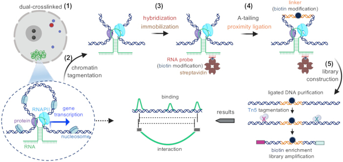
图1:RDD文库构建流程示意图。RDD文库构建的核心流程步骤按顺序展示,并配有相应的示意图。该流程首先进行双重交联和染色质标签化,随后与生物素修饰的RNA探针杂交,并通过链霉亲和素固定。后续步骤包括A尾修饰、使用生物素修饰接头进行邻近连接以及文库构建。在完成DNA连接后,进行纯化、Tn5介导的片段化、生物素富集和文库扩增。请点击此处查看该图的放大版本。
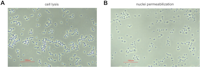
图2:代表性显微镜图像 果蝇 S2细胞展示用于细胞裂解和细胞核通透性的视觉质量控制标准。 (A) 成功的细胞裂解表现为细胞膜通透性增加和胞质内容物释放,使细胞核清晰可见。 (B) 细胞肿胀、质膜部分脱离以及细胞核完整且清晰可见,表明细胞核成功实现了通透化。比例尺 = 100 µm。 请点击此处以查看此图的放大版本。
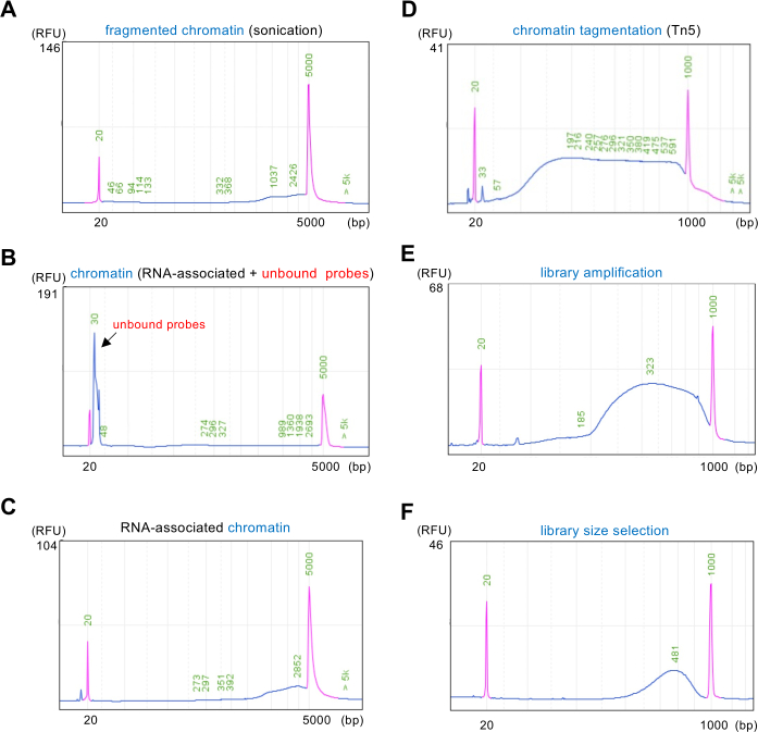
图3:染色质样品的电泳图谱分析。 毛细管电泳系统对多种染色质样品进行电泳图谱分析的截图。(A) 显示经超声处理后的染色质片段,片段大小范围约为 1000 至 3000 bp,表明片段化成功。(B) 展示与RNA结合的染色质(RNA相关+未结合探针),其中较小的峰代表未结合的探针,如箭头所示。(C) 显示RNA相关染色质,其浓度范围同样在1000至3000 bp之间,反映出RNA-蛋白质复合物的特征。(D) 描绘经Tn5转座酶标记的染色质,其片段分布范围反映了标签化(tagmentation)效率。(E) 显示扩增后测序文库的片段大小分布,跨度约为180-1000 bp。(F) 显示经片段大小选择后的测序文库片段分布,片段主要富集于约250-600 bp范围内。各样品的RFU(相对荧光单位)值均已标出,表明对染色质完整性和片段大小的有效定量。紫色峰代表分子量标记物。请点击此处查看该图的放大版本。
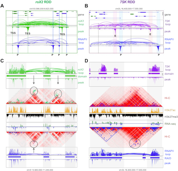
图4:ncRNA相关染色质相互作用的RDD代表性结果。(A-B)展示ncRNA roX2(绿色)和7SK(紫色)结合的基因组位置,以及相应的远端染色质相互作用环。RNAPII ChIA-PET(蓝色)数据显示这些ncRNA与基因表达之间存在明确的调控关系,峰值表示相互作用强度。缩写:TSS = 转录起始位点;TES = 转录终止位点;P = 启动子;E = 增强子。(C-D)提供ncRNA roX2和7SK相关染色质环与TADs(红色)之间关系的高维视图,以及活性与非活性染色质状态和基因表达水平的差异。圆圈标示出跨越TADs具有显著相互作用的区域。请点击此处查看该图的放大版本。
表1:RDD反应试剂。 请点击此处下载该表格。
表2:RDD缓冲液的组成。 请点击此处下载该表格。
讨论
方法中的关键步骤
准确估算RNA探针的数量及其在染色质中对应的RNA水平至关重要。过量的探针可能导致未结合的探针在固定步骤中占据链霉亲和素位点,从而阻碍目标RNA相互作用的有效捕获。此外,加入过多的链霉亲和素会造成试剂浪费,并需要更多的变性IBP来有效封闭空余位点。封闭不充分会负面影响后续邻近连接的效率,尤其是邻近连接接头本身也带有生物素修饰。如果封闭不充分,部分接头可能直接结合到链霉亲和素磁珠上,使结果复杂化。同时,必须谨慎估算接头与染色质的比例——过量的接头可能导致染色质DNA的两端均被标记,从而抑制有效的邻近连接。
方法的改进
在本研究中,我们以 RDD 方法为例进行说明 果蝇 S2 细胞与 roX2 以ncRNA为例。试剂用量需根据细胞类型和ncRNA表达水平进行调整;若ncRNA表达水平较高,可减少细胞用量或提高RNA探针浓度,反之亦然。尽管本方案采用将染色质固定在磁珠上的方法,另一种替代方案则涉及在溶液中进行 原位 邻近连接后通过RNA探针进行富集。
特异性、对照与质量控制
我们此前已描述并验证了RDD技术,并对多种RNA进行了平行RDD实验,结果显示出高度特异性;读者可参考先前的研究4,16。RDD要求依赖探针的、位点特异性的富集,且该富集应具有可重复性,并在可能的情况下通过正交方法进行验证。真正的相互作用需满足三个标准:与邻近的非结合区域相比,在探针靶向位点出现富集;依赖探针(在阴性对照中缺失或显著降低);以及在生物学重复样本中具有可重复性,并经过独立验证(例如通过RT-qPCR)。反义探针使用Stellaris设计,通过BLAST/BLAT筛选以去除脱靶序列,订购时带有3'-生物素标记,并通过RT和qPCR进行验证(包括DNA污染检测以及靶位点与非靶位点的富集比较)。我们建议设置三种对照——无探针对照、随机/非靶向探针对照以及无关RNA或非结合区域探针对照——并通过比较靶标样本与对照样本中的qPCR或测序覆盖度来评估特异性,报告富集倍数及背景水平。对于低丰度RNA,可增加探针密度(在脱靶限制范围内)、提高测序深度,并进行正交验证。
方法的故障排除
如果捕获的 RNA-染色质相互作用显著偏低,可能需要通过调整接头和染色质的用量来优化邻近连接的效率。此外,自动毛细管电泳图谱显示存在大量未结合探针的高峰。结果表明探针添加过多,可通过减少使用量来纠正。
该方法的局限性
RDD 方法的一个关键优势在于其能够高效捕获特定 RNA 相关的染色质相互作用,这意味着每次实验只能针对一种特定的 RNA。此外,由于邻近连接技术旨在检测成对相互作用,因此无法同时评估多点位的相互作用。
该方法的意义
RDD 方法不仅能够揭示 RNA 结合位点,还能阐明这些位点之间的相互作用关系,从而加深我们对染色质结构的理解。
RDD 的实际应用与未来潜力
RDD 适用于多种与染色质相关的 RNA,包括内源性宿主 RNA 以及来自病毒或其他微生物的外源性 RNA,凸显了其在不同研究领域的广泛应用前景。
披露
一项与本方案中所述RDD技术相关的专利(中国专利号:ZL202210182253.X)已获授权。
致谢
本工作得到了国家重点研发计划项目&中国国家重点研发计划(2022YFC3400400 和 2022YFC3400401)及国家自然科学基金(32170644)。
材料
| 姓名 | 公司 | 目录编号 | 评论 |
|---|---|---|---|
| 0.5 M EDTA | Invitrogen | AM9260G | |
| 1 M 醋酸镁 | Sigma-Aldrich | 63052-100ML | |
| 1 M Tris-HCl pH 7.0 | Invitrogen | AM9851 | |
| 1 M Tris-HCl pH 8.0 | Invitrogen | AM9856 | |
| 1.5-mL 离心管 | Eppendorf | 30108051 | |
| 10 mM dATP | Invitrogen | 18252015 | |
| 10% SDS (wt/vol) | Invitrogen | AM9822 | |
| 10× A-tailing buffer | NEB | B7002S | |
| 10× End-repair buffer | NEB | B6002S | |
| 10× T4 DNA ligase buffer | NEB | B0202S | |
| 10× TBE buffer | Invitrogen | AM9863 | |
| 14-mL 圆底试管 | Corning | 352057 | |
| 2× PCR master mix | NEB | M0541 | |
| 20× SSC buffer | Invitrogen | AM9770 | |
| 200-μL PCR 管 | Sangon Biotech | F611541-0010 | |
| 250-mL 方形 PET 储存瓶 | Corning | 431531 | |
| 2-巯基乙醇 (β-ME) | Sigma-Aldrich | M6250-100mL | ! 注意事项:β-ME 具有毒性;务必在通风橱中操作。 |
| 2-mL 管 | Eppendorf | 22431048 | |
| 3 M 醋酸钠 pH 5.5 | Invitrogen | AM9740 | |
| 5 M NaCl | Invitrogen | AM9759 | |
| 5 M 醋酸钾 | Sigma-Aldrich | 95843-100ML-F | |
| 500-mL 方形 PET 储存瓶 | Corning | 431532 | |
| 分析天平 | Sartorius | BP211D | |
| 自动细胞计数仪 | Counterstar | IC1000 | |
| 自动毛细管电泳系统 | Bioptic | C100100 | |
| 生物素标记探针 | GENEWIZ | 参见 表 1 | |
| 封闭粉 | Invitrogen | T2015 | |
| 桥接连接子寡核苷酸 | GENEWIZ | 参见 表 1 | |
| CO2 培养箱 | Thermo Scientific | 51033549 | |
| 二甲基亚砜 (DMSO) | Sigma-Aldrich | D2650-100mL | |
| Illumina 用 DNA 文库构建试剂盒 V2 | Vazyme | TD501 | |
| DNA 纯化试剂盒 | Zymo | D4013 | |
| Dulbecco’s 磷酸盐缓冲液 (DPBS) (1×) | Gibco | 14190250 | |
| 不含 EDTA 的蛋白酶抑制剂混合物 | Roche | 11836170001 | |
| EGS (乙二醇双[琥珀酰亚胺基琥珀酸酯]) | Thermo Scientific | 21565 | |
| 洗脱缓冲液 | Qiagen | 19086 | |
| 纯乙醇 | Sigma-Aldrich | E7023-500mL | |
| 甲醛溶液 | Sigma-Aldrich | 47608-250mL-F | ! 注意事项:甲醛具有毒性;务必在通风橱中操作。 |
| 甲酰胺 | Invitrogen | AM9342 | |
| 凝胶成像系统 | Tanon | 3500R | |
| 甘氨酸 | Invitrogen | 50046-1kg | |
| 含蓝色染料的糖原 | Invitrogen | AM9516 | |
| 高灵敏度检测盒 | Bioptic | C105105 | |
| 高密度相分离管 (1.5 mL) | Qiagen | 129046 | |
| 高灵敏度双链 DNA 荧光定量检测试剂盒 | Invitrogen | Q32851 | |
| Illumina 用 Index 试剂盒 V2 | Vazyme | TD202 | |
| 碘乙酰基-PEG2-生物素 (IPB) | Thermo Scientific | 21334 | |
| 异丙醇 | Sigma-Aldrich | I9030 | ! 注意事项:异丙醇具有毒性;务必在通风橱中操作。 |
| 千碱基检测盒 | Bioptic | C105106 | |
| Klenow 片段 (3'→5' 外切酶缺陷型) | NEB | M0212L | |
| 磁珠 | Beckman | A63881 | |
| 适用于 15-mL 管的磁力分离架 | Invitrogen | 12301D | |
| 适用于 2-mL 管的磁力分离架 | Invitrogen | 12321D | |
| 适用于 PCR 管的磁力分离架 | NEB | S1515S | |
| 微生物培养箱 | Thermo Scientific | IMH180 | |
| 显微镜 | Nikon | Eclipse TS2 | |
| 混匀仪 | ELMI | RM-2L | |
| N,N-二甲基甲酰胺 (DMF) | Sigma-Aldrich | 227056-100ML | ! 注意事项:DMF 具有毒性;务必在通风橱中操作。 |
| 非制冷型微量离心机 | Eppendorf | 5405000204 | |
| 无核酸酶水(非 DEPC 处理,1000 mL) | Invitrogen | AM9932 | |
| 无核酸酶水(非 DEPC 处理,50 mL) | Invitrogen | AM9937 | |
| 核酸定量仪 | Invitrogen | Q33226 | |
| 旋转摇床 | Crystal | SYC-2102 | |
| 苯酚:氯仿:异戊醇 | Solarbio | P1012-100mL | |
| 蛋白酶 K 溶液 | Invitrogen | AM2548 | |
| 制冷型离心机 | Eppendorf | 22628180 | |
| 制冷型培养箱 | Thermo Scientific | IMP180 | |
| 制冷型微量离心机 | Eppendorf | 5404F1621754 | |
| 冰箱 | Haier | DW-25L262 | |
| RNase 抑制剂 | Ambion | AM2696 | |
| 无 RNase 的 15-mL 管 | Invitrogen | AM12500 | |
| 无 RNase 的 50-mL 管 | Invitrogen | AM12502 | |
| 超声破碎仪 | Sonics & Materials | VCX130 | |
| C1 型链霉亲和素包被磁珠 | Invitrogen | 65001 | |
| M-280 型链霉亲和素包被磁珠 | Invitrogen | 11205D | |
| T4 DNA 连接酶 | NEB | M0202S | |
| T4 DNA 聚合酶 | NEB | M0203S | |
| TE 缓冲液 pH 8.0 | Invitrogen | AM9849 | |
| PCR 仪 | Bio-Rad | 1851148 | |
| 恒温混匀仪 | Eppendorf | 5382000023 | |
| Triton X-100 | Acros Organics | 327371000-100mL | |
| 超低温冰箱 | Thermo Scientific | 995 | |
| 真空浓缩仪 | TOMY | MV-100 |
参考文献
- Rao, S. S. P., et al. A 3D map of the human genome at kilobase resolution reveals principles of chromatin looping. Cell. 159 (7), 1665-1680 (2014).
- Fullwood, M. J., et al. An oestrogen-receptor-α-bound human chromatin interactome. Nature. 462 (7269), 58-64 (2009).
- Li, X., et al. GRID-seq reveals the global RNA-chromatin interactome. Nat Biotechnol. 35 (10), 940-950 (2017).
- Tian, S. Z., et al. 3D chromatin structures associated with ncRNA roX2 for hyperactivation and coactivation across the entire X chromosome. Sci Adv. 10 (30), eado5716(2024).
- Rinn, J., Guttman, M. RNA and dynamic nuclear organization. Science. 345 (6202), 1240-1241 (2014).
- Chu, C., Qu, K., Zhong, F. L., Artandi, S. E., Chang, H. Y. Genomic Maps of Long Noncoding RNA Occupancy Reveal Principles of RNA-Chromatin Interactions. Mol Cell. 44 (4), 667-678 (2011).
- Simon, M. D., et al. The genomic binding sites of a noncoding RNA. Proc Natl Acad Sci. 108 (51), 20497-20502 (2011).
- Yan, Z., et al. Genome-wide colocalization of RNA-DNA interactions and fusion RNA pairs. Proc Natl Acad Sci. 116 (8), 3328-3337 (2019).
- Quinodoz, S. A., et al. RNA promotes the formation of spatial compartments in the nucleus. Cell. 184 (23), 5775-5790 (2021).
- Li, X., et al. Long-read ChIA-PET for base-pair-resolution mapping of haplotype-specific chromatin interactions. Nat Protoc. 12 (5), 899-915 (2017).
- Fang, R., et al. Mapping of long-range chromatin interactions by proximity ligation-assisted ChIP-seq. Cell Res. 26 (12), 1345-1348 (2016).
- Mumbach, M. R., et al. HiChIP: efficient and sensitive analysis of protein-directed genome architecture. Nat Meth. 13 (11), 919-922 (2016).
- Lee, B., et al. ChIA-PIPE: A Fully Automated Pipeline for Comprehensive ChIA-PET Data Analysis and Visualization. Sci Adv. 6 (28), eaay2078(2020).
- Meller, V. H., et al. Ordered Assembly of roX RNAs into MSL Complexes on the Dosage-Compensated X Chromosome in Drosophila. Curr Biol. 10 (3), 136-143 (2000).
- Egloff, S., Studniarek, C., Kiss, T. 7SK small nuclear RNA, a multifunctional transcriptional regulatory RNA with gene-specific features. Transcription. 9 (2), 95-101 (2018).
- Tian, S. Z., et al. Landscape of the Epstein-Barr virus-host chromatin interactome and gene regulation. EMBO J. 44 (13), 3872-3915 (2025).
重印与许可
