ELISPOT 分析:检测 IFN-- 分泌性细胞
1. 设置
缓冲液和试剂
- 不含钙或镁的无菌磷酸盐缓冲盐 (PBS)
- 涂层缓冲液 - 无菌 PBS 或碳酸盐缓冲器
- PBS 中测定稀释 - 10% 胎儿牛血清 (FBS)
- 细胞培养介质-RPMI 1640,带10%FBS、青霉素/链霉素和L-谷氨酰胺
- 洗涤缓冲液 - 含有 0.05% Tween20 的 PBS
- 双蒸馏水 (ddH2O)
- 检测基质- 100mg AEC(3-氨基-9-乙基-卡巴佐尔),在10mL DMF(N,N,二甲基甲酰胺)。
设备
- 拉米纳尔流量罩
- 加湿培养箱(设置在37°C,5%CO2)
- 自动 ELISPOT 阅读器或解剖显微镜
材料
- 埃利SPOT板
- 无菌和非无菌储液罐
- 移管器和提示
- 无菌血清学移液器
- 无菌、锥形聚丙烯管
- 两个挤压瓶,用于洗板
测定特异性试剂
- 细胞- 原细胞或细胞系(这里,使用C57BL/6小鼠的血细胞)
- 兴奋剂 - 线粒素或抗原(此处,使用磷脂 12-米精酸盐 13-醋酸酯 (PMA, 50 ng/mL) 和碘霉素 (1 μM)
- 原发抗体-生物氨酸抗细胞因子检测抗体(在测定稀释剂中稀释至2微克/mL)
- 二次抗体-链球菌-马萝卜过氧化物酶(SAv-HRP)
2. 程序
涂层
- 保持无菌状态,在层流罩内,将纯化的抗细胞因子捕获抗体稀释至无菌涂层缓冲液中最终浓度为0.5-4.0 μg/mL。(注:对于IFN-α和IL-6使用5μg/mL)。
- 将捕获抗体溶液 100 μL/孔转移到 ELISPOT 板。
- 用板盖盖住板并密封,以防止蒸发。
- 在4°C下孵育板过夜。
阻塞
- 第二天,在层流罩中揭开 ELISPOT 板。快速将板倒置到无菌湿巾上,从每个孔中取出捕获抗体溶液。
- 然后,在每个孔中加入200μL的细胞培养基。此步骤将阻止在测定期间进行非特定绑定。
- 更换板盖,在 37°C 下孵育板 2 小时。
电镀和激活细胞
- 在培养板时,在细胞培养基中制备含有50纳克/mL PMA和1μM碘霉素的2X线粒体溶液。
- 然后,将目标细胞悬浮液制备到2 x 10⁶细胞/mL的库存浓度。
- 孵育完成后,通过快速将板倒置到层流罩内的无菌湿巾上,从每个孔中取出细胞培养基。
- 接下来,生成库存单元悬架溶液的 2 倍串行稀释。为此,首先将200 μL的制备蜂窝悬浮液添加到ELISPOT板顶行的井中。
- 然后,在包含细胞库存溶液的行下方的板的下五行中添加 100 μL 的普通细胞培养基。
- 之后,通过将细胞悬浮液的 100 μL 从顶行移入正下方的行,执行 2X 串行稀释。通过上下轻轻移液,确保正确混合,确保均匀分布电池。
- 对其余四行重复此过程。
- 仅保留第六行的区域性介质。它将作为实验控制。
- 接下来,将制备的线粒体溶液的100μL添加到板的前五行的实验井中。在对照井和第六行中,添加100μL的细胞培养基,不含线粒体。
- 在 37°C、5% CO2的培养箱中更换板盖和孵育板 20-48 小时。(注:20-24小时通常足以检测IL-2和TNF-α,而48小时对于IL-4和IFN-*是最佳选择。
检测
原抗体
- 制备生物仿当化抗细胞因子检测抗体,在测定稀释剂中浓度为2μg/mL。
- 此时通过在 PBS 中混合 0.05% 补间-20 来制备 20-25 mL 的洗涤缓冲液。
- 孵育完成后,解盖板,并迅速将其倒置在水槽上,以清除井中的所有液体。(注意:在此之后,板不再需要保持无菌)。
- 然后,通过在每个孔中加入±200 μL洗涤缓冲液来洗板。通过快速反转和轻拂水槽上的盘子来排出这种液体。重复此过程,共进行五次处理。
- 接下来,在每个井中加入100μL的稀释生物氨酸抗细胞因子检测抗体溶液。在室温下孵育2小时,室温下孵育,或在4°C下孵育。
二次抗体
- 孵育完成后,通过反转和轻拂水槽上的板来排出检测抗体。
- 与之前一样,用+200 μL洗涤缓冲液清洗板5次,每次洗涤之间排出液体。
- 接下来,在每口井中加入100 μ0μL的稀释链球蛋白-马萝卜过氧化物酶溶液(稀释到其预先确定的最佳浓度测定稀释剂中)。
- 更换板盖,在室温下在37°C下孵育1.5-2小时。
衬 底
- 孵育后,使用前不超过15分钟,根据制造商的说明首先激活AEC基板溶液。
- 接下来,丢弃井内的物品,像以前那样用洗涤缓冲液洗板五次。
- 然后,立即将100μL的制备AEC基板溶液加入每个井中。
- 在室温下孵育板约10-20分钟,同时监测现场发育。
- 用清水将盘子浸在水槽上,从而停止反应。
- 将盘子放在纸巾上,让盘子在一夜之间晾干,或直到完全干燥。取出板下的塑料托盘有助于干燥。
3. 数据采集和分析
- 干燥后,这些斑点已准备好使用自动板读取器计数。在这里,使用CTL免疫点读取器,但该协议可以适用于任何读取器。
- 首先打开仪器,然后打开计算机。然后,打开 CTL 程序并单击"扫描计数"。
- 推动"弹出"托盘从机器中伸出。然后,拆下塑料适配器,并在 ELISPOT 板和适配器上对齐行"A"。
- 选择要保存的文件的文件名和位置,并将板加载到托盘上。
- 然后,单击软件上的"加载"并关闭机器侧面的门。
- 按"计数后开始"。确保保存文件,然后打开质量控制"QC"软件来分析数据并计算点数。
笔记:
- 在初步实验中应确定细胞的最小数量。最佳点数为 +50/井。如果加载的单元格太多,将很难检测出不同的斑点。此外,细胞会重叠,可能不会在膜上形成单层,因此检测水平可能会降低。
- 优化实验时,考虑目标蛋白的预期表达水平。表达式越低,每口井所需的单元格数就越多。
- 与 ELISA 不同,最好用手洗盘子,而不是使用洗盘。ELISPOT板更细腻,应避免刺穿PVDF膜。
- 在孵育期间,应限制板块的运动,因为它可能导致斑点弄脏。
- 板应储存在黑暗中,因为暴露在直光下会导致斑点褪色。
资料来源:托尼娅·韦伯1
1马里兰大学医学院微生物学和免疫学系,马里兰州巴尔的摩的马琳和斯图尔特·格林鲍姆综合癌症中心 21201
ELISPOT 是一种标准化的、可重复的检测,用于检测细胞免疫反应。该测定利用一种基于酶链接的免疫吸附测定(ELISA)方法检测单细胞免疫反应,这些反应可以通过斑点可视化,因此命名为ELISPOT。1983年,Czerkinsky首次将ELISPOT描述为一种列举产生抗原特异性免疫球蛋白(1)的B细胞杂交瘤数量的方法。同一组进一步开发了测定,以测量细胞因子产生T淋巴细胞的频率。现在,ELISPOT已成为在临床试验和疫苗候选中测量抗原特异性T细胞免疫的黄金标准。例如,在接种疫苗后或感染期间,血浆细胞和记忆B细胞分泌抗体,提供保护。通常,通过测量抗原特异性抗体的血清位子来评估这些B细胞反应。然而,这种类型的分析,通常由ELISA测量,可能不包括记忆B细胞,即使在没有可检测的血清抗体水平的情况下,也可能存在记忆B细胞。此外,循环记忆B细胞对于病原体重新暴露后观察到的快速保护性抗体反应非常重要,因此能够检测这些细胞至关重要。因此,为了清楚地评估抗原特异性记忆B细胞反应,应同时使用ELISA和ELISPOT(2)。
ELISPOT 测定使用含有膜衬里孔的板,这些孔被涂有抗体,以捕获感兴趣的分泌蛋白质。然后,板加载细胞和刺激,以诱导蛋白质生产。分泌的蛋白质被表面涂覆的抗体捕获。在适当的孵育时间后,细胞被移除,并且使用生物缩化抗体检测分泌的分子,该抗体是特定为不同表位,与捕获抗体相比。接下来,加入链球菌蛋白过氧化物酶,然后添加基板,允许检测斑点(图1)。这种测定的用量是,它允许定量出产生感兴趣的蛋白质的细胞数量。重要的是,人们可以评估产生特定蛋白质的细胞总数是否有变化,或者种群中的单个细胞是否产生更多的蛋白质。此外,它可以提供有关动力学的信息,并可用于评估与抗原特异性反应(抗原模拟)相关的整体免疫活化(米托根刺激)。ELISPOT测定将允许在线生成或抗原特异性激活后在300,000个细胞中检测一个活化细胞。
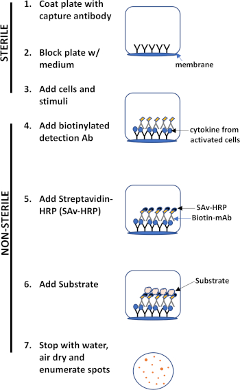
图 1:ELISPOT 协议概述。
这种测定的主要优点是它简单,协议相对简单明了。它不需要技术专长,b. 敏感性 - 它允许在单个细胞水平上检测免疫细胞,并且与其他方法(如流式细胞测量、c. 功能)相比,它只需要很少的细胞,它提供有关免疫的定量数据功能。
本实验演示了用于检测IFN-分泌-血细胞的ELISPOT协议,但如上所述,此测定也可用于评估B细胞(3)的抗体分泌。
1. 设置
缓冲液和试剂
- 不含钙或镁的无菌磷酸盐缓冲盐 (PBS)
- 涂层缓冲液 - 无菌 PBS 或碳酸盐缓冲器
- PBS 中测定稀释 - 10% 胎儿牛血清 (FBS)
- 细胞培养介质-RPMI 1640,带10%FBS、青霉素/链霉素和L-谷氨酰胺
- 洗涤缓冲液 - 含有 0.05% Tween20 的 PBS
- 双蒸馏水 (ddH2O)
- 检测基质- 100mg AEC(3-氨基-9-乙基-卡巴佐尔),在10mL DMF(N,N,二甲基甲酰胺)。
设备
- 拉米纳尔流量罩
- 加湿培养箱(设置在37°C,5%CO2)
- 自动 ELISPOT 阅读器或解剖显微镜
材料
- 埃利SPOT板
- 无菌和非无菌储液罐
- 移管器和提示
- 无菌血清学移液器
- 无菌、锥形聚丙烯管
- 两个挤压瓶,用于洗板
测定特异性试剂
- 细胞- 原细胞或细胞系(这里,使用C57BL/6小鼠的血细胞)
- 兴奋剂 - 线粒素或抗原(此处,使用磷脂 12-米精酸盐 13-醋酸酯 (PMA, 50 ng/mL) 和碘霉素 (1 μM)
- 原发抗体-生物氨酸抗细胞因子检测抗体(在测定稀释剂中稀释至2微克/mL)
- 二次抗体-链球菌-马萝卜过氧化物酶(SAv-HRP)
2. 程序
涂层
- 保持无菌状态,在层流罩内,将纯化的抗细胞因子捕获抗体稀释至无菌涂层缓冲液中最终浓度为0.5-4.0 μg/mL。(注:对于IFN-α和IL-6使用5μg/mL)。
- 将捕获抗体溶液 100 μL/孔转移到 ELISPOT 板。
- 用板盖盖住板并密封,以防止蒸发。
- 在4°C下孵育板过夜。
阻塞
- 第二天,在层流罩中揭开 ELISPOT 板。快速将板倒置到无菌湿巾上,从每个孔中取出捕获抗体溶液。
- 然后,在每个孔中加入200μL的细胞培养基。此步骤将阻止在测定期间进行非特定绑定。
- 更换板盖,在 37°C 下孵育板 2 小时。
电镀和激活细胞
- 在培养板时,在细胞培养基中制备含有50纳克/mL PMA和1μM碘霉素的2X线粒体溶液。
- 然后,将目标细胞悬浮液制备到2 x 10⁶细胞/mL的库存浓度。
- 孵育完成后,通过快速将板倒置到层流罩内的无菌湿巾上,从每个孔中取出细胞培养基。
- 接下来,生成库存单元悬架溶液的 2 倍串行稀释。为此,首先将200 μL的制备蜂窝悬浮液添加到ELISPOT板顶行的井中。
- 然后,在包含细胞库存溶液的行下方的板的下五行中添加 100 μL 的普通细胞培养基。
- 之后,通过将细胞悬浮液的 100 μL 从顶行移入正下方的行,执行 2X 串行稀释。通过上下轻轻移液,确保正确混合,确保均匀分布电池。
- 对其余四行重复此过程。
- 仅保留第六行的区域性介质。它将作为实验控制。
- 接下来,将制备的线粒体溶液的100μL添加到板的前五行的实验井中。在对照井和第六行中,添加100μL的细胞培养基,不含线粒体。
- 在 37°C、5% CO2的培养箱中更换板盖和孵育板 20-48 小时。(注:20-24小时通常足以检测IL-2和TNF-α,而48小时对于IL-4和IFN-*是最佳选择。
检测
原抗体
- 制备生物仿当化抗细胞因子检测抗体,在测定稀释剂中浓度为2μg/mL。
- 此时通过在 PBS 中混合 0.05% 补间-20 来制备 20-25 mL 的洗涤缓冲液。
- 孵育完成后,解盖板,并迅速将其倒置在水槽上,以清除井中的所有液体。(注意:在此之后,板不再需要保持无菌)。
- 然后,通过在每个孔中加入±200 μL洗涤缓冲液来洗板。通过快速反转和轻拂水槽上的盘子来排出这种液体。重复此过程,共进行五次处理。
- 接下来,在每个井中加入100μL的稀释生物氨酸抗细胞因子检测抗体溶液。在室温下孵育2小时,室温下孵育,或在4°C下孵育。
二次抗体
- 孵育完成后,通过反转和轻拂水槽上的板来排出检测抗体。
- 与之前一样,用+200 μL洗涤缓冲液清洗板5次,每次洗涤之间排出液体。
- 接下来,在每口井中加入100 μ0μL的稀释链球蛋白-马萝卜过氧化物酶溶液(稀释到其预先确定的最佳浓度测定稀释剂中)。
- 更换板盖,在室温下在37°C下孵育1.5-2小时。
衬 底
- 孵育后,使用前不超过15分钟,根据制造商的说明首先激活AEC基板溶液。
- 接下来,丢弃井内的物品,像以前那样用洗涤缓冲液洗板五次。
- 然后,立即将100μL的制备AEC基板溶液加入每个井中。
- 在室温下孵育板约10-20分钟,同时监测现场发育。
- 用清水将盘子浸在水槽上,从而停止反应。
- 将盘子放在纸巾上,让盘子在一夜之间晾干,或直到完全干燥。取出板下的塑料托盘有助于干燥。
3. 数据采集和分析
- 干燥后,这些斑点已准备好使用自动板读取器计数。在这里,使用CTL免疫点读取器,但该协议可以适用于任何读取器。
- 首先打开仪器,然后打开计算机。然后,打开 CTL 程序并单击"扫描计数"。
- 推动"弹出"托盘从机器中伸出。然后,拆下塑料适配器,并在 ELISPOT 板和适配器上对齐行"A"。
- 选择要保存的文件的文件名和位置,并将板加载到托盘上。
- 然后,单击软件上的"加载"并关闭机器侧面的门。
- 按"计数后开始"。确保保存文件,然后打开质量控制"QC"软件来分析数据并计算点数。
笔记:
- 在初步实验中应确定细胞的最小数量。最佳点数为 +50/井。如果加载的单元格太多,将很难检测出不同的斑点。此外,细胞会重叠,可能不会在膜上形成单层,因此检测水平可能会降低。
- 优化实验时,考虑目标蛋白的预期表达水平。表达式越低,每口井所需的单元格数就越多。
- 与 ELISA 不同,最好用手洗盘子,而不是使用洗盘。ELISPOT板更细腻,应避免刺穿PVDF膜。
- 在孵育期间,应限制板块的运动,因为它可能导致斑点弄脏。
- 板应储存在黑暗中,因为暴露在直光下会导致斑点褪色。
酶联免疫斑点 (ELISPOT) 测定是一种分析对病原体或细胞损伤的免疫反应的方法。它允许通过检测不同免疫细胞分泌的特定蛋白质来量化它们的激活。例如,ELISPOT 通常用于通过检测分泌的细胞因子来测量暴露于外来抗原时的 T 细胞反应。
对于基于细胞因子的 ELISPOT 检测,该过程从用捕获抗体包被 ELISPOT 微孔板开始,捕获抗体对目标细胞因子具有特异性。抗体包被后,T 细胞被添加到孔中,并受到外部试剂(例如抗 CD3 抗体)的刺激。然后细胞分泌靶细胞因子,该细胞因子立即被捕获抗体固定。由于蛋白质在从活细胞分泌后立即捕获,无需稀释或降解,因此该测定具有很高的准确性。固定目标细胞因子后,加入检测抗体,该抗体也与捕获的细胞因子结合。
ELISPOT 技术还可用于通过分析特异性抗体的产生来量化感染或疫苗接种后的记忆 B 细胞。在基于抗体的酶联免疫斑点免疫斑点法中,在捕获步骤中,抗原将与板结合,或在检测步骤中,抗原在捕获后检测靶抗体,使用特异性抗原代替抗体。在该过程的所有变体中,对于 T 细胞或 B 细胞,检测抗体或抗原被生物素化,这使得它能够与链霉亲和素偶联的检测酶(如辣根过氧化物酶)结合。然后,在加入过氧化物酶的底物 AEC 后,产生深色、不溶性沉淀。该沉淀物标记捕获蛋白质的位置,每个分泌细胞产生一个可见斑点,可以使用 ELISPOT 读数仪或显微镜对其进行定量。斑点的大小是每个细胞分泌的蛋白质量的相对估计值。该检测可以检测来自单个细胞的免疫反应,即使在相对较小的分泌细胞亚群中,也可用于研究细胞水平的免疫反应。
在本视频中,您将学习如何进行 ELISPOT 检测,然后定量代表分泌细胞的斑点。
在整个实验过程中,通过在层流罩中工作并戴上手套来确保无菌条件。
该方案中的所有计算均基于一个 96 孔板所需的体积。
首先,稀释抗细胞因子捕获抗体。为此,将 10 mL 缓冲液转移到无菌 15 mL 锥形管中。然后,使用移液器向缓冲液中加入 10 微升每毫升 1 毫克的单克隆抗体,制成最终浓度为 1 微克/毫升的溶液。接下来,将捕获抗体溶液倒入无菌储液槽中,并使用多通道移液器将 100 μL 分配到 96 孔 ELISPOT 板的每个孔中。
用板盖住板,密封以防止蒸发,并在 4 摄氏度下孵育过夜。第二天,在层流罩中揭开 ELISPOT 板。将板快速倒置到无菌湿巾上,以从每个孔中去除捕获抗体溶液。接下来,使用多通道移液器向每个孔中加入 200 μL 细胞培养基。此步骤将在检测过程中阻断非特异性结合。装回板盖,在 37 摄氏度的培养箱中孵育 2 小时。
在板孵育时,通过在 10 mL 细胞培养基中加入 1 μL PMA 和 20 μL 离子霉素来制备 2X 有丝分裂原溶液,以达到 15 ng/mL PMA 和 1 μmol 离子霉素的最终浓度。
此时也应在无菌罩中制备小鼠脾细胞的细胞悬液。使用显微镜和血细胞计数器测量细胞浓度并调整总体积,直到达到每毫升 200 万个细胞的储备浓度。
孵育完成后,将板快速倒置到无菌湿巾上,以从每个孔中去除细胞培养基。接下来,将 200 微升制备好的细胞悬浮液添加到 ELISPOT 板顶行的孔中。一式三份设置实验,以便将测试的每种细胞类型接种在一组三个分组列中。在此之下,将 100 微升普通细胞培养基添加到板的接下来五行中,在含有细胞储备液的行下方。
接下来,将 100 μL 细胞悬液从顶行移液到正下方的行中,轻轻上下移液以均匀分布细胞,进行连续稀释。对其余行重复此过程,在每个步骤中将 100 μL 从前一行移动到下一行,继续直到第五行被连续稀释。仅将第六行与细胞培养基一起留下,作为对照。为了刺激板实验孔中的细胞,将 100 微升制备的有丝分裂原溶液添加到第 1 行至第 5 行每个孔中的细胞悬液中。确保第六行(将用作对照)不受刺激。盖上盖子,将板在 37 摄氏度和 5% CO2 下孵育 24 至 48 小时。
制备稀释的生物素化抗细胞因子检测抗体。首先,将 5 mL 10% 胎牛血清添加到 45 mL PBS 中,制备 50 mL 检测稀释剂。接下来,在检测稀释剂中将检测抗体稀释至 2 μg/mL 的浓度。此外,此时通过混合 .05% Tween-20 和 PBS 来制备 20 至 25 毫升洗涤缓冲液。
孵育完成后,打开板盖并快速倒置以去除孔中的所有液体。通过向每个孔中加入约 200 微升洗涤缓冲液来洗涤板。通过在水槽上快速倒置和轻弹板来排出这种液体。再重复此过程四次,总共洗涤五次。接下来,向每个孔中加入 100 μL 稀释的检测抗体溶液,盖上盖子,并在室温下孵育 2 小时。孵育后,通过在水槽上倒置并轻弹板,将检测抗体溶液从板孔中排出。
和以前一样,用洗涤缓冲液洗涤板五次,在每次洗涤之间排出液体。最后一次洗涤后,根据制造商的说明稀释链霉亲和素-辣根过氧化物酶溶液,制备该溶液。接下来,在板的孔为空的情况下,向每个孔中加入 100 微升稀释的链霉亲和素-辣根过氧化物酶溶液。将盖子放回板上,在室温下孵育 2 小时。
孵育后,使用前不超过 15 分钟,激活预制的 AEC 底物溶液。丢弃孔中的内容物,像以前一样用洗涤缓冲液洗涤板五次。然后,立即向每个孔中加入 100 μL 制备的 AEC 底物溶液。将板在室温下显影约 10 至 20 分钟,同时监测斑点的发展。这些斑点将在孔表面显示为小而暗的圆圈。然后,用水冲洗板子并将其轻弹到水槽上以停止反应。用纸巾吸干板子,让其风干过夜或直至完全干燥。取下板下方的塑料托盘将有助于干燥。干燥后,可以使用自动读板器对斑点进行计数。
这里使用 CTL ImmunoSpot 阅读器,但该协议可以适用于任何阅读器。然后,打开 CTL 程序并单击 Scan Count.推动弹出,使纸盒从设备中伸出。然后,取下塑料适配器,将 ELISPOT 板和适配器上的 A 行对齐。选择要保存的文件的文件名和位置,然后将板和适配器加载到托盘上。单击软件上的加载并关闭机器侧面的门。然后,按 Start 开始 计数后。确保文件已保存,然后打开 Quality Control QC 软件分析数据并计算斑点数量。将此数据导出为 Excel 文件。分析完成后,单击 Eject (弹出) 以检索板。
在本实验中,对野生型和荷瘤小鼠的细胞进行铺板并分析 IFN γ。请注意,斑点的数量随着细胞浓度的降低而减少。通常,ELISPOT 数据表示为每接种细胞数的斑点计数。在此示例中,斑点的数量显示在条形图中,每个相应的细胞浓度列在 x 轴上。请注意,斑点数表示给定群体中每细胞总数的活化细胞数。
在本次ELISPOT测定中,对野生型和肿瘤小鼠的脾白细胞进行了分析,以检测IFN-α。图 2 A 显示了检测结果的可视图像。绿色数字表示每口井的点数(TNTC 表示"太多,无法计数")。请注意,斑点的数量随着细胞浓度的降低而减少。

图2A:肿瘤小鼠免疫反应下降。请点击此处查看此图的较大版本。
通常,ELISPOT 数据以每镀电池数的点计数数的形式显示。在图 2 B 中,点数显示...
ELISPOT测定允许一个人通过确定分泌特定Anlyat的细胞数量来评估免疫细胞的激活。斑点的大小和强度提供有关每个细胞产生的麻醉剂量的信息。上述协议详细说明了单个细胞因子的检测。然而,最近的事态发展增强了这种测定的效用。目前,人们可以使用荧光检测染料来检测井内多个分析物。这允许检测分泌一种或两种分析物的细胞的不同亚群。
Chapters in this video
0:01
Concepts
3:01
Coating and Blocking the Plate
4:42
Plating and Activating Cells
7:05
Detection
9:03
Substrate
10:01
Data Acquisition and Analysis
11:07
Results
Videos from this collection: