This video demonstrates the process of performing a fluorescent plaque assay using recombinant vaccinia virus encoding red fluorescent protein (RFP) in fibroblast cells, followed by plaque isolation and purification for further analysis.
Ein Abonnement von JoVE ist erforderlich, um diesen Inhalt anzuzeigen. Melden Sie sich an oder beginnen Sie noch heute mit einer kostenlosen Testphase.
Methodenartikel
Fluorescence-Based Detection and Purification of Recombinant Vaccinia Virus
152 Aufrufe
⸱
31. Juli 2026
In diesem Artikel
Zusammenfassung
Protokoll
- Purification of the Modified vaccinia virus (VV) (First Round)
- On day 1, seed 15 6-well plates with 3 x 10⁵ CV-1 cells in each well.
- On day 2, thaw the frozen cell suspension in a water bath at 37 °C for 3 min and vortex the cryovials vigorously for 30 s to obtain cell lysate without removing cell debris.
- On day 1, seed 15 6-well plates with 3 x 10⁵ CV-1 cells in each well.
- For the infection of one 6-well plate of CV-1 cells, dilute 1 µL of cell lysate with 3 mL of Dulbecco's modified eagle medium (DMEM) and add 0.5 mL of the diluted cell lysate into each well of the 6-well plate, on top of the media already present. Infect all the 6-well plates prepared.
NOTE: Removal of cell debris is not required. - After two days of infection (i.e., on day 4), identify red fluorescence protein (RFP)-positive plaques under a fluorescence microscope using a 10X objective lens and label the plaques underneath the plate using a marker pen by circling the plaque location.
NOTE: See Figure 1 for a representative RFP-positive plaque.- Prepare six labeled cryovials containing 200 µL serum-free DMEM cell culture medium to pick up six different RFP-positive plaques. Aspirate the cell culture medium from the well with labeled plaque(s). Attach a 200 µL tip to a P200 pipette, set the volume to 30 µL, and take ~ 10 µL cell culture medium from one cryovial.
- Hold the pipette push button without releasing the medium and use the tip to scratch the area of one labeled plaque. Release the pipette push button to lift up the detached cells and transfer them into the cryovial containing 190 µL of serum-free DMEM.
- Take another 10 µL medium from the vial with the scratched cells and repeat the procedure of picking up the same RFP-positive plaque three times to collect as many scratched cells as possible. Transfer all cells into the same vial and store the vial at -80 °C.
NOTE: Alternatively, freeze the cryovials on dry ice for 10 min if the second round of purification is performed immediately.
Zugriff eingeschränkt. Bitte melden Sie sich an oder starten Sie eine Testversion, um diesen Inhalt anzuzeigen.
Ergebnisse
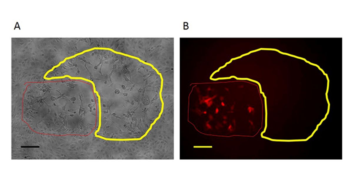
Figure 1: An RFP-positive plaque in the first round of purification of the mutant virus. In the first-round purification of the mutant virus, the RFP-positive plaque (circled with red line) with the target region deletion is surrounded by plaques forme...
Zugriff eingeschränkt. Bitte melden Sie sich an oder starten Sie eine Testversion, um diesen Inhalt anzuzeigen.
Materialien
| Name | Unternehmen | Katalognummer | Kommentare |
|---|---|---|---|
| Plasmid #41824 | 41824 | Addgene | gRNA cloning vector |
| pST1374 | 13426 | Addgene | Cas9 cloning vector |
| pGEMT easy | A1360 | promega | repair donor vector cloning |
| One Shot TOP10 Chemically Competent E. coli | C4040-10 | Invitrogen | transformation |
| QIAprep Spin Miniprep Kit | 27106 | Qiagen | plasmid extraction |
| Dulbecco’s Eagle’s Medium (DMEM) | 11965-092 | Life Technologies | cell culture medium |
| Fetal bovine serum (FBS) | SH30088.03HI | Hyclone | cell culture serum |
| Penicillin, Streptomycin | 15070-063 | Thermo Scientific | antibiotics |
| Effectene | 301425 | Qiagen | transfection |
| Thermo Scientific Nalgene Cryogenic vial; 2.0 mL | W-06754-96 | Thermo Scientific | collect virus |
