Ein Abonnement von JoVE ist erforderlich, um diesen Inhalt anzuzeigen. Melden Sie sich an oder beginnen Sie noch heute mit einer kostenlosen Testphase.

Methodenartikel

Immunofluorescence Assay to Detect Hepatitis C Virus Replication Complexes in Infected Cells

156 Aufrufe

31. Juli 2026

In diesem Artikel

Zusammenfassung

Source: Ren, S., et al. A Protocol for Analyzing Hepatitis C Virus Replication. J. Vis. Exp. (2014).

This video demonstrates an immunofluorescence-based method to detect Hepatitis C virus replication in infected hepatoma cells. The protocol involves cell fixation, antibody staining for viral protein and double-stranded RNA, and visualization using fluorophore-tagged secondary antibodies. Dual signal detection under a fluorescence microscope confirms active viral replication within the host cells.
Protokoll

1. Immunofluorescence Assay (IFA)

  1. Fix the Hepatitis C virus (HCV)-infected and transfected cells using methanol for 30 min at -20 ºC for the immunofluorescence assay.

  2. Wash the cell with phosphate-buffered saline (PBS) three times and block with IFA blocking buffer.

  3. Use rabbit polyclonal anti-NS5A primary antibody or mouse monoclonal anti-double-stranded RNA (dsRNA) antibody J2 at a dilution of 1:200 (1 μg/ml) and incubate for 5 hr to overnight at 4 ºC.

  4. Wash the cells with PBS three times after the primary antibody.

  5. Add goat anti-rabbit Immunoglobulin G (IgG)-488 polyclonal secondary antibody or goat anti-mouse IgG-594 polyclonal secondary antibody at a 1:1,000 dilution (1 μg/ml) and incubate for 1 hr at room temperature.

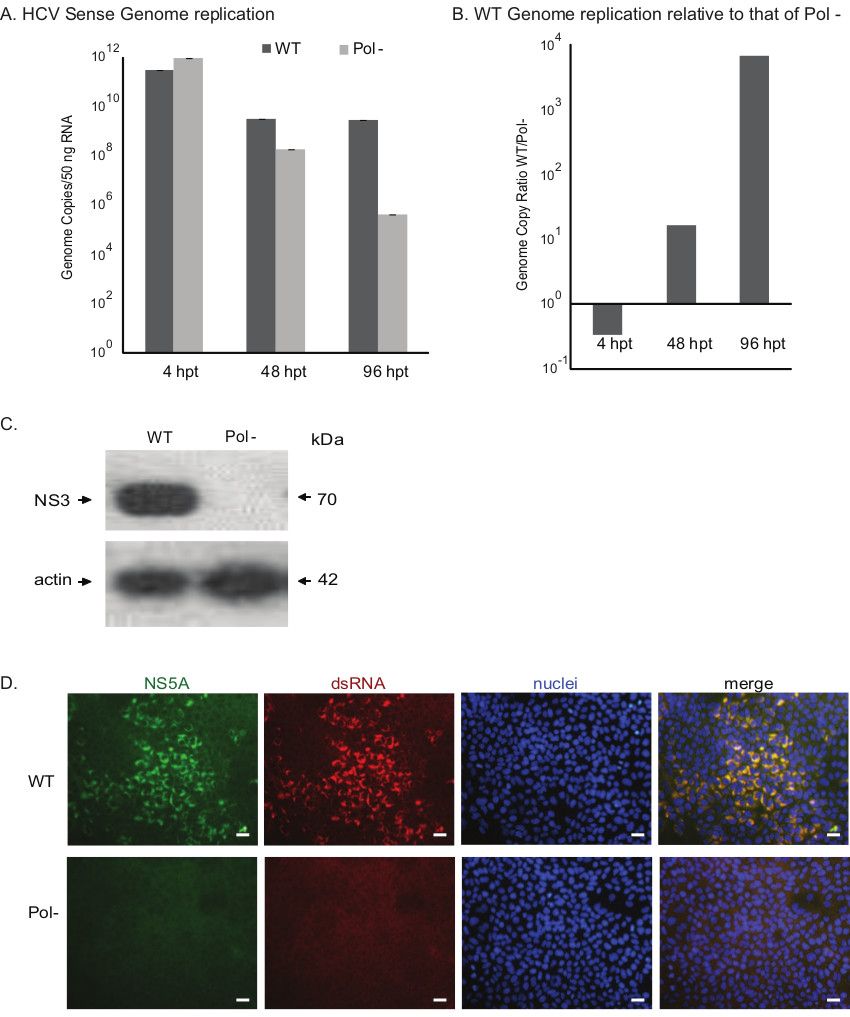
  6. Wash cells with PBS three times and stain nuclei using Hoechst dye and view using a fluorescence microscope (Figure 1D).

Zugriff eingeschränkt. Bitte melden Sie sich an oder starten Sie eine Testversion, um diesen Inhalt anzuzeigen.

Ergebnisse

figure-results-1

Figure 1. Assessing the growth phenotypes of HCV constructs. A) Evaluating the genome replication kinetics of wild-type (WT) and polymerase null (Pol-) viruses. The genome copy numbers of the sense RNA strand assessed by RT-qPCR are present...

Zugriff eingeschränkt. Bitte melden Sie sich an oder starten Sie eine Testversion, um diesen Inhalt anzuzeigen.

Materialien

Liste der in diesem Artikel verwendeten Materialien
NameUnternehmenKatalognummerKommentare
Dulbecco’s modified Eagle’s medium (DMEM)Fisher Scientific10-017-CV 
Non essential amino acidFisher ScientificMT25025CI 
HEPESLife Technologies15630080 
GlutamaxLife Technologies35050061 
Opti-MEM Reduced Serum Medium,no Phenol RedLife Technologies11058-021 
Huh-7.5.1The Scripps Research Institute The cell line was kindly provided by Dr. Francis Chisari to Dr. Arumugaswami under executed MTA between The Scripps Research Institute and Cedars-Sinai Medical Center
mouse monoclonal anti-dsRNA antibody J2English & Scientific Consulting Kft.10010200 
Goat anti-rabbit IgG Alexa Fluor 488Life TechnologiesA11008 
Goat anti-rabbit IgG Alexa Fluor 594Life TechnologiesA11020 
Blotting Grade Blocker Non Fat Dry MilkBio-Rad170-6404XTU 
Tween-20Bio-Rad170-6531XTU 
Anti-Hepatitis C Virus NS3 antibody [8 G-2]Abcamab65407 
Anti-Hepatitis C Virus NS3 antibody [H23]Abcamab13830 

Tags

HCV-Replikationinfizierte Hepatomzellendoppelstrangige RNAAntikörperfärbungFluoreszenzmikroskopievirale ProteinndetektionZellfixierungSekundärantikörper