The squid giant axon model allows for the real-time assessment of axonal transport in a neurodegenerative disease model. The presented protocol details the preparation of the isolated axoplasm, perfusion of mutant proteins of interest, and measurement of vesicle trafficking using VEC-DIC microscopy to identify pathogenic mechanisms in amyotrophic lateral sclerosis.
Methodenartikel
Isolated Axoplasm from Squid and Pathogenic Mechanisms in Amyotrophic Lateral Sclerosis
In diesem Artikel
Zusammenfassung
Zusammenfassung
The squid giant axon is an experimental model that has been instrumental in providing answers to many fundamental questions in cell biology, including the discovery of the motor protein kinesin and probing molecular mechanisms of neurodegenerative diseases such as amyotrophic lateral sclerosis (ALS). The giant axon offers scale and ease of access that is not possible in mammalian models. The power of this model stems from the fact that one can introduce reagents and proteins that would not cross a plasma membrane at defined concentrations in a buffer environment that is closely modeled on normal components of the axoplasm. By leveraging the unique biology of the squid axon, early studies were able to define the channels and ion specificities of the action potential. Subsequent studies using video-enhanced contrast- differential Interference contrast microscopy (VEC-DIC) and the isolated axoplasm led to the identification of kinesins, a class of motor proteins responsible for intracellular vesicle trafficking toward the plus end of microtubules. This combination of isolated axoplasm and advanced imaging continues to provide insights into pathogenic mechanisms for adult-onset neurodegeneration, including ALS, and regulation of fast axonal transport, a process disrupted in motor neuron disease. Here, procedures for dissecting the giant axon, isolating axoplasm, and perfusing defined amounts of proteins and/or reagents into the axoplasm are detailed, as well as methods for visualizing and interactively analyzing vesicle traffic in real time. This model represents a singular avenue to understand the critical function of neurons, shedding light on the mechanisms driving ALS pathology and other neurodegenerative diseases. This system also offers a tractable platform for drug testing, where candidate therapeutic compounds can be introduced directly into the axoplasm, and their effects on vesicle trafficking can be assessed in real time, providing a rapid and physiologically relevant means of identifying potential ALS therapeutic targets.
Einleitung
The longfin squid Doryteuthis pealeii has provided multiple critical models for understanding how a neuron functions. In nature, squid must escape predators to survive, yet the squid's giant axon lacks a myelin sheath. To compensate, the axon must have a larger diameter to increase conduction speed. The large size of the axon allowed physiologists to determine how action potentials were generated and the currents involved. Triggering a giant axon requires a giant presynaptic terminal. Unlike vertebrate synapses, the giant synapse is easily imaged and suitable for intracellular recording and microinjections, which have provided critical insights into synaptic mechanisms1 and provided an opportunity to visualize endogenous vesicular trafficking without selective labeling. Furthermore, this model is valuable because foundational transport mechanisms are conserved across eukaryotic cells, including motor proteins, cytoskeletal structures, and signaling pathways, allowing for the translation of findings to research in mammals, including humans2,3. A number of other groups have adapted the axoplasm model for the study of aspects of vesicle trafficking2,4,5,6,7,8,9,10,11.
Live cell imaging allows investigators to answer questions surrounding the intracellular trafficking of cellular proteins. Although high-resolution fluorescence imaging systems are growing in popularity, VEC-DIC microscopy is a powerful tool that minimizes limitations in spatial resolution secondary to diffraction at cell edges. Additionally, high-resolution fluorescence imaging requires dyes or labels that can create artifacts and potentially alter protein conformation, and are subject to fluorophore degradation. By combining this technology with the squid giant axon, VEC-DIC enables visualization of intracellular organelles at rates of 2–4µm/s without the need for confounding protein tagging3. Many contemporary studies using fluorescent tags are limited by acquisition speed due to the exposure time needed to get a fluorescent signal from low-abundance tagged molecules. In addition, overexpression of fluorescently tagged proteins can affect trafficking. VEC-DIC technology avoids these issues and allows visualization of fast-moving vesicles in the axoplasm with as few alterations to conditions as possible. Although VEC-DIC microscopy does not permit visualization of specific cargo-containing vesicles, it provides the most accurate description available of overall vesicle traffic.
The isolated axoplasm model combines analysis of vesicle trafficking while granting unprecedented access to the interior of an axon12. This combination led to the discovery of the kinesin family of molecular motors responsible for axonal transport, the trafficking of vesicles in the neuron13 and identified signaling pathways that may regulate motor protein functions. The goal of this protocol is to use the axoplasm to assess the effects of pathogenic proteins associated with ALS on neuronal function, focusing on vesicle trafficking14,15. Gaining a better understanding of pathogenic mechanisms allows for exploration of novel therapeutic strategies.
Protokoll
This study was completed in compliance with institutional regulations. All experiments are conducted according to principles established by the Marine Biological Laboratory Animal Care and Use Committee in Woods Hole, Massachusetts, consistent with NIH rules and regulations.
1. Dissection of the giant axon
NOTE: Isolated axoplasm is obtained from giant axons of longfin squid (Doryteuthis pealeii) found in the coastal areas of Cape Cod, MA. A detailed description of the procedure has been published previously3, so the focus here is on the concepts and laboratory protocols. Wild-caught squid suitable for axoplasm isolation should be translucent (indicating health) with mantles typically 25–30cm in length.
- Use large, freshly wild-caught squid with mantles 20cm or more, as these have axons large enough for routine extrusion. Use squid collected through the MBL program from April through October.
NOTE: Typically, use large male squid, but female squid can also be used. Consider that females run smaller than males; however, their axons may be comparable to those of larger male squid. Exclude male squid less than 20cm in length, as they will have axons too small for effective extrusion. Exclude squid with damaged mantles (opaque spots or cloudy muscle), as they are dying and will not yield reliable results. - Hold the squid with the mantle and decapitate it with a large pair of scissors. Place the mantle, with the fins and pen facing down, on the dissection table, with a continuous flow of ambient seawater and illumination from below.
- Cut the mantle along the midline with surgical scissors, starting from the base.
- Pin the mantle in place with 18-G needles and remove the viscera by separating the connective tissues from the mantle. This exposes the pen, a stiff, quill-shaped, clear structure, and the interior surface of the mantle.
CAUTION: Exercise caution when handling sharps needles, scissors, etc.
- Identification and exposure of the giant axons
NOTE: Each squid has multiple giant axons emanating from the stellate ganglion, each one scaled in size so that the longest axons are the largest. This allows the coordinated contraction of all muscles in the mantle. Only the two largest axons (roughly 500µm in diameter) located on either side of the pen are used. Axons must be at least 200–300µm in diameter for a successful extrusion.- Remove the pen by gently pulling from the top to avoid damaging the adjacent giant axons, then the mantle is flipped so that the skin is exposed.
- Peel the skin from the base toward the top using forceps, teasing with surgical scissors as needed. Remove the fins during this procedure. Removing the skin leaves the translucent muscle and facilitates dissection.
- Flip the mantle over so the interior surface is exposed.
- Using a dissecting microscope, dissect the two giant axons free from connective tissue and smaller axons starting from near the stellate ganglion and working toward the distal axon.
- Free up 2–3cm of each axon, which is carefully freed up. At this stage, the giant axon is intact with plasma membrane, glial sheath, and associated connective tissue/small axons still present.
- Tie off each axon with standard sewing thread. Use black thread to tie off the distal end of the axon and white thread for the proximal end (adjacent to the stellate ganglion) to indicate orientation during handling.
2. Preparation of isolated axoplasm
- Handle the axon by the threads and transfer to a Petri dish with chilled (2–4°C), filtered seawater for fine dissection using a dissecting microscope under dark field illumination.
- Gently tease away the remaining connective tissue and small axons using Vannas style iris scissors and No. 5 Dumont forceps.
- A properly cleaned axon appears as a smooth cylinder and is almost clear under dark field illumination.
- Axons are now ready for extrusion. Blot the axon on filter paper to remove excess seawater, then rinse in buffer X (Table 1) to remove remaining seawater. Blotting again to remove excess fluid, cut the axon adjacent to the white, proximal thread.
NOTE: The composition of buffer X is provided in Table 1. The 7.2pH is critical as acidic conditions as low as pH6.8 will have a deleterious effect on transport. Combine aliquots of stock reagents as indicated in the table and check pH. Adjust pH to 7.2 with KOH if needed. Bring to the final volume of 25ml with 20mM HEPES, pH 7.2, and filter on a 45µm Millipore filter or equivalent. Store as 0.5ml aliquots at -20°C until ready for use. - Place the axon on a clean 24mm × 50mm No. 0 coverslip on the fine dissecting microscope stage with dark field illumination.
- Extrude the axoplasm by holding the black thread and pressing gently just proximal to the ligature on the axon with a piece of PE190 polyethylene tubing. Pull the axon through the compression point, leaving a cylinder of isolated axoplasm on the coverslip.
NOTE: If the axoplasm does not have a gel-like consistency, this is a sign that cytoskeletal integrity has been compromised, and the axoplasm should not be used. - Assemble a simple perfusion chamber with two No.0 coverslip spacers secured with a thin coat of Molykote Compound 111 food-grade silicone lubricant (Figure 1) and top with a 22 mm × 22 mm No. 0 coverslip to create a chamber with open ends for perfusion. Apply Molykote 111 sparingly to both sides of the spacers.
- Secure the top coverslip at each corner with a small amount of VALAP (1:1:1 by weight of vaseline, lanolin, and paraffin).
NOTE: VALAP is a formulation developed by Robert Allen4, which is liquid at 50°C and solid at room temperature (Figure 1). This is a rapid way to secure the coverslip without compromising transport and leaving the ends of the chamber open for perfusion.
CAUTION: VALAP should be kept at 50°C in a temperature block or on a hot plate to maintain a liquid state, and care should be taken around hot objects. - Place the axoplasm in the chamber in a Petri dish with a circle of moist filter paper to keep the axoplasm from drying out. Use plastic shims to prevent the coverslip from contacting the damp filter paper.
- Keep the axoplasm in the coverslip chamber at 4°C for at least 15 min in a humidified environment.
NOTE: This step yields more consistent results. - At that point, the axoplasm can be returned to room temperature. Place it on the microscope for analysis.
NOTE: Alternatively, the axoplasm in the coverslip chamber can be held at room temperature for up to 2h in a chamber humidified as above with a damp filter paper round to prevent drying out. Vesicle trafficking continues at control levels for more than three hours in a large axon when stored in this manner. - Select axons larger than 250µm and extrude them to produce clear axoplasm cylinders approximately 20mm long that can be handled with forceps16.
NOTE: Properly extruded axoplasm displays linear striations corresponding to cytoskeletal elements and abundant bidirectional organelle trafficking under VEC-DIC microscopy. It maintains control transport rates for more than 4h when unperfused or perfused with control buffer. Axons smaller than 250µm generally do not extrude cleanly and produce disorganized axoplasm that is difficult to measure. Low-pH buffers (<pH 6.80) or incompatible contaminants, such as ethanol or dimethyl sulfoxide, rapidly degrade the axoplasm and stop transport within 5–10 min.
3. Setting up the microscope and VEC-DIC microscopy
NOTE: Video Enhanced Contrast Differential Interference Contrast (VEC-DIC) microscopy is critical for real-time (≥30 fps) analysis of vesicle trafficking. This microscopy method, originally described by Allen et al.17,18, was used to document fast axonal transport in the giant axon and isolated axoplasm4,19. A detailed description of the methods and equipment used in these assays has been published3, so the focus will be on the principles and practice of these studies to evaluate signaling pathways affected by mutant proteins associated with familial ALS.
- Image vesicle trafficking in the axoplasm using a Zeiss Axiomat microscope equipped with full DIC optics and a 100 ×, 1.3 NA objective at zoom setting 2. Capture images using a high-resolution CCD video camera with an adjustable dynamic range at a frame rate of ≥30 fps. Use a DIC-optimized objective with an NA of at least 1.3 and a magnification of 63× or 100×.
NOTE: The Axiomat provides an unusually large field of view; however, any microscope with suitable high-resolution DIC optics and a stable platform may be used. At lower frame rates, small organelles moving at 1.5–2µm/s become blurred and can no longer be detected. - Determine the video field of view using a reticle scale and use this measurement to calibrate the cursor speed. Perform measurements interactively on real-time video images acquired at 30fps.
- Real-time video processing and contrast enhancement.
NOTE: Real-time video enhancement of contrast on a live image is necessary for visualization of subresolution structures. Processing involves three stages:- Adjust the black level of the video image to minimize background scattered light.
- Focus just outside the plane of the axoplasm to obtain an image of the optical noise in the light path, and digitally store this image. Subtract the stored image from all subsequent frames of the live video to minimize optical noise.
- After minimizing scattered light and optical noise, expand the contrast range of the image to increase its dynamic range and amplify the signal.
NOTE: These three processing steps enhance the detection and real-time tracking of subresolution cytoskeletal and vesicular structures in living cells without tags or molecular manipulation. The Rayleigh diffraction limit still applies; therefore, objects separated by less than 200nm may be detected but cannot be resolved as individual objects in the XY plane. Nevertheless, this approach provides structural and dynamic information from living cells or isolated axoplasm.
4. Interactive measurement of vesicle trafficking
NOTE: Visualization of the small, transported vesicles (50–100nm) is complicated further by the number of organelles moving and the density of the microtubule and neurofilament network. Resolution in the x-y plane is roughly 200nm, and the depth of focus is approximately 400nm. The field of view is 25µm, but organelle movement is tracked in a volume that is 25µm × 25µm × 0.4µm. The focal plane is flat, but the axoplasm is not. Since the axoplasm is 500µm thick, microtubules and the organelles moving along them will often enter and leave the optical section, so it is not feasible to accurately track individual vesicles for any distance. Even organelles as large as mitochondria will enter and leave the field of view, so do not track individual organelles; instead, match the speed of calibrated cursors to the general flow of organelles, which is apparent in the video image. The readout is given as a rate, but this parameter reflects a combination of the speed at which vesicles are moving and the number of vesicles being trafficked. As the interval between a vesicle entering the focal plane and a similar vesicle leaving the plane increases, the apparent rate slows, analogous to changing the frame rate of a film.
- Match the speed and direction of cursor movement to the visible organelle flow. Alternate the cursor direction to measure the anterograde and retrograde transport components. Calibrate the cursors using a scaled reticle insert that defines the field of view. Train observers using control axoplasms until they consistently obtain values within the control range.
- Perform measurements using teams of two or three observers blinded to the experimental conditions whenever possible. Record a measurement when the observers reach a consensus. Conceal the cursor speed until a match with the organelle flow has been achieved to reduce observer bias.
- Begin transport measurements before perfusion to establish baseline anterograde and retrograde transport values at the 0 min time point. Repeat a divergent measurement once if it is challenged. If the repeated measurement produces the same value, record it.
5. Perfusion of mutant proteins and identification of pathogenic mechanisms
- Prepare perfusates containing the proteins of interest using Buffer X as the base. Refer to Table 1 for the composition and preparation of Buffer X. Adjust 20mM HEPES to pH 7.2 using KOH. Prepare Buffer X/2 by bringing an aliquot of Buffer X to the required final volume with 20mM HEPES, pH 7.2.
NOTE: Buffer X is modeled on the small-molecule composition of axoplasm to preserve protein structure and cellular compartments 3,20,21. Amino acids are its primary components, with potassium aspartate present at the highest concentration. EGTA buffers the Ca²⁺ level, while Na⁺ and Cl⁻ concentrations are maintained at low levels. Maintaining the specified pH is critical. - Use Buffer X to maintain the axoplasm as a compact cytoplasmic column with minimal dispersion of its cytoskeletal elements. Use Buffer X/2 when slight separation of the cytoskeletal elements is required to improve the detection of vesicle movement.
NOTE: Reducing the ionic strength through dilution with Buffer X/2 slightly increases the separation of cytoskeletal elements without altering vesicle movement. Buffer X/2 also facilitates the preparation of perfusates containing proteins or reagents at defined concentrations. Buffer X and Buffer X/2 alone do not affect axonal transport and can therefore serve as buffer controls. - Place 20–25µL of the protein-containing Buffer X/2 perfusate at one open end of the chamber using a pipettor. Allow surface tension to draw the perfusate into the chamber. Use a protein concentration of 1–5µM, depending on its relative abundance and solubility. Compare the post-perfusion transport measurements with the baseline anterograde and retrograde transport values recorded before perfusion.
NOTE: A 2cm-long axoplasm has an approximate volume of 5µL. A perfusion volume of approximately 20µL fills the empty chamber space, prevents light-scattering air–buffer interfaces, and minimizes the dilution of axonal components. Small molecules, such as ATP or pharmacological agents, generally equilibrate within 2–5min, whereas larger proteins may require up to 10 min16. - After introducing the perfusate, alternately measure anterograde and retrograde transport for 50min to allow the perfusate to equilibrate and axoplasmic signaling pathways to be initiated.
- Plot the raw measurements over time, as shown in Figure 2A,B, and fit the data using linear regression. Bin all measurements obtained between 30 and 50min for each condition and calculate the mean ±; SD. Compare a test condition with a buffer control using a t-test, or compare multiple conditions using ANOVA.
Ergebnisse
Measurement of anterograde and retrograde flow in the isolated axoplasm typically generates 25–50 individual transport measurements, which can be plotted to show change over time (Figure 2A,B) and binned from a given time segment (Figure 2C) for statistical comparisons across experimental conditions.
With >40 years of experience running these assays involving hundreds of different observers and different experimental manipulations, the assay is highly reproducible when healthy squid of suitable size are used. Squid with obvious damage to the mantle, such as gaps in the skin, opaque sections of the mantle, and cloudy muscles, should not be used. Properly extruded axoplasm is translucent and gel-like in consistency, so it can be picked up with a pair of surgical forceps. Axoplasms that are too liquid and do not hold their form are likely to be poorly organized and give variable rates. Regions of an axoplasm that are cloudy are an indication that seawater contamination with elevated Ca2+ has contaminated the axoplasm and activated endogenous proteases.
Buffer controls or proteins that do not affect transport give anterograde rates of 1.5–2.0µm/sec and retrograde rates of 1.0–1.4µm/sec in squid axoplasm at ambient temperatures. This is consistent with rates of vesicle movement calculated using other methods, including direct measurement of vesicles moving along individual microtubules. Retrograde rates are typically slower than anterograde transport due to more frequent pauses.
The apparent changes in rate for either direction are not a simple measurement of velocity movements. As noted above, the rate measurements using this method are a function of both the rate of movement and the number of moving structures entering and leaving the plane of focus. The difference can be seen when comparing rates of transport in the axoplasm to movement on individual microtubules in the periphery of perfused axoplasm. If the effect is on the number of vesicles moving, then there will be fewer vesicles moving on the individual microtubules. If there is a direct effect on the rate of a vesicle moving, there will be abundant vesicles on individual microtubules moving at the slower rate. This has been tested directly by looking at the effects of a high ratio of ADP/ATP22, which slows the rate of movement.
This assay protocol has proven invaluable for characterizing molecular mechanisms of fast axonal transport, leading to the discovery of a new class of molecular motor, the kinesins13,23, based on the action of the nucleotide analog AMP-PNP on vesicle movement in axoplasm24,25. Subsequently, analysis of kinases that regulate fast axonal transport, such as GSK3b, became possible26.
Similarly, the axoplasm model makes it possible to evaluate potential pathogenic mechanisms associated with neurological diseases such as ALS. Given that mutations in more than 40 unrelated genes have been associated with familial forms of ALS, the critical unanswered question is to understand how mutant genes are toxic to motor neurons. Starting with mutant superoxide dismutase type 1 (SOD1), the wild-type protein was perfused into the axoplasm and showed no effect (Figure 2). In contrast, mutant SOD1 (G93A) specifically inhibited anterograde fast axonal transport as did misfolded SOD1 or SOD1 isolated from patient spinal cords27. Subsequent studies based on pharmacology, biochemistry, and immunochemistry demonstrated that the effect of pathological SOD1 was due to activation of a p38 MAP kinase14 as mediated by activation of the MAP kinase kinase kinase ASK115. Analogous studies have evaluated other pathogenic proteins that produce ALS, as well as proteins associated with other adult-onset neurodegenerative diseases28.
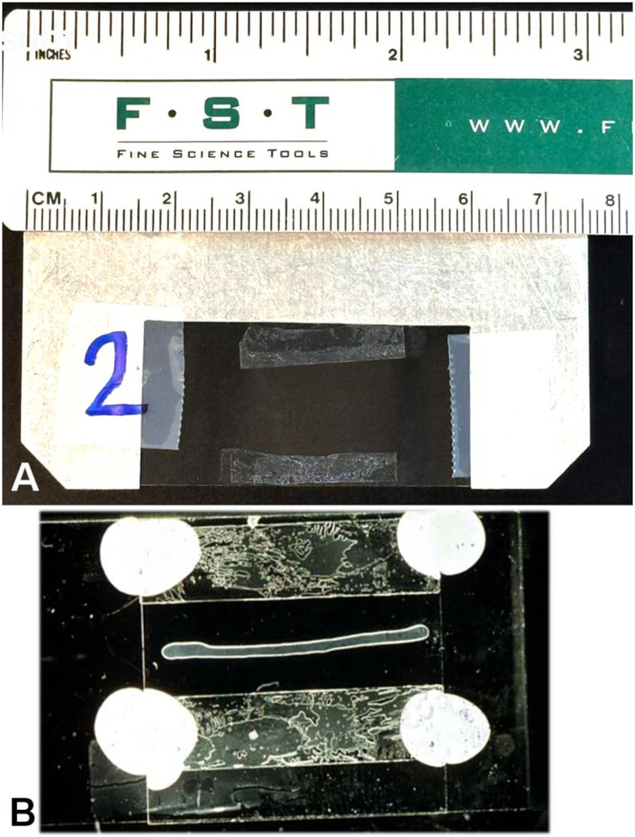
Figure 1: Perfusion Chambers. (A) Assembled the perfusion chamber prior to extrusion. The slide holders are custom-machined from aluminum to fit into standard slide holders with a cutout of 20 mm by 40 mm. The larger coverslip is held in place by tape, and the spacers are made from 22 × 22 No. 0 coverslips cut into narrow rectangles before coating with Molykote Compound 111. They are positioned to allow extrusion initially and adjust to 3 mm–4 mm apart to form a chamber with the 22mm × 22mm coverslip placed on top of the extruded axoplasm. The top coverslip is secured at the corners with VALAP, leaving the ends open for perfusion. (B) Assembled chamber with extruded axoplasm and top coverslip. Please click here to view a larger version of this figure.
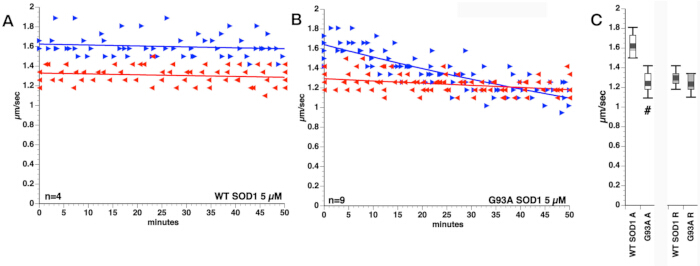
Figure 2: Effects of perfusing wild-type and mutant superoxide dismutase into axoplasm. (A) Perfusion of wild-type human superoxide dismutase type 1 (WT SOD1) had no effect on fast axonal transport in either direction. (B) In contrast, perfusion with a mutant form of SOD1 that is associated with familial ALS (G93A SOD1) has a dramatic effect on anterograde transport but not on retrograde transport. (C) The difference in anterograde transport for G93A SOD1 relative to WT SOD1 in the interval of 30–50 min was significant at p ≤ 0.01 using a t-test. Please click here to view a larger version of this figure.
| Chemical | MW | M (in stock) | grams per 25 ml stock | mM in X | ml stock in 25 ml Buffer X |
| Potassium aspartate | 171.2 | 1 | 4.28 | 350 | 8.75 |
| Taurine | 125.1 | add by wt. | add by wt. | 130 | 0.407 g |
| Betaine | 135.2 | 1 | 3.38 | 70 | 1.75 |
| Glycine | 75.07 | 1 | 1.88 | 50 | 1.25 |
| MgCl2·6H2O | 203.31 | 1 | 5.08 | 12.92 | 0.323 |
| K2·EGTA (adjust stock to pH 7.2 with KOH before bringing to final volume) | 380.4 | 0.1 | 0.951 | 10 | 2.5 |
| HEPES (adjust stock to pH 7.2 with KOH before bringing to final volume) | 238.3 | 1 | 5.96 | 20 | 0.5 |
| CaCl2·2H2O | 147.02 | 1 | 3.68 | 3 | 0.075 |
| Glucose | 180.2 | 0.1 | 0.4505 | 1 | 0.25 |
| K2·ATP (adjust stock to pH 7.2 with KOH before bringing to final volume) | 583.4 | 0.2 | 2.92 (0.1167/ml) | 2 | 0.25 ml stock, 0.0292 g or dry powder |
Table 1: Preparation of Buffer X. This physiological buffer is modeled after the composition of axoplasm, simplified for use in axoplasm experiments19,21.
Diskussion
VEC-DIC microscopy is critical as it allows real-time imaging of the movements of cellular structures smaller than the diffraction limit of light microscopy (~200 nm depending on the wavelength of light used) without labeling in live cells. Objects as small as individual microtubules (25 nm) can be detected, and 50 nm–80 nm diameter vesicles can be visualized moving on a single microtubule. In the isolated axoplasm, the density of microtubules, neurofilaments, and vesicular structures is too great to resolve and track individual particles. Nonetheless, the trafficking of vesicles in both directions can be visualized and average speeds measured interactively in real time.
The isolated axoplasm perfusion model uses axonal transport as a reporter of specific disease-relevant cellular functions and can be used to define specific targets and mechanisms. The isolated axoplasm model in combination with VEC-DIC microscopy avoids many of the limitations of transfection and injection methods, as it provides unparalleled control of experimental conditions, including concentration of mutant proteins, buffer conditions, and cell physiology. Unlike transfection or injection paradigms, the axoplasm perfusion model introduces a protein at concentrations comparable to physiological levels uniformly throughout the axoplasm. Transfected proteins cannot be expressed at a defined concentration and maintained at a specific concentration, particularly in axonal domains that can be quite distant from sites of synthesis. Newly synthesized proteins must be assembled into complexes that are transported to the axon. Injections are present as a bolus that will be at a high local concentration and then must redistribute either by diffusion or transport, also making it difficult to estimate effective concentrations and specific actions. Studies using lysates are further limited by disruption of normal compartmentation and dilution by orders of magnitude in non-physiological buffers. Transfection, injection, and in vitro lysates are indirect as they do not provide a simple biological readout or identify a unique axonal target, such as the pathogenic cytoplasmic proteins in ALS.
The reproducibility of these transport assays allows us to see the acute effects of pharmacological inhibitors, enzymes, and pathological proteins. As a result, axoplasm has proven to be a powerful tool in dissecting pathogenic mechanisms of mutant and modified proteins. For example, more than 40 different proteins have been shown to cause familial forms of amyotrophic lateral sclerosis (ALS). These proteins (including SOD127, Fus29, profilin, and TDP43) are not obviously related in function or sequence. Further, many of the proteins produced by these mutant genes are widely expressed in both neuronal and non-neuronal tissues, yet the mutant forms of these proteins selectively affect upper and lower motor neurons.
Using the axoplasm model, it was demonstrated that pathological forms of multiple different familial ALS proteins preferentially inhibit kinesin-based fast axonal transport. This was first documented using multiple different mutants of SOD114,27, showing that they all activated P38 MAP Kinase, which phosphorylated kinesin and inhibited transport. Subsequent experiments showed that this effect was not due to a direct action of SOD1 on P38 MAP kinase but instead was mediated through interactions with the MAP kinase kinase kinase ASK115 as part of a MAP kinase signaling pathway. The same pathway was activated by mutant Fus29 and has now been demonstrated with profilin, TDP43, and one of the dipeptides produced by mutant C9ORF7230. The importance of this pathway in familial and sporadic forms of ALS has subsequently been confirmed in mouse models, IPSC-derived models, and in human patient postmortem tissue14,15,29,30.
The axoplasm model has several key advantages. For example, the lack of a plasma membrane permits precise control over the concentration of perfused elements, either singly or in combination. The experimental conditions are near native with respect to the organization of axoplasmic structures and buffer conditions. Effects are measured interactively in real time. Pathological and physiological effects that are unique to the axon can be evaluated without the confound of cell body effects and protein synthesis. Similarly, axonal events are separated from synaptic actions30. Some of these advantages also impose limitations on experiments. The axoplasm model allows analysis of a single subcellular compartment, separating cell body and axonal events. As a result, one cannot tag vesicles containing specific cargoes. Effects of experimental manipulations are limited to the axon, and there may be different effects on the cell body or synapse-specific processes30. Similarly, the absence of a plasma membrane means that membrane-based activities cannot be evaluated, although downstream signaling pathways can be assessed. However, most proteins implicated in adult-onset neurodegeneration are cytoplasmic proteins that can be evaluated in the axoplasm. There are many experiments that can only be done in the isolated axoplasm model, but results obtained with the axoplasm facilitate the design of studies in intact neurons and organisms by generating information on mechanisms that can be tested in other models.
In recent years, some have argued that human diseases are unique and can only be studied in humans and human tissues/cells. However, over a hundred years of research on adult-onset neurodegenerative diseases like ALS has not yielded effective therapies or even a consistent pathogenic mechanism to explain the disease. The complexity of humans and the limited types of experimental approaches feasible with human samples have frustrated many outstanding scientists. Use of well-designed model systems, such as the one provided by the isolated axoplasm preparation, can answer specific questions related to disease pathology and provide insights that can then be validated in human tissue and potentially translated into therapies. The key is to ask the right questions and interpret the data using complementary model systems.
Offenlegungen
The authors have no conflicts of interest to declare.
Danksagungen
The authors would like to acknowledge Robert Day Allen and Raymond Lasek for their roles in developing the squid axoplasm model. Gerardo Morfini and Daryl Bosco played critical roles in refining the axoplasm model for assaying proteins relevant to familial and sporadic ALS. Hannah Brown and Robert Brown provided the first samples of SOD1 to test. Finally, numerous students assisted in measuring axonal transport using this approach. Funding for development of the axoplasm model was provided by grants from the NIH (NS023868 and NS082730) to STB and NIH AG072516, Pape Adams ALS Transformative Scholar Award, AARG grant from the Alzheimer’s Association, Healey Center ALS Scholar Award, and Mass General Hospital Mussallem Transformative Scholar Award in ALS Research to YS.
Materialien
| Name | Unternehmen | Katalognummer | Kommentare |
|---|---|---|---|
| 22 mm x 22mm No. 0 coverglass | Electron Microscopy Sciences | 63750-01 | |
| 24 mm X 50mm No. 0 coverglass | Thor Laboratories | N/A | |
| Argus | N/A | Video processor | |
| Argus 29 video processor | Hamamatsu | N/A | The capabilities may be repicated by conte,prray video processors and analyzeers combination of hardware and software |
| C2400 CCD camera | Hamamatsu | N/A | High-resolution CCD with adjustable dynamic range and the ability to acquire images at 30fps would be suitable. |
| COHAN-VANNAS SPRING SCISSORS | Fine Science Tools | 15000-01 | |
| Dark field dissecting microscope | Zeiss | 47 50 52 - 9901 | |
| DUMONT 5 MEDICAL FORCEPS straight | Fine Science Tools | 11254-20 | |
| DUMONT 7 MEDICAL FORCEPS | Fine Science Tools | 11274-20 | |
| DUMONT AA FORCEPS | Fine Science Tools | 11210-20 | |
| Light source | Schott | KL1600 LED | |
| Photonics Microscopy C2117 video manipulator | Hamamatsu | N/A | The capabilities may be replicated by a combination of hardware and software |
| Squid table dissecting microscope | Zeiss | STEMI DV4 | |
| STANDARD SURGICAL SCISSORS - SHARP/BLUNT | Fine Science Tools | 14001-14 | |
| SURGICAL STANDARD FORCEPS - 1X2 TEETH | Fine Science Tools | 11021-14 | |
| TRIDENT II SPRING SCISSORS Straight | Fine Science Tools | 15110-08 |
Referenzen
- Llinás RR. The Squid Giant Synapse: A Model for Chemical Transmission. Oxford University Press; New York; 1999. 224 p.
- DeGiorgis JA, Jang M, Bearer EL. The giant axon of the squid: a simple system for axonal transport studies. Methods Mol Biol. 2022;2431:3–22.
- Song Y, Kang M, Morfini G, Brady ST. Fast axonal transport in isolated axoplasm from the squid giant axon. Methods Cell Biol. 2016;131:331–48.
- Allen RD, et al. Fast axonal transport in squid giant axon. Science. 1982;218:1127–9.
- Gilbert SP, Allen RD, Sloboda RD. Translocation of vesicles from squid axoplasm on flagellar microtubules. Nature. 1985;315:245–7.
- Bearer EL, et al. Evidence for myosin motors on organelles in squid axoplasm. Proc Natl Acad Sci U S A. 1993;90:11252–6.
- Seamster PE, et al. Quantitative measurements and modeling of cargo-motor interactions during fast transport in the living axon. Phys Biol. 2012;9(5):055005.
- Kuznetsov SA, Langford GM, Weiss DG. Actin-dependent organelle movement in squid axoplasm. Nature. 1992;356:722–5.
- Weiss DG, Langford GM, Seitz-Tutter D, Maile W. Analysis of the gliding, fishtailing and circling motions of native microtubules. Acta Histochem Suppl. 1991;41:81–105.
- Schnapp BJ, Vale RD, Sheetz MP, Reese TS. Single microtubules from squid axoplasm support bidirectional movement of organelles. Cell. 1985;40:455–62.
- Vale RD, Hotani H. Formation of membrane networks in vitro by kinesin-driven microtubule movement. J Cell Biol. 1988;107:2233–41.
- Leopold PL, et al. The nervous system of Loligo pealei provides multiple models for analysis of organelle motility. In: Abbott NJ, Williamson R, Maddock L, editors. Cephalopod Neurobiology: Neuroscience Studies in Squid, Octopus and Cuttlefish. Oxford University Press; Oxford; 1995. p. 15–34.
- Brady ST. A novel brain ATPase with properties expected for the fast axonal transport motor. Nature. 1985;317:73–5.
- Morfini GA, et al. Inhibition of fast axonal transport by pathogenic SOD1 involves activation of p38 MAP kinase. PLoS One. 2013;8(6):e65235.
- Song Y, et al. Molecular chaperone Hsp110 rescues a vesicle transport defect produced by an ALS-associated mutant SOD1 protein in squid axoplasm. Proc Natl Acad Sci U S A. 2013;110(14):5428–33.
- Morris J, Lasek RJ. Monomer-polymer equilibria in the axon: direct measurement of tubulin and actin as polymer and monomer in axoplasm. J Cell Biol. 1984;98:2064–76.
- Allen RD. New observations on cell architecture and dynamics by video-enhanced contrast optical microscopy. Annu Rev Biophys Biophys Chem. 1985;14:265–90.
- Allen RD, Allen NS, Travis JL. Video-enhanced contrast, differential interference contrast (AVEC-DIC) microscopy: a new method capable of analyzing microtubule-related motility in the reticulopodial network of Allogromia laticollaris. Cell Motil. 1981;1(3):291–302.
- Brady ST, Lasek RJ, Allen RD. Fast axonal transport in extruded axoplasm from squid giant axon. Science. 1982;218:1129–31.
- Brady ST, Lasek RJ. Axonal transport: a cell biological method for studying proteins which associate with the cytoskeleton. Methods Cell Biol. 1982;25:365–98.
- Brady ST, Lasek RJ, Allen RD. Video microscopy of fast axonal transport in extruded axoplasm: a new model for study of molecular mechanisms. Cell Motil. 1985;5:81–101.
- Leopold PL, Snyder R, Bloom GS, Brady ST. Nucleotide specificity for the bidirectional transport of membrane-bounded organelles in isolated axoplasm. Cell Motil Cytoskeleton. 1990;15:210–9.
- Vale RD, Reese TS, Sheetz MP. Identification of a novel force-generating protein, kinesin, involved in microtubule-based motility. Cell. 1985;42:39–50.
- Lasek RJ, Brady ST. Adenylyl imidodiphosphate (AMPPNP), a nonhydrolyzable analogue of ATP, produces a stable intermediate in the motility cycle of fast axonal transport. Biol Bull. 1984;167:503.
- Lasek RJ, Brady ST. Attachment of transported vesicles to microtubules in axoplasm is facilitated by AMP-PNP. Nature. 1985;316:645–7.
- Morfini G, et al. Glycogen synthase kinase 3 phosphorylates kinesin light chains and negatively regulates kinesin-based motility. EMBO J. 2002;21(3):281–93.
- Bosco DA, et al. Wild-type and mutant SOD1 share an aberrant conformation and a common pathogenic pathway in ALS. Nat Neurosci. 2010;13(11):1396–403.
- Brady ST, Morfini GA. Regulation of motor proteins, axonal transport deficits and adult-onset neurodegenerative diseases. Neurobiol Dis. 2017;105:273–82.
- Sama RR, et al. ALS-linked FUS exerts a gain of toxic function involving aberrant p38 MAPK activation. Sci Rep. 2017;7(1):115.
- Okekenwa S, et al. Divergent molecular pathways for toxicity of selected mutant C9ORF72-derived dipeptide repeats. bioRxiv [Preprint]. 2023. PMID: 37808871. Available from: https://doi.org/10.1101/2023.09.28.558663.
Nachdrucke und Genehmigungen
Dieser Artikel wurde veröffentlicht
Video demnächst verfügbar